FMTVDM/BEST Imaging©℗, Breast Cancer, Breast Inflammation, Quantification, AI, Nuclear Camera Quantitative Calibration, Patent protected, Male Breast Cancer
Prior to FMTVDM/BEST Imaging, diagnostic testing was limited to looking for breast cancer either using "qualitative" imaging defining disease as either present or absent, or semiquantitative methods [1-10] which is also limited to the same yes you have breast cancer or no you don’t interpretation. These imaging methods include mammography, ultrasound, CT, MRI, as well as other SPECT/Planar and PET imaging approaches and as such are associated with sensitivity and specificity problems. These tests are further limited when imaging dense breast tissue or when breast implants exist either for augmentation, following mastectomy or when looking for breast cancer in men.
The ability of FMTVDM/BEST to differentiate transitional changes in tissue, associated with regional blood flow differences (RBFDs) and increasing tissue metabolism, makes it possible for FMTVDM/BEST to measure these tissue changes across an entire spectrum of changes; recognizing a "Health-Spectrum" shown by Figure 1.
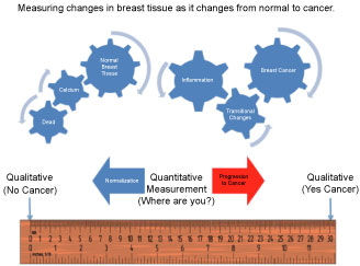 Figure 1: FMTVDM/BEST "Health-Spectrum" Measurements.
View Figure 1
Figure 1: FMTVDM/BEST "Health-Spectrum" Measurements.
View Figure 1
The interaction between cellular genetics and environmental factors consequently influence changes in tissue resulting in transitional changes including regional blood flow differences and metabolism. At one end of the spectrum is the absence of life, which may or may not be associated with a significant accumulation of cellular debris, inter alia, calcium, which may occur prior to the actual loss of cellular "life". In order of increasing regional blood flow and cellular metabolism are what is considered to be the "normal" state of a given tissue; resulting changes occurring due to an increased signaling and consequential accumulation of an inflammatory state; sequentially followed by further changes in tissue transitioning through increasingly metabolically active tissue with increased metabolic activity and demands for increased regional blood flow, until arriving at a tissue state where the expected controls and functionality of tissue no longer exist; what has classically been defined as "cancer". Implicit in this understanding is the appreciation that this continuum across transitional states reflects the interrelationship between these various "Health-Spectrum" states, which (1) Are not defined by some arbitrary absolute cutoffs between tissue states, (2) Demonstrate the ability of tissues to transition in either direction towards "normalization" or towards "cancer" and (3) Allow for the absolute measurement of the effect of treatment upon any of these tissue states across the "Health-Spectrum" providing the ability to direct care at the "patient-specific" level, improving outcomes across these transitional states; saving time, money and most importantly lives.
Because FMTVDM/BEST measures the RBFDs and metabolism of tissue and does not require tissue to compress for imaging and measurement, FMTVDM/BEST can be used to evaluate breast cancer in men as well as women. The following study represents a man referred for evaluation of a breast lump using FMTVDM/BEST.
A wife discovered a lump in the breast of her husband on Valentine’s Day. There was no family history and he was referred by his primary care physician for FMTVDM/BEST imaging, which was performed via protocol [11,12].
The results revealed a well-localized mass near the nipple of the left breast with a FMTVDM/BEST measurement of 310 consistent with breast cancer. The excision biopsy with clean margins confirmed the cancer Figure 2.
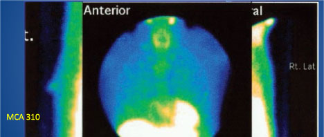 Figure 2: FMTVDM/BEST Imaging of a male patient.
View Figure 2
Figure 2: FMTVDM/BEST Imaging of a male patient.
View Figure 2
Research presented at the 2018 European Society for Medical Oncology (ESMO) meetings in October of 2018 revealed an increased incidence in male breast cancer of 1.9% per year between 2000 and 2015. The increased incidence was associated with a median age of 68.1 years, with 66.3% of the cases in the left breast.
Recommendations from the ESMO conference was for men to be treated with the same treatment modalities as women; however, diagnostic methods for finding the breast cancer are limited. FMTVDM/BEST imaging does not require breast tissue for compression as mammography does. Measurement of RBFDs and metabolism of male breast tissue is equally applicable in men as women and provides a diagnostic tool for the evaluation of men as well as women.