Barrett's esophagus requires surveillance to monitor for dysplasia and esophageal adenocarcinoma. New methods have been developed to detect dysplasia and esophageal adenocarcinoma. One of these methods, the wide area transepithelial sampling 3D brush biopsy (WATS 3D) is a computer-assisted brush-biopsy technique that uses an abrasive sampling instrument to detect dysplasia or esophageal adenocarcinoma compared to the standard four quadrant biopsy method. This case illustrates a patient with esophageal adenocarcinoma diagnosed by WATS 3D initially diagnosed as high-grade dysplasia and treated appropriately with HALO procedure during esophagogastroduodenoscopy. We aim to demonstrate the clinical utility of WATS 3D in Barrett's surveillance.
Barrett's, Wide area transepithelial sampling, Esophageal adenocarcinoma
Barrett's esophagus requires surveillance to monitor progression for esophageal adenocarcinoma. The development of esophageal adenocarcinoma occurs in about 0.5% of patients with Barrett's esophagus (BE) [1,2]. Unfortunately, dysplasia and even adenocarcinoma can be missed during the current method of surveillance for Barrett's [3]. The current method of surveillance involves taking four quadrant biopsies every 1-2 cm through the suspected area of Barrett's [4]. In contrast, wide area transepithelial sampling 3D brush biopsy (WATS 3D) is a computer-assisted brush-biopsy technique that uses an abrasive sampling instrument that obtains a sample of the entire thickness of the squamous or glandular epithelium being tested down to the lamina propria [4]. The sample is analyzed via a computer algorithm as well as a pathologist. The aim of our case is to demonstrate the clinical utility of this tool in detecting esophageal adenocarcinoma.
Patient is a 66-year-old with a past medical history of lifelong heartburn and regurgitation. He was on Esomeprazole daily and denied family history of colon or esophageal cancer. The patient had a screening endoscopy for surveillance of BE three months ago, which showed a 3-5 cm area of full circumferential salmon colored mucosa suspicious for BE. Biopsies revealed squamocolumnar mucosa with intestinal metaplasia and focal high-grade glandular dysplasia without malignancy. Repeat endoscopy confirmed a Prague C5M5 lesion (Figure 1). Inspection of white light endoscopy and NBI showed no nodules or mucosal irregularity that might have required endoscopic mucosal resection. Repeat biopsies read by two different pathologists confirmed the diagnosis of high grade dysplasia (Figure 2). Wide area transepithelial sampling 3D brush biopsy was also performed at that time. The WATS pathology report stated that patient has at least high-grade dysplasia in his cytopathology and was suggestive of esophageal adenocarcinoma (Figure 2 and Figure 3). Patient was successfully managed with two radio frequency ablation sessions with obliteration of the BE and replacement of the Barrett's mucosa with a neosquamous epithelium.
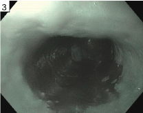 Figure 1: Narrow band image of a C5M5 area of Barrett's seen on endoscopy. View Figure 1
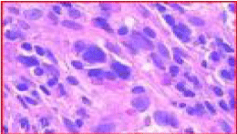 Figure 2: Esophagus 35-40 cm, few foci of highly dysplastic epithelium, seen singly and within apparent small vascular spaces, strongly suggestive of adenocarcinoma, in a background of Barrett's metaplasia. View Figure 2
 Figure 3: Cell block shows columnar epithelial fragments containing a few small clusters of cells with marked atypia characterized by atypical large cells, singly and in apparent small vascular channels, with large irregular nuclei, architectural distortion, and loss of polarity in a background of benign goblet cell metaplasia. View Figure 3
Esophageal adenocarcinoma has a high mortality rate and a poor prognosis with a relative 3-year survival rate of only 20% in the United States from 1995-1998 [5]. A significant risk factor in these patients in their initial presentation is the presence of GERD with over 25% of the Western population having reflux symptoms at least once a month [6]. As such, treatment of GERD and screening for Barrett's are critical in preventing progression. As illustrated in our case, the transepithelial brush biopsy addresses the sampling error inherent in random forceps biopsy testing of the esophagus. In just a few minutes, gastroenterologists can easily obtain a wide area, full-thickness transepithelial tissue sample for computer-assisted 3D laboratory analysis.
The case was discussed with experts in Barrett's and felt there was small foci of adenocarcinoma in the setting of high grade dysplasia. Without evidence of invasion, lesion was safe to be treated with RFA. This focus of esophageal adenocarcinoma is likely missed with the traditional sampling methods. If these patients are not properly identified during endoscopic screening, these patients may be lost to follow up and present at last stages. This is critical as early diagnosis and staging directly affects survival in esophageal adenocarcinoma.
Misdiagnosis can occur in Barrett's due to either inadequate sampling or poor determination of landmarks [2]. In our case, the WATS 3D brush biopsy allows the practitioner to obtain tissue through abrasive sampling instrument and also of the entire thickness of the squamous or glandular epithelium being tested down to the lamina propria [4]. In contrast, current method of surveillance involves taking four quadrant biopsies every 1-2 cm through the suspected area of Barrett's. In a multicenter study of 151 subjects with a prior history of Barrett's dysplasia, who underwent both forceps and brush biopsy, the brush biopsy added an additional 16 positive cases increasing the yield of dysplasia detection by 42% stressing the role of wide epithelial biopsy as a key tool in dysplasia detection [4]. Even further, another study assessed the inter-observer agreement among pathologists in the diagnosis of Barrett's-associated dysplasia using the WATS computer-assisted analysis technique which showed high kappa values indicating high agreement [7].
Overall, WATS 3D brush biopsy has an important role in the surveillance of Barrett's which has high mortality rate with progression to esophageal adenocarcinoma. This case illustrates that cancers arising in high grade dysplasia in Barrett's can be very small and easily missed on 4-quadrant forceps biopsies. These small lesions can be detected using WATS-3D as the brushing samples a much greater surface area. Prospective studies have assessed its ability to detect dysplasia at higher rates as well as high inter-observer variability between pathological specimens. Overall, institutions should consider adding WATS 3D as an adjunct to standard forceps biopsy for detecting dysplasia.