Accurate localisation of the cricoid cartilage is a key step in the successful application of cricoid pressure during rapid sequence induction. Poorly localised pressure is unlikely to confer any protective benefit to our patients and may have deleterious effects on laryngoscopy. We postulated that the use of ultrasound would greatly improve the accuracy of cricoid cartilage localisation prior to the application of cricoid pressure. 20 anaesthetic nurses attempted to locate the cricoid cartilage in 4 healthy, low BMI volunteers using a landmark technique and then repeated their attempt using ultrasound. Identification of the cricoid cartilage to within 5 mm increased from 44% to 90% with the use of ultrasound (p < 0.0001). None of our anaesthetic nurses had prior experience in using ultrasound before taking part in this study.
The application of cricoid pressure during rapid sequence induction to protect against aspiration of regurgitated gastric content was first described by Sellick in 1961 [1]. Compression of the hypopharynx between the cricoid cartilage ring and the cervical vertebral bodies and prevertebral musculature has since became an integral component of the induction of anaesthesia in patient groups thought to be at high risk of aspiration [2,3]. Conflicting evidence relating to the safety and effectiveness of the technique and its inherent potential to distort airway anatomy have been raised by a number of individuals with some abandoning the practice altogether despite possible legal ramificiations [4-7]. Perceived and realised failings of the technique may in part be due to the huge variability in how practitioners apply or misapply cricoid pressure [8,9]. Amidst this confusion what remains intuitively clear is that any potential benefit from the technique will only be gained through its correct application in terms of location as well as the amount of pressure applied. Application of pressure at alternative locations in the neck will not achieve the desired occlusion of the hypopharynx and thus will not prevent regurgitation of gastric content. Should such applied pressure also lead to distortion of airway anatomy to such an extent that intubation is made more difficult then no protection has been conferred to the patient and additional difficulty is delivered to the anaesthetist in a situation that by its nature may be challenging and stressful [10,11]. Driven by a desire to increase patient safety, the aim of this two part study was firstly to determine the accuracy with which operating department assistants and anaesthetic nurses are able to identify the cricoid cartilage using landmark techniques. We then asked our operating department assistants and anaesthetic nurses to identify the cricoid cartilage using ultrasound rather than a landmark technique to see if any increase in accuracy was achieved.
Written informed consent was obtained from all participants in this study. Local research ethics committee approval was sought but deemed unnecessary.
Four healthy volunteers from our anaesthetic department were enrolled in the study. The subject's characteristics were measured and recorded. The subjects were positioned on powered, adjustable patient trolleys with a head up tilt of 25 degrees. All subjects then underwent neck ultrasonography by a consultant anaesthetist using a SonoSite S-nerve™ (SonoSite Inc. Bothell, WA, USA) ultrasound machine using a 25-mm linear array high frequency probe (Figure 1 and Figure 2). The cricoid cartilage was identified and measurements were made of the cartilage width and height in all subjects. The cricoid cartilage was then marked with a horizontal line using an invisible ultraviolet marker pen (Compact Pro®, UK). The anterior neck was then covered using a clear occlusive Tegaderm™ dressing (3M™, St Paul, MN, USA). Operating department assistants and anaesthetic nurses received a written invitation to take part in our study. 20 gave written informed consent and were enrolled as candidates. Each candidate completed a prestudy questionnaire recording details of their level of experience, training and experience in the application of cricoid pressure. Candidates were then invited individually to examine the necks of each volunteer and mark where they thought the cricoid cartilage was using a non-permanent marker (Figure 3 and Figure 4). No time constraints were applied. The necks of all 4 volunteers were then examined sequentially using a hand held 4 watt ultraviolet light (Compact Pro®, UK) to reveal the true location of the cricoid cartilage. The distance from the true cricoid cartilage to the mark applied by the candidate was then measured and recorded. The mark drawn by the candidate was then removed from the neck of the volunteer using a Clinell® 70% alcohol/2% chlorhexidine wipe (GAMA healthcare, London, UK). Data were collected and analysed. Comparisons of accuracy between anaesthetic assistants of differing experience was carried out using nonparametric analysis (Kruskal-Wallis one-way analysis of variance) on Microsoft® Excel®.
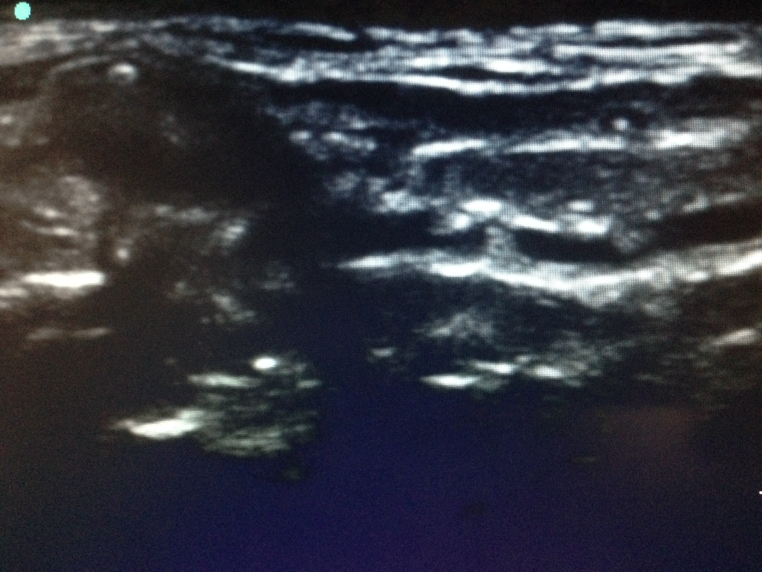 Figure 1: Ultrasound of the anterior neck at the level of the cricoid cartilage. View Figure 1
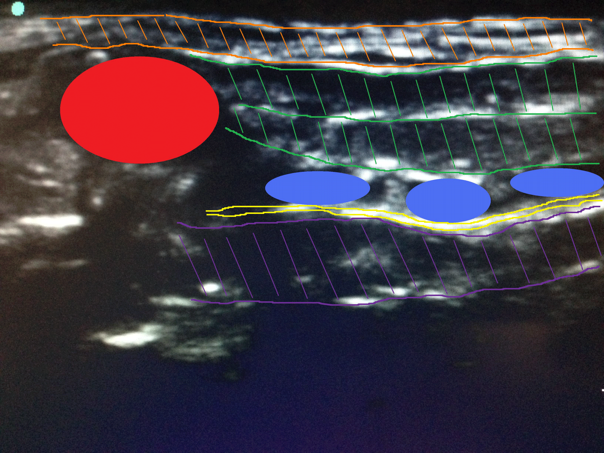 Figure 2: Annotated ultrasound image. Cricoid cartilage (red). Subcutaneous fat (orange). Strap muscles (green). Tracheal rings (light blue). Tissue/air interface (yellow). Tracheal lumen (purple). Thyroid cartilage (pink). Cricothyroid membrane (dark blue). View Figure 2
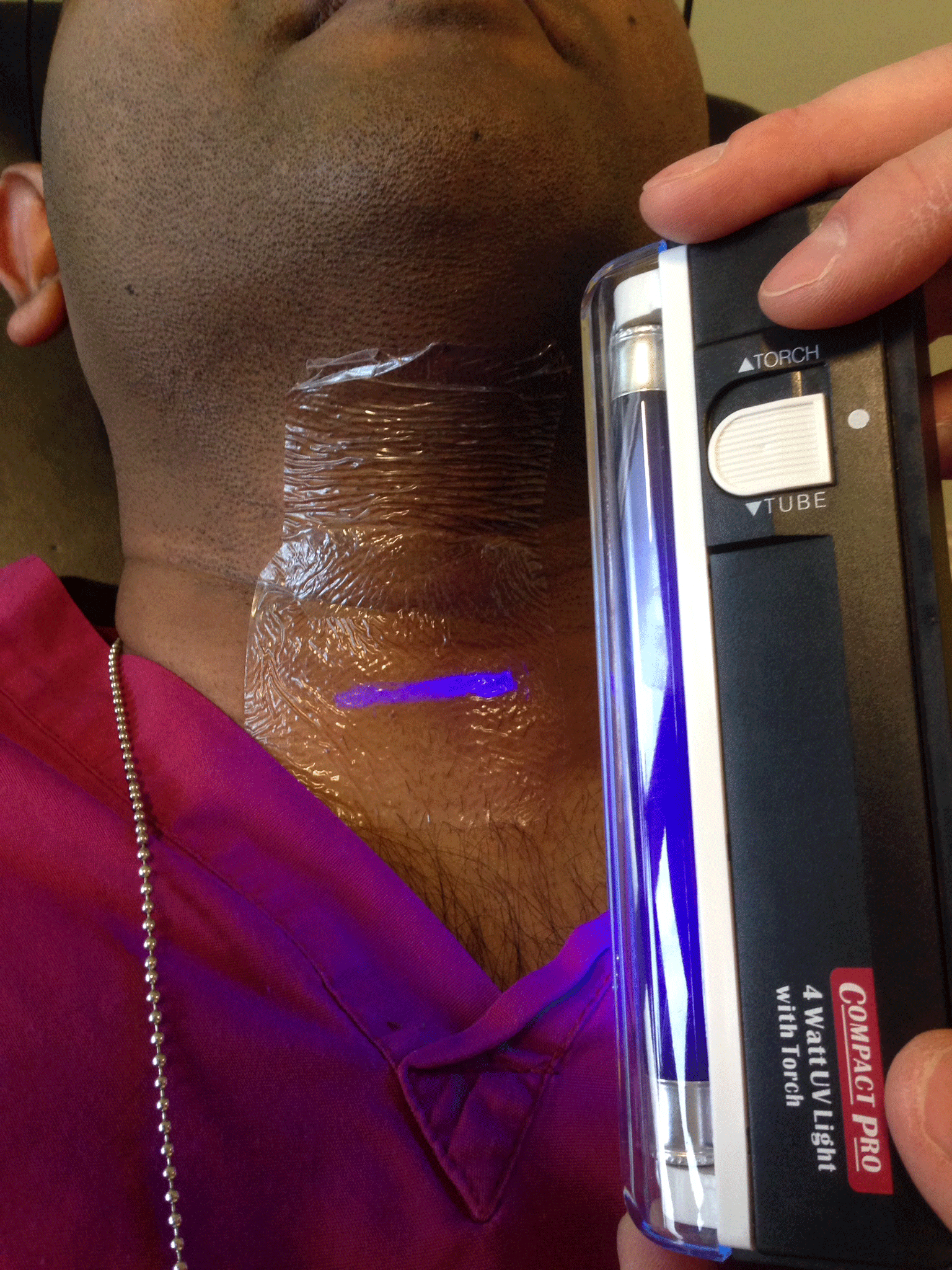 Figure 3: Location of cricoid cartilage marked with invisible UV marker revealed under UV light.
View Figure 3
Figure 3: Location of cricoid cartilage marked with invisible UV marker revealed under UV light.
View Figure 3
 Figure 4: Estimated location of cricoid cartilage by anaesthetic nurse using landmark technique compared to actual location as revealed under UV light. View Figure 4
An introductory guide to cricoid cartilage identification using ultrasound was emailed to our operating department assistants and anaesthetic nurses. A 20 minute practical tutorial on the use of ultrasound to identify the cricoid cartilage was given to our 20 candidates. Enlarged laminated ultrasound images of the cricoid cartilage and associated structures were distributed amongst the candidates to aid pattern recognition. None of the candidates had received prior training in the use of ultrasound. 4 healthy volunteers were once again recruited from our anaesthetic department. The subject's characteristics were measured and recorded. Subject positioning, neck ultrasonography and identification and marking of the cricoid cartilage in each subject was performed in an identical manner to that used in the first part of the study. Each candidate was then invited in turn to perform neck ultrasound on each volunteer and identify and mark the cricoid cartilage using a non-permanent marker pen. No time constraints were applied. Once again, the necks of all 4 volunteers were then examined sequentially using a hand held ultraviolet light to reveal the true location of the cricoid cartilage. The distance from the true cricoid cartilage to the mark applied by the candidate was then measured and recorded. The mark drawn by the candidate was then removed from the neck of the volunteer using a 70% alcohol/2% chlorhexidine wipe. Data were collected and analysed. Comparisons of accuracy between anaesthetic assistants of differing experience and between landmark and ultrasound techniques was carried out using nonparametric analysis (Kruskal-Wallis one-way analysis of variance and Mann-Whitney independent samples test) on Microsoft® Excel®.
Our 4 subjects were aged between 30 and 41 years of age. 2 males and 2 females were enrolled (Table 1). Body mass indices ranged from 21.2 to 26.6 kg.m-2 (mean 24.8 kg.m-2). Neck circumferences at cricoid level ranged from 30.5 to 36.5 cm (mean 33.9 cm). Our 4 subjects were examined by the following candidates: 13 anaesthetic nurses, 5 operating department practitioners and 2 student operating department practitioners. Experience ranged from 3 months to 20 years (mean 3 years 9 months, median 2 years). All candidates acknowledged receiving formal training in the application of cricoid pressure. Based upon our ultrasound measurements of the width of the cricoid cartilage in our subjects we concluded that application of pressure at a point within 5 mm of the midline of the cartilage would constitute application of cricoid pressure. Of the 80 neck examinations that were carried out in our study 21% resulted in accurate identification of the cricoid cartilage (Table 2). 44% of attempts were within 5 mm. 21% of attempts were more than 15 mm above or below the cricoid cartilage. There was no statistically significant difference in accuracy of localisation between anaesthetic assistants of less than 1 year experience (median (IQR [range]) 3.5 (-3-8.3 [-31-24]) mm vs. 1 to 2 years' experience 9 (-6.8-9.8 [-32-43]) mm vs. 2 to 5 years' experience 5.5 (-5.5-4.5 [-15-19]) mm vs. greater than 5 years' experience 5 (-17.5-0 [-21-4]) mm, p = 0.16.
Table 1: Summary of subject's characteristics - part 1 and 2. View Table 1
Our 4 subjects were aged between 27 and 39 years of age. 2 male and 2 females were enrolled (Table 1). Body mass indices ranged from 19.2 to 23.4 kg.m-2 (mean 21.9-24.8 kg.m-2). Neck circumference at the cricoid level ranged from 32.4 to 38.5 cm (mean 35.1 cm). Our 4 subjects were examined by the following candidates: 14 anaesthetic nurses, 5 operating department practitioners and 1 student operating department practitioner. Experience ranged from 3 months to 28 years (mean 5 years 2 months, median 3 years). No candidates had any prior experience of using ultrasound. Of the 80 neck examinations that were carried out using ultrasound 49% of attempts resulted in accurate identification of the cricoid cartilage. 90% of attempts were within 5 mm. No attempts were more than 15 mm above or below the cricoid cartilage (Table 2). There was no statistically significant difference in accuracy of localisation between anaesthetic assistants of less than 1 year experience (median (IQR [range])) 0 (-0.25-2 [-15-4]) mm vs. 1 to 2 years' experience 0.5 (0-2.5 [-10-10]) mm vs. 2 to 5 years' experience 0 (0-0.25 [-2-3]) mm vs. greater than 5 years' experience 0 (0-0.25 [-2-4]) mm, p = 0.48. Identification to within 5 mm of the cricoid cartilage increased from 44% using to a landmark technique to 90% with the use of ultrasound p < 0.0001 (Mann-Whitney independent sample test).
Table 2: Distance from cricoid cartilage with varying experience of the candidate using landmark and ultrasound guided techniques. View Table 2
The application of cricoid pressure has become a routine step in rapid sequence induction following its introduction in 1961 [1]. Historically, the anaesthetic community has awarded the technique a significant reduction in morbidity and mortality associated with the induction of anaesthesia in patient groups at high risk of regurgitation and subsequent aspiration [2,3]. Despite this assertion, a number of studies have questioned the validity of the technique [4,7] as evidence has emerged showing anatomical variations in hypopharyngeal and oesophageal anatomy that could render the manoeuvre ineffective in some patients [12-14]. Further concerns have been raised about the deleterious effect the technique may have on laryngoscopy in an emergency situation and its association with reductions in lower oesophageal tone [15-18]. What remains logical is that any benefit that may be conferred by the technique will only be realised if the technique is performed correctly. Pressure applied to the anterior neck in a posterior direction at any location other than directly over the cricoid cartilage will not compress the hypopharynx and upper oesophagus against the cervical vertebral bodies and prevertebral musculature. Previously published studies have consistently demonstrated that anaesthetists, anaesthetic nurses and operating department assistants apply cricoid pressure poorly both in terms of location and force [8,9,19]. It is entirely possible that poor performance of the manoeuvre in addition to variations in the anatomical position of the hypopharynx and upper oesophagus in relation to the cricoid cartilage are responsible for reported incidences of regurgitation during rapid sequence induction with applied cricoid pressure [7,9,20]. The first part of our study confirms that cricoid cartilage localisation in our institution by those individuals that routinely provide cricoid pressure during rapid sequence induction is performed poorly. Only 44% of candidates were able to localise the cricoid cartilage to within 5 mm using a landmark technique in the 4 subjects in our study, all of whom had body mass indices under 30 kg.m-2. This is comparable with previously published studies analysing the accuracy with which anaesthetists identify the cricothyroid membrane [21]. This inaccuracy was unaffected by the level of experience of the candidate. Our study suggests that in our institution more than 50% of attempts to provide cricoid pressure during rapid sequence induction may be ineffective due to inaccurate identification of the cricoid cartilage. This exposes patients not only to the risk of regurgitation and aspiration of gastric content but may also impair the anaesthetists view at laryngoscopy. The second part of our study shows that the use of ultrasound significantly improves the accuracy of localisation of the cricoid cartilage. Identification to within 5 mm of the cricoid cartilage increased from 44% to 90% with the use of ultrasound (p < 0.001). Furthermore, our results indicate that the ability to use ultrasound in this way can be rapidly acquired by the very members of the anaesthetic team that routinely apply cricoid pressure and that this can be achieved through a very short programme of training and education. Further increases in accuracy may be realised with additional training and experience in this technique.
The application of cricoid pressure during rapid sequence induction is unlikely to be an infallible method of protecting our patients against regurgitation and aspiration of gastric contents. The possible role for a compression device for the accurate and reproducible application of force during cricoid pressure has been proposed [22]. Whilst we applaud a means of ensuring that the appropriate force is applied, this device still has to be applied at the correct anatomical site. Our study would suggest that this is achieved in less than 50% of attempts when relying upon a landmark based approach. In our experience the identification of the cricoid cartilage with ultrasound is a rapid, non-invasive procedure that can be mastered with minimal training using equipment that is now widely available within most operating theatre complexes, accident and emergency departments and intensive care units. Therapid and accurate identification of this important anatomical landmark is likely to maximise the efficacy of cricoid pressure during rapid sequence induction whilst minimising any deleterious effects the technique may have on direct laryngoscopy. For these reasons we would advocate its use and intend to introduce ongoing training to our anaesthetic staff in an attempt to improve performance of a manoeuvre that when performed correctly may prevent morbidity and mortality amongst our patients but when performed badly may jeopardise laryngoscopy without conferring any protective benefit.
No external funding and no competing interests declared.