Background: A chronic, hemispheric, calcified subdural hematoma, referred to as an 'Armored Brain', is a rare complication of hydrocephalus shunting that can significantly alter intracranial dynamics.
Methods: We report the case of a 27-year-old male with a history of ventriculoperitoneal shunt placement at 8 months of age for congenital hydrocephalus. He presented to the emergency department two days after a minor cervical trauma, exhibiting signs of intracranial hypertension and a rapid decline in consciousness. On examination, his Glasgow Coma Scale score was 12/15. Cervical spine radiography revealed a disconnection of the shunt tubing at the cervical level.
Results: Emergency brain CT revealed a hemispheric chronic calcified subdural hematoma without ventricular dilatation. Given the acute shunt disconnection and the chronic nature of the hematoma, shunt revision was performed, and the hematoma was managed conservatively. The patient recovered fully and immediately postoperatively.
Conclusion: This case highlights the diagnostic challenge of shunt malfunction in the presence of a chronic calcified subdural hematoma. The “armored brain” can mask typical radiological signs of ventricular dilatation. Clinical context and careful interpretation of imaging are crucial in guiding appropriate management.
Subdural hematoma, Calcified, Armored brain, Shunt dysfunction, Ventricular dilatation
Calcified chronic subdural hematoma is a rare entity, accounting for approximately 0.3% to 2.7% of all chronic subdural hematomas [1-3]. When the calcified or ossified subdural mass extensively covers the cerebral cortex, it is referred to as an "armored brain" [1,4]. The first case of this condition was reported in 1884 [2]. However, its management remains controversial, with treatment decisions often guided by clinical presentation and radiological findings.
We report the case of a 27-year-old male with a history of ventriculoperitoneal (VP) shunt placement at the age of 8 months for congenital hydrocephalus. His postoperative course had been favorable, with normal psychomotor development. He was brought to the emergency department by his parents due to the onset of headaches and vomiting two days prior, followed by a deterioration in his level of consciousness.
On examination, the patient had a Glasgow Coma Scale (GCS) score of 13/15. His pupils were mid-sized and reactive, and no motor deficits were noted. The parents also reported a minor cervical trauma two days before symptom onset.
An urgent brain CT scan revealed bilateral calcified chronic subdural hematomas, consistent with the radiological appearance of an "armored brain." No other intracranial lesions were identified, and there was no evidence of ventricular dilation (Figure1).
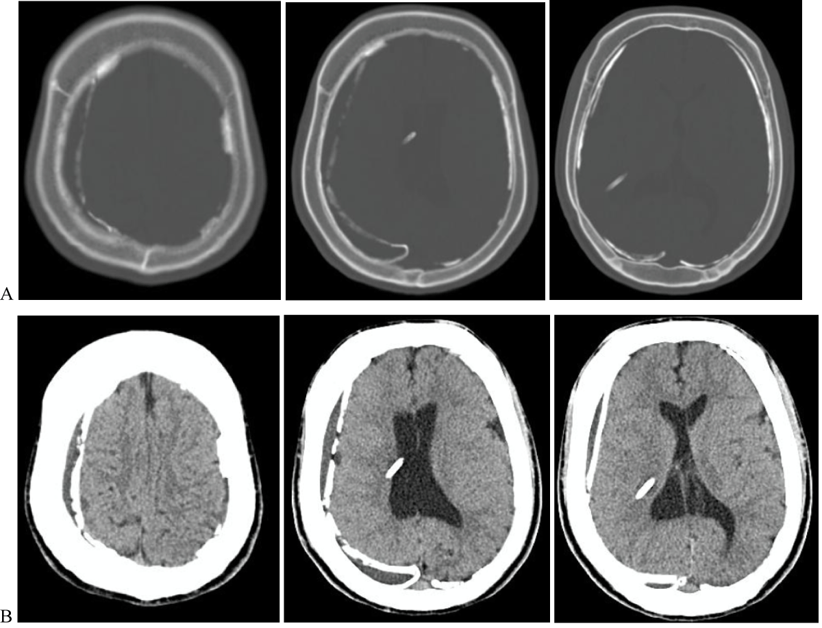 Figure 1: Non-contrast head computed tomography, A-Bone and B-Brain windows on axial computed tomograph imaging of the head demonstrating billateral calcified chronic subdural hematomas, and a non-dilated ventricular system.
View Figure 1
Figure 1: Non-contrast head computed tomography, A-Bone and B-Brain windows on axial computed tomograph imaging of the head demonstrating billateral calcified chronic subdural hematomas, and a non-dilated ventricular system.
View Figure 1
Further imaging, including a shunt series X-ray, demonstrated a disconnection of the VP shunt along the cervical tract (Figure 2), and most likely related to the recent cervical trauma.
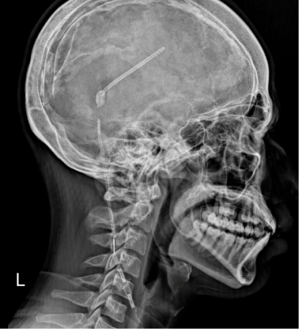 Figure 2: Lateral skull X-ray (right profile) showing a disconnection of the shunt system.
View Figure 2
Figure 2: Lateral skull X-ray (right profile) showing a disconnection of the shunt system.
View Figure 2
Given the acute onset of symptoms, shunt dysfunction was considered the most probable cause, despite the absence of radiological signs of acute hydrocephalus in this rare context of an armored brain.
The patient subsequently underwent surgical revision of the ventriculoperitoneal shunt. He was extubated on the operating table and showed immediate postoperative improvement, with full recovery of consciousness and complete resolution of symptoms. Given the favorable clinical outcome and the absence of signs of intracranial hypertension or neurological deficits, a conservative approach was chosen for the management of the bilateral calcified chronic subdural hematomas. Follow-up was marked by a good clinical evolution, with no recurrence of symptoms.
Calcified chronic subdural hematoma is a rare complication. While chronic subdural hematomas themselves are commonly encountered in neurosurgical practice, the development of calcification within these hematomas is uncommon and generally occurs in long-standing, unresolved cases.
This entity is most frequently reported as a late complication of traumatic brain injury [5-9], or following post-meningitic subdural effusions [4,10]. Although less commonly, this condition may also arise as a complication of long-term cerebrospinal fluid (CSF) shunting for hydrocephalus [4,11-15].
The exact pathophysiological mechanisms responsible for calcification remain incompletely understood, but several hypotheses have been proposed.
One leading theory suggests that chronic impairment of local circulation within the subdural space, due to inadequate arterial perfusion and/or venous drainage, leads to stasis of blood and proteins, which hinders resorption of the hematoma. This stagnation contributes to the chronic persistence of the collection and promotes fibrosis and membrane formation. Over time, this fibrotic tissue may undergo hyalinization and dystrophic calcification [16,17].
Intravascular thrombosis within the subdural collection may further impede clearance and favor calcium deposition. Additionally, microscopic calcium deposits may initially form within the neomembranes lining the hematoma cavity, serving as a nidus for progressive calcification and, in some cases, ossification.
Other contributing factors may include individual variations in metabolic and calcium homeostasis, which could predispose certain patients to abnormal calcium deposition [18].
Theoretically, the "armored brain", a condition defined by bilateral calcified chronic subdural hematomas encasing the cerebral hemispheres, can significantly alter intracranial dynamics, even though it is often regarded as a chronic, inactive process. One key consequence is a reduction in brain compliance, or decreased intracranial elasticity, due to the rigid calcified membranes limiting the brain's capacity to accommodate fluctuations in intracranial volume [19].
This rigid encasement may also contribute to a paradoxical or masked presentation of hydrocephalus, particularly in patients with cerebrospinal fluid (CSF) diversion systems. To the best of our knowledge, this phenomenon has not been previously documented in the literature. Here, we report what may be the first case of masked hydrocephalus in the setting of an armored brain, where shunt dysfunction was clinically evident despite the absence of typical radiological signs of ventricular dilatation, likely due to the restrictive effect of the calcified subdural encasement.
Chronic calcified subdural hematoma, or "armored brain," is a rare entity that may remain clinically silent for years. However, its rigid encasement of the brain can significantly impact intracranial dynamics. This case highlights an unusual presentation of shunt dysfunction without radiological signs of hydrocephalus, likely due to the restrictive nature of the calcified subdural layers. To the best of our knowledge, this is the first reported case suggesting a possible association between armored brain and masked hydrocephalus. Clinicians should maintain a high index of suspicion for shunt malfunction in symptomatic patients with a history of CSF diversion, even in the absence of classical imaging findings. Early recognition and appropriate management can lead to excellent outcomes.