Background: Low-grade posterior fossa ependymomas are neoplasms that rarely disseminate at the initial presentation or recurrence.
Methods: We describe a case of low-grade posterior fossa ependymoma initially treated with resection and radiation 20 years prior. At the time of presentation, multiple intracranial and spinal lesions were identified, most notably a non-enhancing ventral intradural lesion at C6-7.
Results: The 56-year-old presented with progressive left upper and lower extremity weakness. He underwent resection of the ventral intradural lesion at C6-7. The surgery involved a C5-7 corpectomy for resection and a C2-T3 posterior spinal fusion for stabilization. His weakness resolved postoperatively.
We then present a systematic review of the literature on the dissemination of low-grade posterior fossa ependymoma. Fifteen studies were identified, involving 23 patients.
Conclusion: Late dissemination of low-grade posterior fossa ependymoma could arise decades after the initial surgery despite gross-total resection and adjuvant radiotherapy. Malignant transformation and multicentric tumor development may also occur after treatment of the primary ependymoma. A varied extent of resection and adjuvant therapies have been involved in treating disseminated conventional posterior fossa ependymoma, with surgical decisions largely dependent on a multifactorial basis.
Ependymoma, Metastasis, Dissemination, Intradural-extramedullary, Posterior fossa
WHO: World Health Organization; RT: Radiation Therapy; MRI: Magnetic Resonance Imaging; CT: Computed Tomography; PEEK: Polyetheretherketone; PRISMA: Preferred Reporting Items for Systematic Review Meta-Analyses
Ependymomas account for approximately 1.9% of all primary brain and central nervous system tumors, originating from the ventricular lining, the central canal of the spinal cord, the filum terminale, or the choroid plexus [1,2]. Around 60-70% of intramedullary tumors in adults are ependymomas [3], while intradural extramedullary and extradural ependymomas are less common [4,5]. Historically, ependymomas were classified as subependymomas (grade I), myxopapillary ependymomas (grade II), conventional ependymomas (grade II), and anaplastic ependymomas (grade III) [6,7]. The 2021 World Health Organization (WHO) classification system further categorized ependymomas into 10 types based on tumor location and molecular features [8].
Ependymomas occasionally disseminate along the neuraxis, primarily due to cerebrospinal fluid carriage of exfoliated cells, while extraneural dissemination is less likely to occur [9]. High-grade or myxopapillary histology, younger age, subtotal resections, infratentorial location, high tumor proliferative indices, and genetic predispositions may increase the risk of dissemination [10-12]. Although gross-total resection is associated with improved survival rate and patient outcome, the extent of posterior fossa tumor resection could be limited due to the encasement of cranial nerves and brainstem vasculature [13].
A paucity of disseminated conventional posterior fossa ependymomas has been reported, which can complicate management due to their location and pathophysiological features. We present a case of drop metastases at multiple spinal levels from a prior resected posterior fossa ependymoma, followed by a literature review of disseminated conventional posterior fossa ependymomas.
The patient underwent resection of a low-grade posterior fossa ependymoma in 2002 when he was 36 years old. The resection was followed by radiation therapy (RT). The patient was believed to be in remission and had not had a recent follow-up. The patient was also diagnosed with ulcerative colitis versus Crohn's disease, for which he received a colectomy in 2010, followed by a reversal a year after. In addition, the patient had chronic anemia secondary to gastrointestinal bleeding.
At the age of 56, the patient presented with radicular pain radiating from the left buttock down the left lateral thigh, as well as numbness and tingling in the left posterior thigh, after a fall three months prior. The physical examination demonstrated full strength in bilateral upper and lower extremities. Reflexes were unremarkable. A magnetic resonance imaging (MRI) with and without gadolinium contrast of the entire neuraxis, including brain, cervical, thoracic, and lumbar spines, as well as a computed tomography (CT) with contrast of the chest, abdomen, and pelvis, was ordered to evaluate for metastatic disease. MRI of the brain demonstrated a questionable enhancement in the posterior fossa resection bed (Figure 1a). MRI of the cervical and lumbar spines demonstrated a non-enhancing 1.7 cm lesion anterior to the spinal cord at the C6-7 levels, a 1.6 cm enhancing lesion eccentric to the left in the thecal sac at the L4 level, and a 5.6 cm enhancing lesion in the thecal sac of the sacrum (Figure 1b and Figure 1c). These three intradural extramedullary lesions were suggestive of drop metastases from the patient’s prior ependymoma.
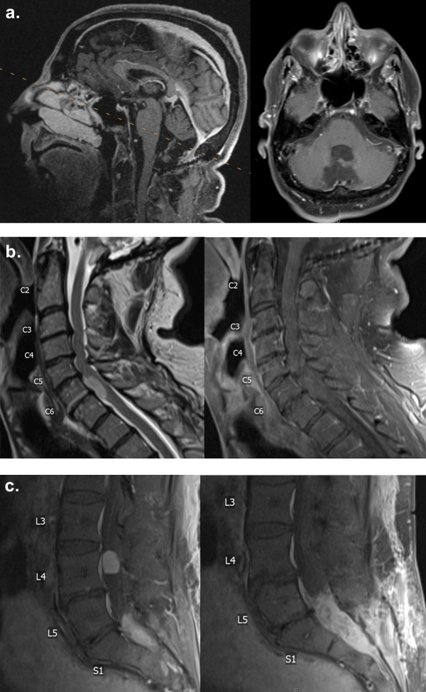 Figure 1: MRI with gadolinium contrast demonstrating. (a): The posterior fossa; (b): A non-enhancing lesion at the C6-7 level; (c): An enhancing lesion at the L4 level and an enhancing lesion in the sacral thecal sac.
View Figure 1
Figure 1: MRI with gadolinium contrast demonstrating. (a): The posterior fossa; (b): A non-enhancing lesion at the C6-7 level; (c): An enhancing lesion at the L4 level and an enhancing lesion in the sacral thecal sac.
View Figure 1
The ventral C6-7 spinal lesion was most concerning. However, the patient did not initially present with symptoms from the ventral C6-7 lesion, and the risks of such an operation were extremely high. The patient’s radiculopathy was most consistent with the L4 lesion. However, resection of the L4 spinal lesion could involve significant intraoperative cerebrospinal fluid loss, leading to inferior migration of the spinal cord with respect to the C6-7 lesion, causing quadriplegia. Therefore, a biopsy of the sacral lesion without loss of cerebrospinal fluid was considered the safest option at the time to determine the etiology of these spinal lesions. Since the patient did not have bowel or bladder problems, no resection of the sacral lesion was planned. An S2 laminectomy was performed, followed by opening of the dura at the midline to expose tumor-like material. A portion of the tumor was sent for frozen and permanent sections, and the dura was sewn and sealed without cerebrospinal fluid leak. Pathology was consistent with grade II ependymoma. Postoperative MRIs showed a non-enhancing lesion at C6-7 that appeared stable, while the L4 and sacral lesions were also grossly stable. In the 1.5-month postoperative follow-up, the patient developed no additional neurological deficits and started receiving RT to his lumbar spine.
Less than two years after the sacral lesion biopsy, when the patient was 58 years old, he reported progressive weakness in his left arm and leg without numbness or tingling, though he endorsed some loss of vibration sensation in his left leg. He denied radiating pain in his extremities, saddle anesthesia, or bowel or bladder problems but reported experiencing urinary incontinence 3-4 months ago. Slight muscle weakness was observed in his left upper extremity in addition to weakness in left hip flexion and knee extension. MRI of the cervical spine revealed an intradural anterior lesion at the C6-7 level, eccentric to the left, measuring 2.2 cm in superior-inferior dimension, representing a 5.4 mm increase from the original size (Figure 2a and Figure 2b). The lesion occupied more of the spinal canal and caused a more significant mass effect on the spinal cord compared to the MRI two years prior. An open biopsy of the C6-7 lesion was proposed, as this lesion did not enhance, in contrast to the L4 and sacral lesions. It was unclear at this time whether the lesion was consistent with disseminated ependymoma. A C6-7 laminectomy was performed, and a small portion of the accessible tumor on the right side was obtained for pathology. We did not believe it was possible or safe to retract the spinal cord to attempt resection of the tumor from a posterior approach. The soft mass was consistent with low-grade ependymoma. No intraoperative or immediate postoperative complications were observed.
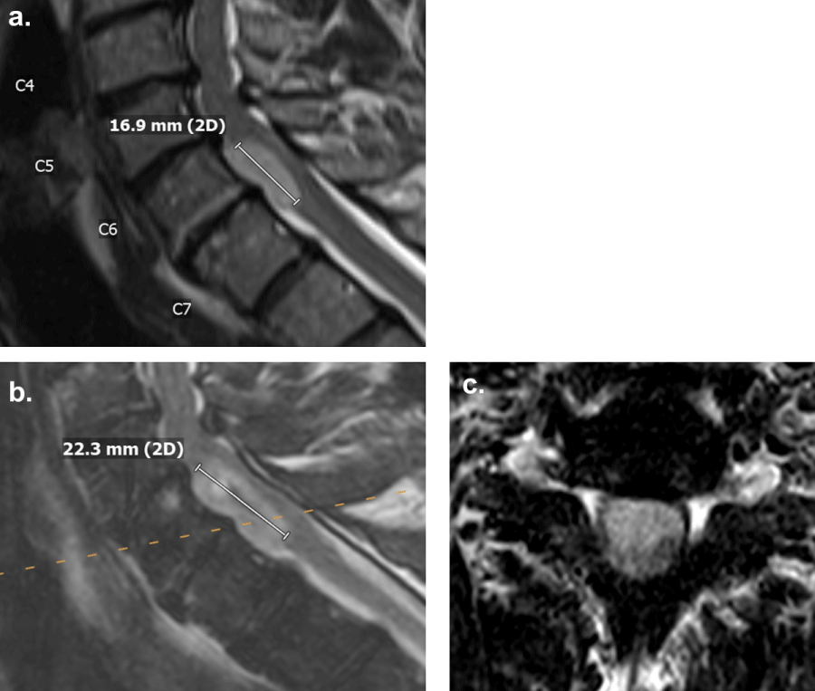 Figure 2: T2-weighted MRI demonstrating a hyperintense lesion without contrast enhancement at the C6-7 level anterior to the spinal cord, (a): At initial diagnosis measuring 16.9 mm in superior-inferior dimension; (b,c): Two years after initial diagnosis measuring 22.3 mm in superior-inferior dimension.
View Figure 2
Figure 2: T2-weighted MRI demonstrating a hyperintense lesion without contrast enhancement at the C6-7 level anterior to the spinal cord, (a): At initial diagnosis measuring 16.9 mm in superior-inferior dimension; (b,c): Two years after initial diagnosis measuring 22.3 mm in superior-inferior dimension.
View Figure 2
The patient returned to the clinic for a 1.5-month postoperative follow-up. MRI of the cervical spine taken after the biopsy continued to show a non-enhancing intradural anterior lesion at the C6-7 level, eccentric to the left. A C5-7 corpectomy for resection of the intradural extramedullary ependymoma would avoid displacement of the spinal cord, allowing for exposure of the tumor while visualizing the anterior vasculature of the spinal cord. It was also unclear whether the tumor was adherent to the anterior aspect of the spinal cord. Due to the cervical spinal cord covering nearly the entire tumor posteriorly, we did not believe a posterior approach was safe (Figure 2c). The operation was high-risk but was considered beneficial to the patient as the progressive weakness in his left upper and lower extremities would likely continue without surgical resection of the tumor. An extensive discussion was conducted with the patient and his wife, who elected to proceed with an anterior approach for cervical ependymoma resection, followed by a stage two, posterior spinal fusion, involving C2 to T3.
The anterior approach was assisted by an otolaryngology-head and neck surgeon. A co-surgeon from neurological surgery also assisted with this operation due to its complexity. The operative field included from the inferior aspect of C4 to the superior component of T1. The C5-7 vertebral bodies were removed. Intraoperative ultrasound visualized the spinal cord and tumor location. The dura was opened to expose the tumor, which was under the arachnoid with tumor-feeding vessels from the spinal cord (Figure 3). Microsurgical techniques created a plane between the tumor and the spinal cord, and gross-total resection was accomplished over six hours through debulking with microsurgical techniques and ultrasonic aspirators while rolling the tumor from the spinal cord. The corpectomy was completed with the placement of a polyetheretherketone (PEEK) cage in the C5-7 defect, then an anterior cervical plate was placed over the PEEK cage. PEEK was chosen to decrease metallic artifacts from future MRIs for tumor surveillance. The patient was kept intubated in preparation for stage two the subsequent day. On the following day, neuronavigation was used to perform a posterior instrumented fusion from C2 to T3.
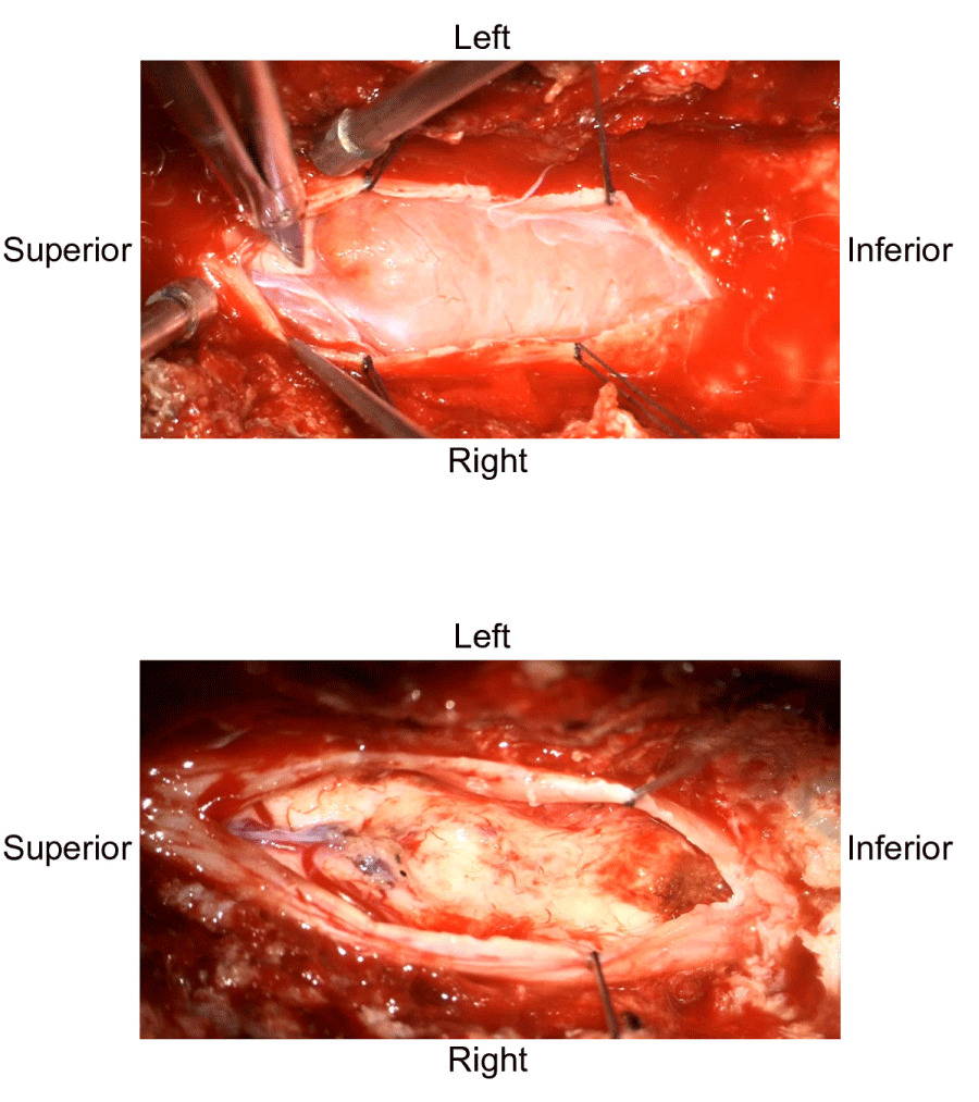 Figure 3: Intraoperative pictures showing the anterior tumor under the arachnoid with tumor-feeding vessels from the spinal cord.
View Figure 3
Figure 3: Intraoperative pictures showing the anterior tumor under the arachnoid with tumor-feeding vessels from the spinal cord.
View Figure 3
The patient experienced full resolution of the weakness in his left upper and lower extremities (Figure 4). He denied neck pain, difficulty swallowing, numbness, tingling, radiating pain, saddle anesthesia, or problems with bowel or bladder. MRI of the cervical spine obtained eleven months after the cervical ependymoma resection showed the prior C5-7 corpectomy procedure and no obvious evidence of tumor recurrence (Figure 5a). Final Pathology confirmed that the cervical lesion was consistent with a spinal metastasis from the patient’s posterior fossa ependymoma, histologic anaplasia not identified. X-rays of the cervical spine taken one year postoperatively demonstrated intact hardware without evidence of complication (Figure 5b). Other lesions, including a stable right anterior temporal lesion shown on the MRI of the brain, a dorsal intradural tumor at the T6-7 level on the MRI of the thoracic spine, and an intradural tumor at the L4 level in addition to the tumor in the sacrum on the MRI of the lumbar spine, are managed nonoperatively as the patient remains asymptomatic.
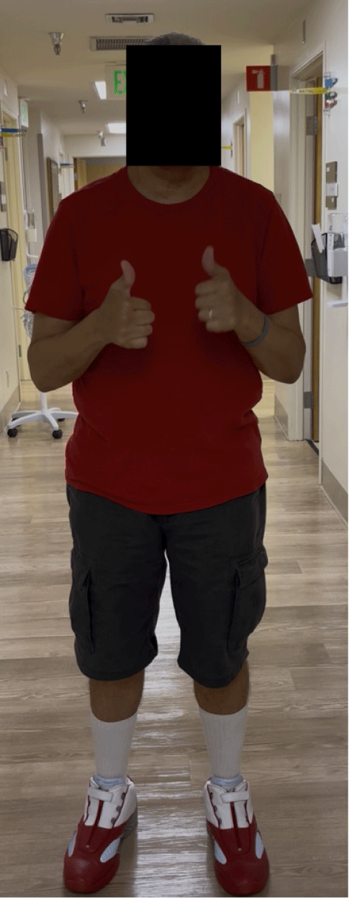 Figure 4: The patient at 1-year postoperative follow-up with resolution of symptoms.
View Figure 4
Figure 4: The patient at 1-year postoperative follow-up with resolution of symptoms.
View Figure 4
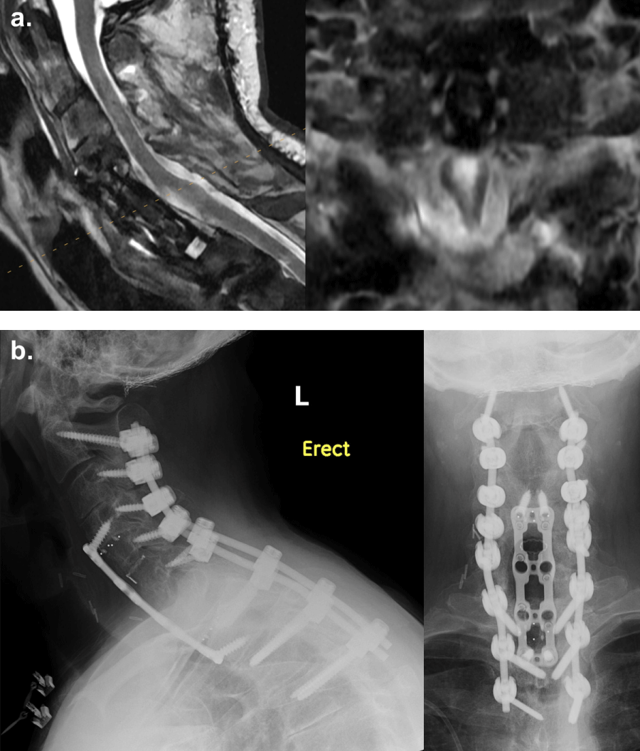 Figure 5: (a): Postoperative MRI demonstrating complete resection of the cervical ependymoma; (b): Radiographs showing intact hardware.
View Figure 5
Figure 5: (a): Postoperative MRI demonstrating complete resection of the cervical ependymoma; (b): Radiographs showing intact hardware.
View Figure 5
We present a case of disseminated conventional posterior fossa ependymoma treated with biopsies and eventual gross-total resection of a cervical ventral intradural ependymoma. A systematic review of posterior fossa grade II ependymoma dissemination was conducted on PubMed, Web of Science, and Scopus databases using the search terms: (“posterior fossa” OR “infratentorial”) AND “ependymoma” AND (“disseminated” OR “dissemination” OR “metastasize” OR metastasis”). 495 results were generated and screened following the Preferred Reporting Items for Systematic Review Meta-Analyses (PRISMA) (Figure 6). 15 studies, including one prospective clinical trial, six retrospective cohort studies, and eight case reports, totaling 23 distinct patients, were identified using the inclusion criteria of neuraxis dissemination, extraneural dissemination, or drop metastases of posterior fossa grade II ependymoma at first presentation and/or recurrence (Table 1) [14-23,11,24-27]. Articles describing other ependymoma subtypes or locations, involving no dissemination, containing no treatment information, or written in non-English languages were excluded. The unique aspects of our illustrative case are the technical considerations involved and the late dissemination. We employed a three-level anterior cervical corpectomy for gross-total resection of an anterior intradural extramedullary tumor, which led to complete symptom resolution and no recurrence. The discovery of drop metastases 20 years after the prior posterior fossa ependymoma resection may represent one of the longest latencies described for dissemination of conventional ependymoma.
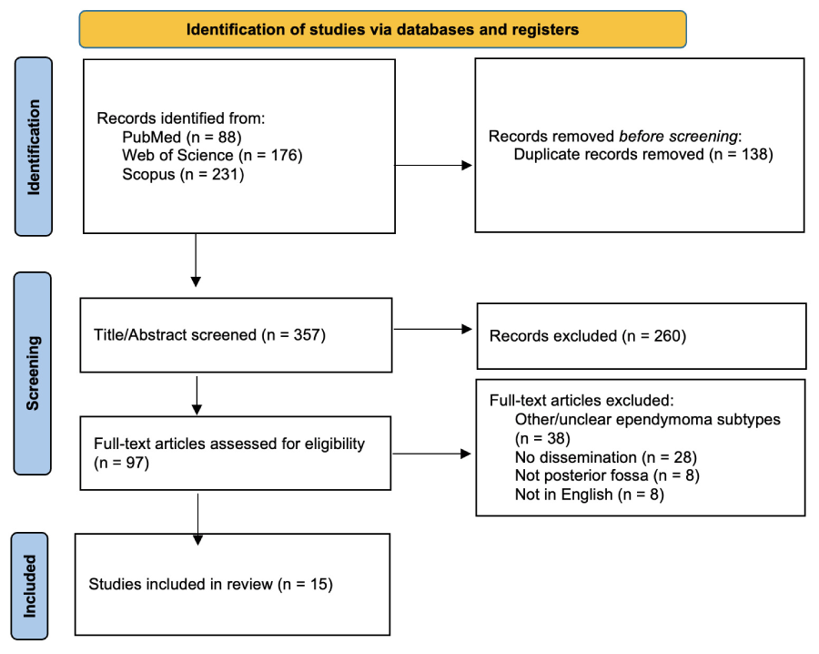 Figure 6: PRISMA flowchart showing the literature review process.
View Figure 6
Figure 6: PRISMA flowchart showing the literature review process.
View Figure 6
Four cases initially presented with disseminated infratentorial conventional ependymoma [16,20,23,24]. Disseminated disease could result from under-screening. The initial diagnosis of all disseminated cases was prompted by symptom presentation [16,20,23,24]. Diagnostic measures, including CT or MRI imaging, were typically not part of the standard of care for asymptomatic patients unless they had a medical history of ependymomas and were within the period of recurrence/dissemination monitoring [22,23,28]. Moreover, the lack of imaging other than the primary tumor site at first diagnosis could misdiagnose a disseminated disease as a focal lesion [11]. Given the possibility of dissemination, the literature has recommended systematic imaging of the entire neuraxis when ependymoma is in the differential diagnosis [29,30].
Disease recurrence following treatment of the initial disseminated ependymoma was reported in three cases. In Antony, et al. a total of four infratentorial relapses occurred involving the cerebellar peduncles and the medulla, each of which was treated with surgical resection, except the last recurrence that preceded the patient’s death [24]. The other two patients developed disseminated recurrent disease. Robertson, et al. reported dissemination to the right temporal lobe after subtotal resection of the primary posterior fossa tumor and adjuvant therapies [16]. Dissemination leading to multiple discrete spinal lesions was reported by Vural, et al. despite gross-total resection of all the initial disseminated ependymomas [23]. The disseminated recurrent disease was asymptomatic; therefore, the patient did not engage in any additional follow-up or treatment [23]. Surveillance imaging has identified 60-70% of recurrent ependymomas prior to symptom onset [24,31]. Given the risk of progression in asymptomatic recurrences, long-term routine surveillance imaging following initial treatment has been recommended regardless of symptom presentation to facilitate timely intervention and optimized outcomes [31-33].
In the 19 cases that presented an initial focal lesion, disseminated disease developed at the first recurrence in 17 cases and by the second recurrence in one case [14-17,19-23,11,24-27]. Exfoliated ependymoma cells were detected in the cerebrospinal fluid in one patient at the fourth recurrence, preceded by three local relapses [18]. The number of recurrences ranged from one to four, with a median of one, in contrast to the median of two and a maximum of nine reported in the retrospective cohort study of intracranial ependymomas by Antony, et al. [24] Among the 22 cases that involved recurrent disease, time to first recurrence was reported in 17 cases, with the median being 67 months compared to 21 months reported by Armstrong, et al., who reviewed 112 cases of conventional intracranial and spinal ependymomas [14,16-19,22,23,11,24-27,34,35]. Time to recurrence ranged from 1.3 months to 19 years, all of which had a shorter interval between initial surgery and first recurrence than the case we present [22,24]. Given the potential for dissemination, long-term monitoring of conventional ependymomas could be critical even in the absence of local recurrence [22,25]. The median time to first recurrence after gross-total resection of the primary tumor was 89 months, compared to 30 months after incomplete resections, suggesting a delayed recurrence with gross-total resection [14,16-19,22,23,11,24-27].
10 studies recorded the primary tumor site within the posterior fossa for 11 patients [14,17-20,22,23,11,25,26]. Among the locations of the initial focal lesion, eight cases involved the fourth ventricle [14,17-19,22,11,25,26]. One case involved the cerebellum, one involved the cerebellopontine angle, and one involved the cervicomedullary junction [14,20,23]. The majority of posterior fossa ependymomas are of the fourth ventricle, with the most likely point of origin being the floor [36,37].
Similar to our case, spinal cord recurrences were reported in 12 cases [14-17,19,22,23,11,24,26]. Spinal metastases have been reported in 10-25% of patients with primary intracranial ependymoma, with posterior fossa locations associated with higher metastatic risks [16,38].
Most recurrent ependymomas, whether local or disseminated, tend to retain the same histologic grade. However, Hong, et al. reported the development of grade III anaplastic ependymoma at disease recurrence [11].
Though quite rare, anaplasia has been observed in ependymoma recurrences [39]. In Armstrong, et al. two out of 79 patients with grade II spinal ependymomas developed grade III lesions at recurrence [35]. “Anaplastic” features, including increased cellularity, mitotic activity, microvascular proliferation, and necrosis, are typically found in grade III tumors, in contrast to the more organized architecture of conventional ependymomas [39]. Management could differ, with adjuvant RT largely recommended for treating anaplastic ependymoma, while its use in treating conventional ependymomas remains controversial and dependent on infratentorial versus supratentorial tumor location [40]. Histopathologic evaluation at recurrence could assist with an accurate diagnosis that impacts treatment planning and prognosis.
Moreover, Nicely, et al. reported an uncommon case of myxopapillary ependymoma arising more than 20 years after resection of the initial grade II posterior fossa ependymoma [41]. However, it was unlikely that the new myxopapillary ependymoma was a drop metastasis from the conventional posterior fossa ependymoma due to their distinct histological differences. Based on our interpretation, we considered the new myxopapillary ependymoma a separate tumor.
Resections are classified based on the extent of tumor removal: biopsy (< 10%), partial resection (10-50%), subtotal resection (51-90%), near-total resection (> 90%), and gross-total resection (~100%) [42]. The literature has largely recommended gross-total resection, with en bloc resection preferred over piecemeal due to the reduced risk of dissemination, when possible under maximal safety [13,34]. Among the 19 reviewed cases of initial focal ependymoma, 9 involved gross-total resection [15,18,19,21,22,11,25 -27]. Despite gross-total resection, dissemination may still occur, sometimes necessitating repeated resection. Similar to our case, Alshaya, et al. and Hong, et al. reported gross-total resection of the primary symptom-causing tumor in disseminated recurrent diseases while sparing the rest for adjuvant therapies and continued monitoring [11,25]. Gross-total resection of the entire disseminated recurrent disease has been achieved when only one or two distinct masses were identified [19,22,26].
Adjuvant therapies, including radiation therapy (RT) and chemotherapy, have demonstrated effectiveness in slowing disease progression and dissemination, particularly after incomplete resections [3,43,44]. RT has been reported following all types of resections and has shown improved local control compared to gross-total resection alone [14-16,18,19,21,22,11,25-27,45]. The National Comprehensive Cancer Network guidelines recommend craniospinal irradiation for upfront management of disseminated ependymoma [46]. In Nakasu, et al. RT prevented disease progression without surgery for the disseminated recurrent lesions [17]. However, local recurrence following adjuvant RT should prompt evaluation for repeat resection and/or re-irradiation [47]. Moreover, chemotherapy following completion of RT has been associated with prolonged progression-free survival, while chemotherapy without RT could also prevent relapse [20,48]. Successful remission was reported in a two-year-old boy after treatment of disseminated conventional posterior fossa ependymoma with partial resection followed by chemotherapy [20]. However, chemotherapy in adults should be considered only when surgery and RT have been exhausted, such as after multiple recurrences or increased malignancy [13].
The generalizability of this case report and literature review is limited by the rarity of disseminated posterior fossa grade II ependymoma in the literature. Although treatment strategies were discussed, we refrained from making recommendations as this was not our focus and would require an extensive review of randomized controlled trials and evidence-based guidelines. Surgical decisions should consider patient-specific risks, which may render gross-total resection less optimal than incomplete resections if maximal safety cannot be ensured. Moreover, management may vary by age, such as the use of adjuvant RT or neoadjuvant chemotherapy in pediatric ependymoma, where the evidence remains conflicting [49,50].
Furthermore, articles written before the 2021 WHO classification system update may have employed different classification schemes. The lack of molecular details in most articles made it challenging to match previous publications with the current taxonomy. Therefore, our literature review emphasized dissemination of posterior fossa grade II ependymomas rather than a specific type in the 2021 WHO classification.
We present a case of drop metastases from posterior fossa grade II ependymoma discovered two decades later that was successfully treated with three-level anterior cervical corpectomy for gross-total resection of the symptom-causing anterior cervical tumor. Although gross-total resection followed by adjuvant therapies has been utilized in focal and disseminated conventional posterior fossa ependymomas, thorough evaluations of risks and benefits remain essential to individualized treatment planning.