The Tension-free Vaginal Tape (TVT) is the most commonly performed procedure for the management of Stress Urinary Incontinence (SUI). There is insufficient data on the occurrence of complications and their management. The incidence of tape erosion and infection is observed to be higher with synthetic non-absorbable slings.
A 50-year-old parous lady with multiple co-morbidities underwent the TVT procedure and no urinary bladder perforation was noted on routine check cystoscopy intraoperatively. She complained of persistent vaginal discharge, urinary frequency and urgency with nocturia, vaginal pain and dysuria from one month postoperatively. Examination findings were normal and she was prescribed tolterodine for overactive bladder symptoms and advised diabetes control.
One year and seven months postoperatively, the patient was referred by the polyclinic to a urology clinic in view of incidental CT (Computed Tomography) scan findings of thickening with calcification in the left lateral aspect of her urinary bladder. A diagnostic cystoscopy revealed tape erosion into the bladder. A few days later, she had gross hematuria. She underwent an open partial cystectomy and the eroded mesh and the adjacent parts of the bladder wall were excised and the bladder was repaired.
She recovered well postoperatively and there was no recurrence of SUI.
Bladder erosion is a rare complication of TVT but should be suspected in a patient who presents with disproportional vaginal pain or hematuria. There should be a low threshold for diagnostic cystoscopy in women who have undergone previous TVT.
The incidence of Stress Urinary Incontinence (SUI) in women is up to twenty percent and it adversely affects their quality of life [1]. Ulmsten and Petros were the first to describe the Tension-free Vaginal Tape (TVT) procedure in 1996 and since then, it has slowly become the most commonly performed procedure for the management of SUI [2]. It is a minimally invasive procedure with a short operative time and fast patient recovery. Ever since its introduction, more than 1,200,000 TVT procedures have been performed worldover [3]. The success rates are 85-90 percent, which is comparable to Burch Colposuspension in the medium term [3]. However, it is preferred over Burch Colposuspension as it is minimally invasive, faster and associated with fewer complications.
Several studies have been conducted to assess the safety and success rates of this procedure, but there is insufficient data on the occurrence of complications and their management [4]. Patients with comorbidities like diabetes and vascular diseases are at a higher risk of complications [4]. The postoperative complications of this procedure include voiding dysfunction, retropubic hematoma formation, new onset urinary urgency and tape erosion into the vagina, urethra or bladder [4]. Erosion of the tape into the bladder occurs in 0.5-0.6% of cases [4]. The incidence of erosion, infection and fistula formation is observed to be higher with synthetic non absorbable slings [4]. The following case report outlines the course of a patient with TVT erosion into the bladder detected one year and seven months after the procedure.
A 50-year-old parous lady presented to our urogynecology clinic with a history of urine leakage on coughing and sneezing for 5 months. She also complained of urgency and urge urinary incontinence (UUI) for 3-4 months. The severity of SUI was greater than that of UUI. She had multiple co-morbidities which included hypertension, type 2 diabetes mellitus, bronchial asthma, gastro-oesophageal reflux disease and chronic depression.
On examination, she leaked 11 g of urine during the erect stress test and had demonstrable SUI. She also had a grade 1 cystocele, grade 2 rectocele and an enterocele. Urodynamics and urethral pressure profile (UPP) were performed during which she leaked a few drops of urine after the second last cough. The maximum cystometric capacity was 318 ml and the UPP peak was 23 cm of water.
She was counselled regarding the management options for SUI and vaginal wall prolapse - pelvic floor exercise or TVT with posterior vaginal mesh placement. The patient opted for surgical management.
She underwent the procedure and no urinary bladder perforation was noted on routine check cystoscopy intraoperatively with a 70 degree scope. There was a large enterocele noted on intraoperative assessment, hence a posterior vaginal mesh was inserted. There were no intraoperative complications and her postoperative stay in the hospital was uneventful. She was managed with sliding scale insulin adjustment in view of high blood glucose level. She was able to pass urine well with minimal residual urine on the third day after surgery and was discharged with an endocrinology referral for better control of blood glucose.
The patient was followed up regularly after the procedure. One month after the procedure, she complained of persistent vaginal discharge, increased frequency and urgency of urination with nocturia, vaginal pain and dysuria. Examination was unremarkable and a low vaginal swab was negative for trichomonas, gardnerella and candida infections. Urine culture was normal. Urinalysis showed 25 red blood cells and 330 white blood cells/µL while her HbA1C was found to be 9.4%. She was advised to optimize her blood glucose control and prescribed tolterodine tartarate for control of her urinary urgency.
Nine months after the procedure, she continued to complain of frequency of urination and nocturia. She was on insulin for diabetes control and her HbA1c then was 10.8%. She was advised regular follow up for blood glucose control and to continue tolterodine. She defaulted subsequent follow up visits.
One year and seven months after the surgery, the patient was referred by the polyclinic to a urology clinic at a different tertiary care hospital in view of CT (Computed Tomography) scan findings of thickening with calcification in the left lateral aspect of her urinary bladder (Figure 1 and Figure 2). She complained of frequency of urination with urgency and pain in the suprapubic region whenever she held her urine.
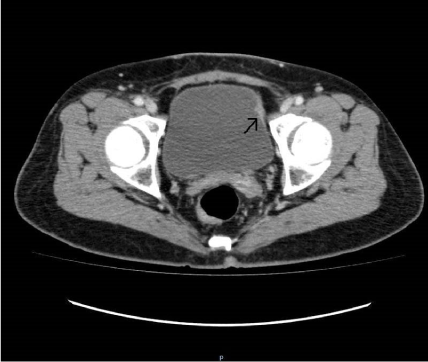 Figure 1: CT scan showing thickening of left lateral aspect of the urinary bladder. View Figure 1
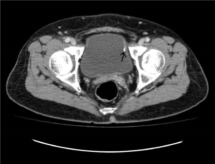 Figure 2: CT scan showing calcification of the left lateral aspect of the urinary bladder. View Figure 2
She underwent a rigid cystoscopy and bladder biopsy. Intraoperatively, the urethra was noted to be normal. A papillo-sessile lesion was noted on the left posterolateral wall of the urinary bladder, near the bladder dome, with adherent stones/debris. Underlying prolene threads were noted after the stones/debris were scraped off. Bilateral ureteric orifices appeared normal. The histology of the biopsy was reported as extensive acute ulceration with granulation tissue. A few days later, the patient complained of gross hematuria. She was admitted and a cystoscopy with evacuation of blood clots from the urinary bladder was performed under general anaesthesia. There was no active bleeding seen from the bladder mucosa and 100 ml of old blood clots were evacuated. An eroded synthetic mesh was noted at the upper part of the left lateral wall of her bladder with surrounding cystitis.
She was electively admitted for surgery the next day. A low abdominal laparotomy with open partial cystectomy was performed. Intra-operatively, the synthetic mesh was noted to have eroded into the left lateral bladder wall, extending for a distance of 3 cm in the bladder wall. The eroded mesh and the adjacent parts of the bladder were excised and the bladder was repaired in layers.
The histology was reported as synthetic fabric material merging into the bladder muscle, with fibrous scarring and chronic inflammation. She was discharged four days after the surgery. She recovered well from the surgery and a postoperative uroflowmetry demonstrated normal urinary voiding profile with minimal post void residual urine. There was no recurrence of SUI.
The use of minimally invasive procedures using synthetic material for the treatment of SUI can cause specific complications that are uncommon with native tissue repair [5]. Urethral and bladder erosion usually present with hematuria, frequency, urgency, urethral or vaginal pain or recurrent urinary tract infections [6,7]. It is possible in the above mentioned case that intraoperative bladder perforation had been missed due to incomplete bladder distention leading to the exposed portion of the tape being concealed by the bladder mucosal folds. Another possibility is pressure necrosis due to high abdominal pressure and unrecognized submucosal placement of the tape leading to gradual penetration [7,8]. Thus, the diagnosis may be delayed. Encrustation of the tape occurs due to contact with urine leading to exacerbation of or new symptoms [8].
Several factors contribute to the wide range of erosion/extrusion rates, including the operative technique, implant size, and the specific properties of the sling material such as small mesh pore size, local ischemia, stiffness involving poor tissue irrigation, elasticity, poor mesh incorporation, subclinical infection and basic tissue compatibility [9]. Complications of the tape, such as erosion and infection, which are due to local inflammatory reactions, reflect its biocompatibility. A sling's biologic characteristics, that is, how well it incorporates within native tissue, is the main factor predicting erosion. TVT has a high biocompatibility, with low inflammatory infiltrate and fibrosis, which are considered parameters of rejection [9].
The recommended treatment is removal of the eroded portion of the tape from the bladder but there is no consensus on the optimal technique of tape removal [10]. Removal of mesh by open suprapubic approach (cystectomy) or transurethrally have been described in literature [11,12]. More recently, endoscopic removal using laparoscopic scissors via a suprapubic trocar and cystoscope to grasp the tape has been used [13]. It avoids the need for an open cystotomy and the associated risk of wound complication, while allowing precise division of the tape flush with the bladder mucosa, but may miss tape erosion close to the bladder neck due to poor visibility. The use of Holmium-YAG laser through an operating cystoscope for cutting the encrusted and eroded TVT tape flush with the bladder has also been described. It melts the suture at the point of application with fragmentation of encrustation and concurrent hemostasis and can be used with a single minimally invasive device like a flexible cystoscope [8,14,15].
Pikaart, et al. described laparoscopic removal of TVT in 3 cases where the retropubic space was entered using an intraperitoneal approach and dissection was completed with a Harmonic scalpel blade and blunt dissection to identify the mesh sling. A cystotomy is required to remove the portion of the mesh that has eroded into the bladder which is later repaired laparoscopically in two layers [16]. O' Sullivan, et al. described a transurethral approach passing laparoscopic endo shears side by side the cystoscope to excise the exposed mesh below the level of the epithelium [17]. Heon Kim, et al. described transvesical laparoscopy with a pneumovesicum approach for mesh removal and bladder reconstruction [18].
The supports of the urethra tend to be maintained with partial tape removal in several cases without the recurrence of SUI [19]. Any urethral injury during the procedure should be repaired in layers with indwelling catheter for 7-10 days. In a 3-year follow up study by Frenkel, et al., 52% of the patients who underwent surgical resection had recurrence of incontinence. Placement of a repeat sling should be delayed for several months [20].
Bladder erosion is a rare complication of TVT but should be suspected in a patient who presents with disproportionate vaginal pain, hematuria, irritative symptoms or recurrent urinary tract infections. There should be a low threshold for diagnostic cystoscopy in women who have undergone previous TVT.
Nil.
None of the authors have any conflicts of interest.