50-year-old woman with end-stage renal failure on haemodialysis presented with chest pain. She was recently treated with methicillin-resistant Staphylococcus aureus bacteraemia secondary to tunnel line infection. Transthoracic echocardiogram demonstrated a linear structure in the descending thoracic aorta suspicion for an aortic dissection (Figure 1). Computed tomography (CT) thoracic aorta (Figure 2) showed a large descending thoracic aortic aneurysmat the T7/T8 level. Because of her comorbidities, a stent was deployed in view of high risk of perforation and she was placed on prolonged antibiotics.Post-stenting CT scan (Figure 3) a few weeks later showed a patent stent and the aneurysm appeared successfully excluded.
Mycotic aneurysms are rare but are associated with a high risk of rupture if not treated promptly. Transthoracic echocardiography provides only a limited view of the proximal ascending aorta and a small portion of the descending aorta and arch, therefore generally not considered an adequate diagnostic tool for assessment of the aorta. CT defines the location, vascular anatomy, and complications of mycotic aneurysms and allows serial monitoring. In addition, it is the imaging modality of choice for both preprocedural planning and post-procedural surveillance of mycotic aneurysm. This case illustrates the role of CT in guiding treatment and management of patients with mycotic aneurysm.
None.
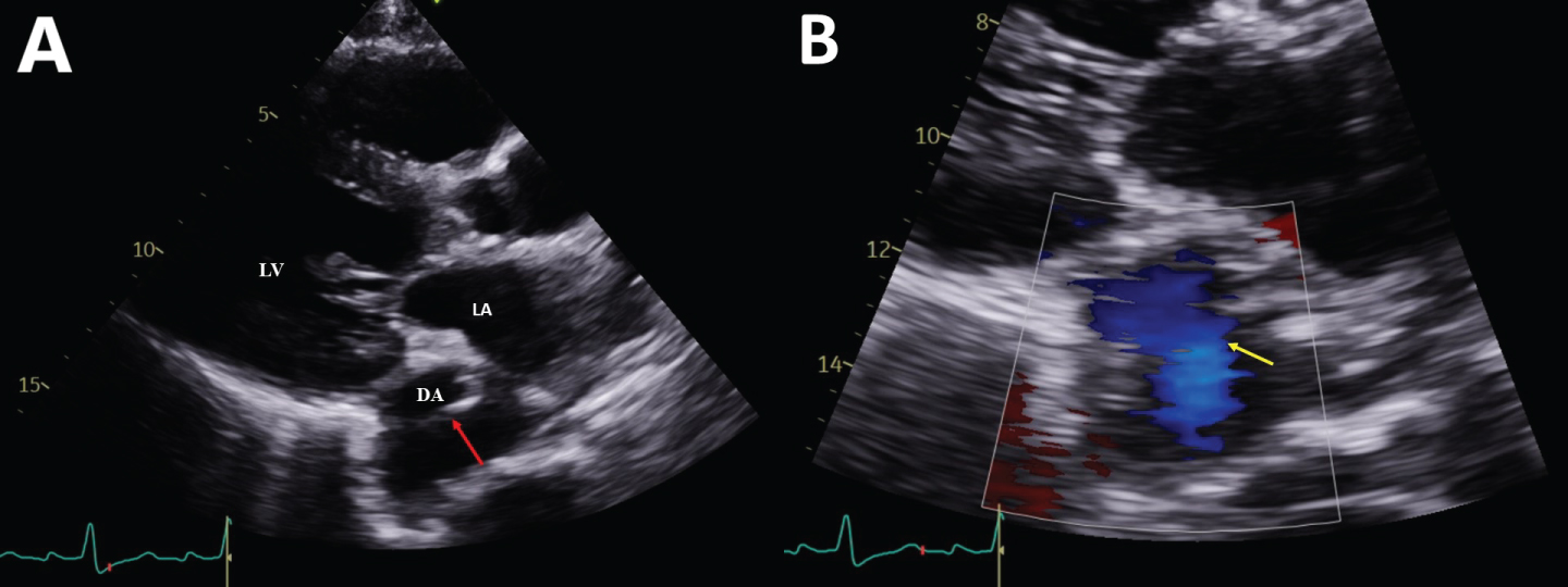
Figure 1: (A & B) Parasternal long-axis view on transthoracic echocardiogram showed a linear structure in the descending thoracic aorta (red arrow) and there was colour flow (yellow arrow) seen across the linear structure, raising the suspicion of a dissection flap.
LV: Left Ventricle; LA: Left Atrium; DA: Descending Thoracic Aorta
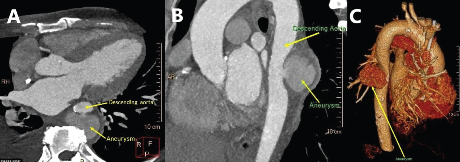
Figure 2: (A & B) CT thoracic aorta shows a descending thoracic aortic aneurysm (up to 5.6 cm maximum transverse diameter in the axial plane) at the T7/T8 level; (C) Volume rendered image of the CT thoracic aorta demonstrating a large aortic aneurysm (arrow).
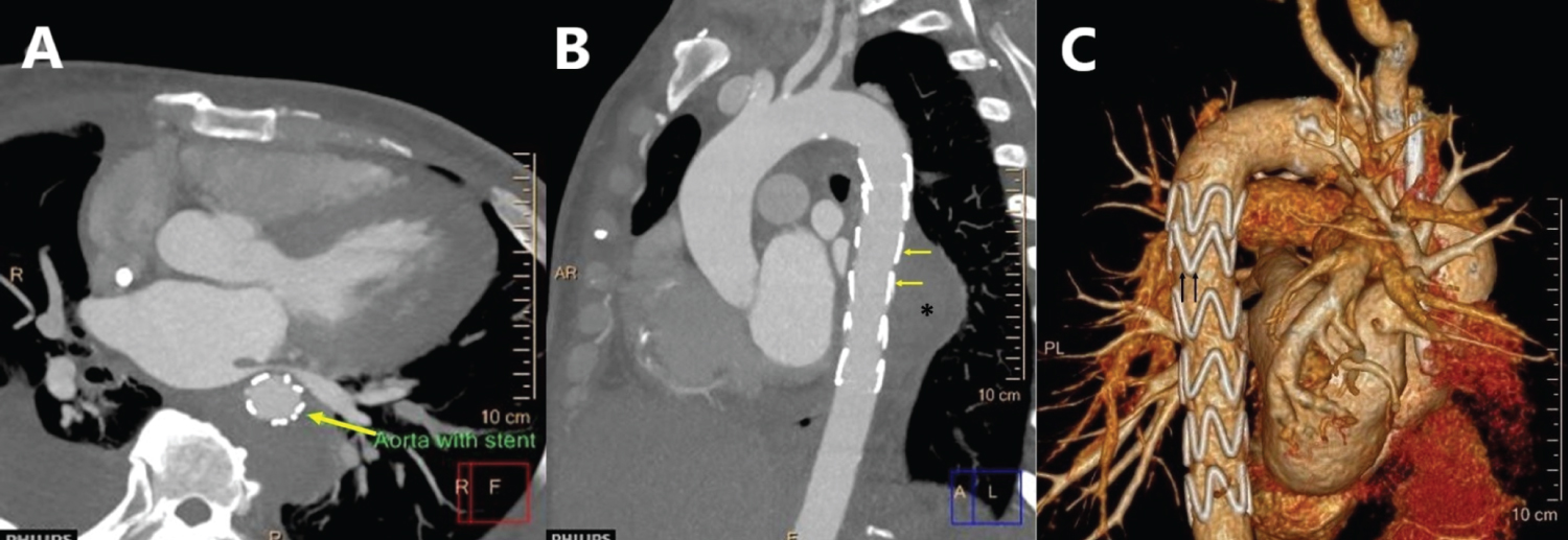
Figure 3: (A & B) Thoracic aortic stent graft demonstrated in situ (yellow arrow). The site of previous aneurysm (asterisk) now appears successfully excluded with no contrast enhancement demonstrated within; (C) Volume rendered image of the CT thoracic aorta demonstrating the endovascular thoracic aortic stent graft (black arrows).