A 8-year-old girl present for 2 months a mass of the oral cavity gradually increasing in volume causing discomfort to the mobilization of the tongue, snoring and sometimes deglutition disorders.
The clinical examination finds an exophytic mass at the expense of the tongue, measuring 3 by 2 cm in diameter. The mass is of muscular consistency, not painful, deeply indurated extending on the left edge of the tongue with infra-centimeter and sub-mandibular cervical lymphadenopathy. The examination also finds 6 coffee-and-milk spots.
A CT scan was immediately performed, showing a lingual process weakly heterogeneously after injection of contrast medium, measuring approximately 35*45 mm locally advanced. The mass was biopsied, which led to the diagnosis of embryonic rhabdomyosarcoma poorly differentiated with immunohistochemical study. Metastatic work-upshowed no metastasis.
Treatment was based on chemotherapy and surgery. She has been followed up for 1 year after termination of treatment with no evidence of disease. The patient is not experiencing any major speech problems.
Oral cavity, Rhabdomyosarcoma, Tongue, Child
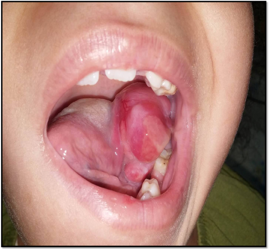
Figure 1: Intraoral aspect showing extensive mass of the tongue.