Pulmonary artery, Spontaneous Echo Contrast, Tricuspid Regurgitation, Atrial Septal Defect
Cases of dilatation of pulmonary artery in medical literature are not very uncommon. However presence of swirling smoke in dilated pulmonary arteries is very rare. Detection and management of such cases are very urgent to prevent fatal complications. Here I have reported one such rare case of dilated pulmonary artery with spontaneous echo contrast/smoke in a case of idiopathic pulmonary artery dilatation. 55-year-old female presented with shortness of breath on minimal exertion for last 4 months. She had no past history of any significant cardiovascular illness. She underwent echocardiography at the age of 40 years as a preoperative requirement before elective cholecystectomy. The examination revealed no abnormality. She was doing apparently well after that. She has been experiencing shortness of breath on climbing stairs and/or walking at more than normal pace of 100 metres for last 1 year, which has progressed at present level in last 4 months. She had no associated history of chest pain, palpitations, syncope, chronic cough, fever, pedal edema, orthopnea, or paroxysmal nocturnal dyspnea. CXR PA revealed dilated pulmonary trunk with dilated right and left pulmonary artery. ECG revealed several premature atrial ectopic. She was planned for TTE (transthoracic echocardiogram). TTE revealed Normal LA, LV, RA, dilated RVOT with normal LV ejection fraction. Main pulmonary artery was dilated 7.6 cm with dilatation of right and left pulmonary artery 4.1 cm and 3.94 cm respectively. There was mild PR and mild TR. There was swirling pattern of Spontaneous Echo Contrast (SEC) noted in the pulmonary arteries. Figure 1 showing the dilated MPA, LPA, RPA. Figure 2 showing dilated pulmonary trunk with normal pulmonary and aortic valve. Video 1 showing swirling pattern of smoke in the pulmonary trunk and its branches. Patient was immediately started on anticoagulation and planned for TEE (Transoesophageal Echocardiography). TEE did not reveal any septal defect. There was no clear cut shunt in any level. Bubble contrast study also failed to prove any communications. Patient was diagnosed as suffering from Idiopathic dilatations of pulmonary artery.
None.
None.

Figure 1: Showing dilated MPA, LPA, RPA with swirling smoke.
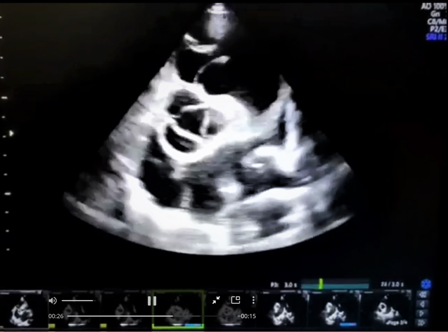
Figure 2: Showing dilated pulmonary trunk, normal aortic and pulmonary valve.
Video 1: Swirling pattern of smoke in the pulmonary trunk and its branches.