A 57-year-old Senegalese woman consulted to us for a pigmented mass inside the oral cavity evolving since 10 months. She had history of repetitive traditional tattoousing thorns on the gingiva. Intraoral examination showed a large pigmented tumour extending from the right maxillary gingiva to the left maxillary gingiva. It was 7 × 4 cm in size, firm in consistency with slight bleeding on touch. The patient also presented sub-mandibular lymphadenopathies and significant weight loss. An incisional biopsy with histopathological examination confirmed the clinical diagnosis of malignant melanoma. Biological examinations revealed anaemia at 6.9 g/dl and hypoalbuminemia at 23 g/l. HIV serology was negative. The patient dies after 2 weeks prior to any therapeutic perspective.
Oral malignant melanoma is a highly rare malignancy accounting for only 0.5% of all oral malignancies and < 1% of all other melanomas. The etiological factors associated with oral melanomas are still very poorly known. Some authors put forward the hypothesis of repeated microtrauma on pre-existing nevi. Thus, the responsibility of the traditional tattoo is evoked in our case. The prognosis for melanomas of the gingiva remains very poor (Figure 1).
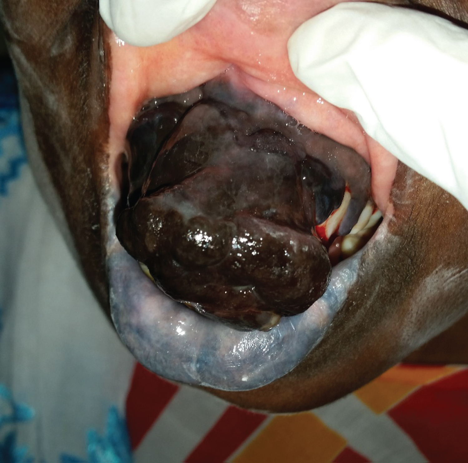
Figure 1: Gingival melanoma image.