Seborrhoeic keratoses are benign tumors that mainly occur in the head and in the trunk. Their size generally varies from a few millimeters up to a few centimeters. Giant lesions are very rare but pose a problem in terms both of treatment and transformation.
We report a case of giant seborrhoeic keratoses in an elderly man remarkable for their chronic evolution and their impressive size [1,2].
We report the case of a 90-year-old man, with no particular past medical history, who presented with asymptomatic axillary lesions evolving over the last twenty years. Clinical examination showed four dented hyperpigmented tumours and very limited cerebriforms measuring 12 cm along its longer axis (Figure 1 and Figure 2). Dermoscopic examination showed cerebriform convolutions appearance suggesting seborrheic keratosis. Histology confirmed the diagnosis, without signs of viral infection or malignant transformation. The patient underwent surgical resection resulting in permanent scar.
Seborrhoeic keratosesis a benign skin tumor, which mainly occurs in people aged over 50 years. It is common on the trunk and face [2,3]. Despite the frequency of seborrheic keratosis, little is known about the epidemiology of the disorder. It is believed to be more prevalent among Caucasians and to affect roughly equal numbers of men and women. It is also generally acknowledged that its prevalence increases with advancing age. One Australian study reported a 100% prevalence of seborrheic keratoses among people over age 50. On average, 69 seborrheic keratoses were found per patient in those over age 75. Seborrheic keratosis is also known by various synonyms: Verruca senilis, senile wart, verruca seborrhoica, seborrheic wart, basal cell acanthoma, benign acanthokeratoma, and basal cell papilloma. The present paper uses the internationally prevalent term "seborrheic keratosis" to describe this highly common, benign epithelial skin tumor [4,5]. Lesions are sharply demarcated, round or oval-shaped, usually elevated and stuck on the skin with a verrucous, uneven, dull, or punched-out surface (Figure 3). Flat seborrheic keratoses often have a smooth, velvety surface and are barely elevated above the surface of the skin. Whether these flat forms are merely initial seborrheic keratoses, which grow thicker over time, or whether they are a sub-type is still unclear [6]. Some seborrheic keratoses have a surface which appears rather oily and shiny, hence the misnomer "seborrheic" (i.e., greasy) keratosis. Seborrheic keratoses typically appear as if stuck on the skin. Lesions range in size from a few millimeters to several centimeters, with a typical diameter of 0.5-1 cm. Lesions vary in color and may be skincolor/yellowish, gray-brown, or black [7]. Pediculated forms have also been observed in intertriginous areas. Seborrheic keratoses are often penetrated by invaginations of keratotic plugs and horn cysts (filled with concentrically attached keratin). On dermatoscopy these appear as "pseudofollicular openings" and "horn pseudocysts" with light, sharply bordered, round plugs. Inflammation around the margin of the lesion can occasionally occur (Meyerson phenomenon). Seborrheic keratoses can appear anywhere on the body with the exception of the palms or soles. The mucus membranes are also generally spared. The most commonly affected sites are the chest, back, head (near the temples), and neck [8]. The main differential diagnoses are condyloma, pigmented basal cell carcinoma and melanoma. The etiology of Seborrhoeic keratosesis unknown, although there may be some familial predisposition, which was not demonstrated in our patient. They could also occur after inflammatory dermatosis or at a site of repeated rubbing [9]. In fact, Seborrhoeic keratoses which are located on sites of chronic friction, such as shaving areas, or the folds of obese patients, can reach large dimensions, up to ten centimeters in diameter. This is illustrated in our patient who presented Seborrhoeic keratoses initially of small dimensions gradually increasing in size to reach 12 cm [10].
Histologically, Seborrhoeic keratoses most commonly manifests as epidermal hyperplasia with acanthus is and papillomatosis [9,10].
The treatment of giant Seborrhoeic keratoses can combine several means at the same time, in particular cryotherapy and electrocoagulation-curettage; in some cases, it may require surgery [10].
Our observation clearly illustrates the role of repeated rubbing in the increase in the size of Seborrhoeic keratoses, hence the value of an anatomo-pathological examination of any Seborrhoeic keratoses of unusual site or atypical clinical manifestations.
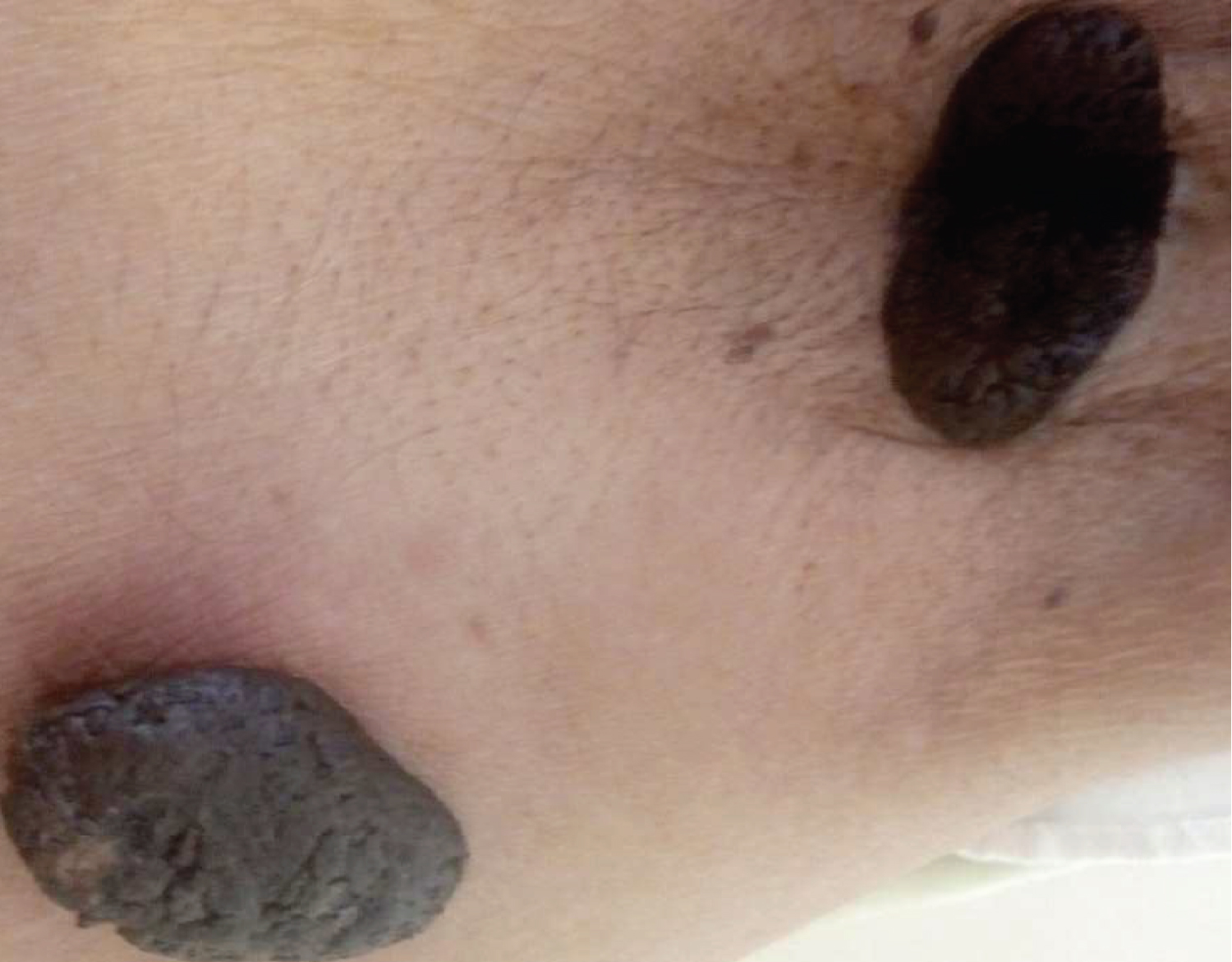
Figure 1: Dented hyperpigmented tumours and very limited cerebriforms.
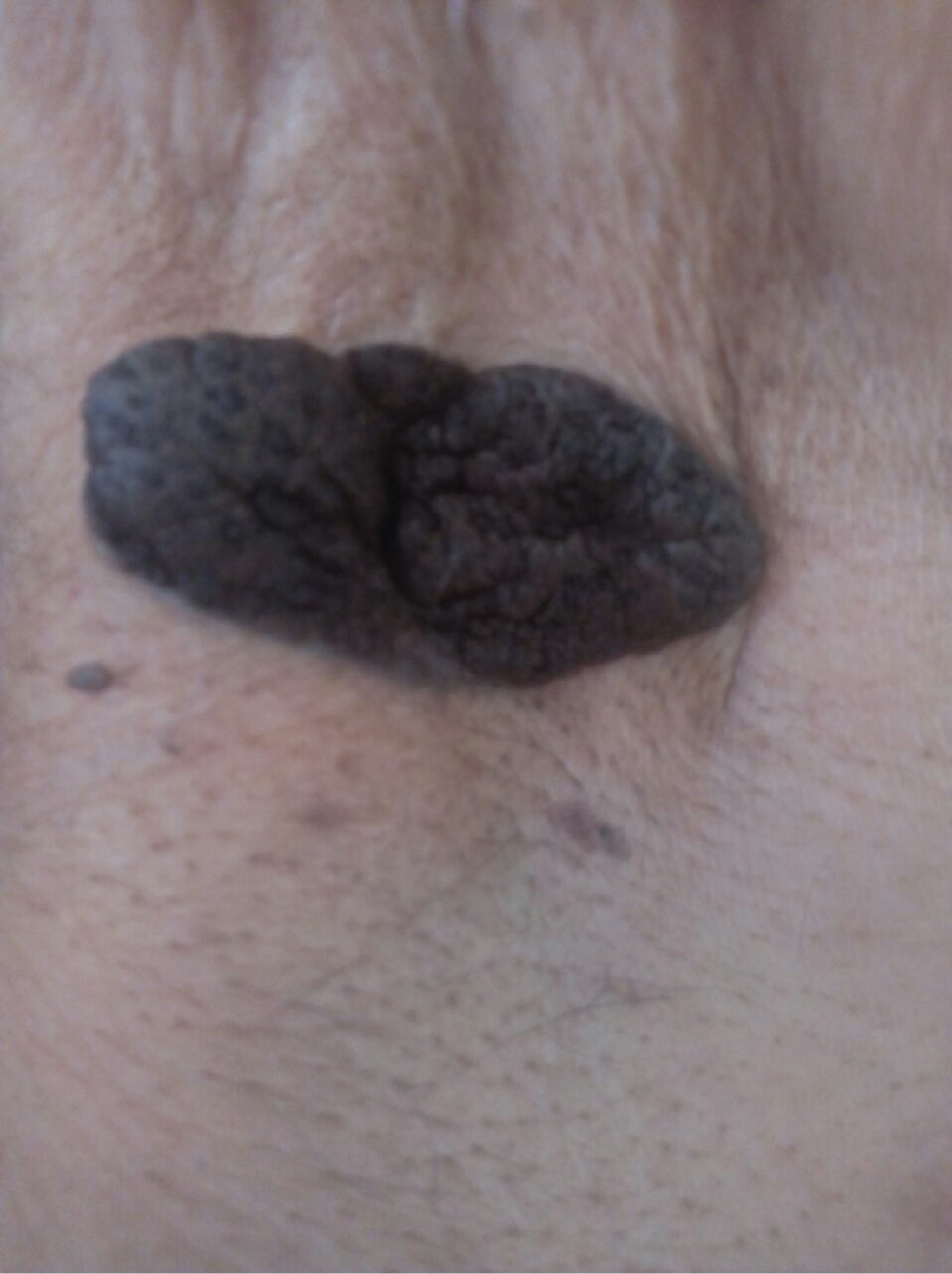
Figure 2: Dented hyperpigmented tumours and very limited cerebriforms.
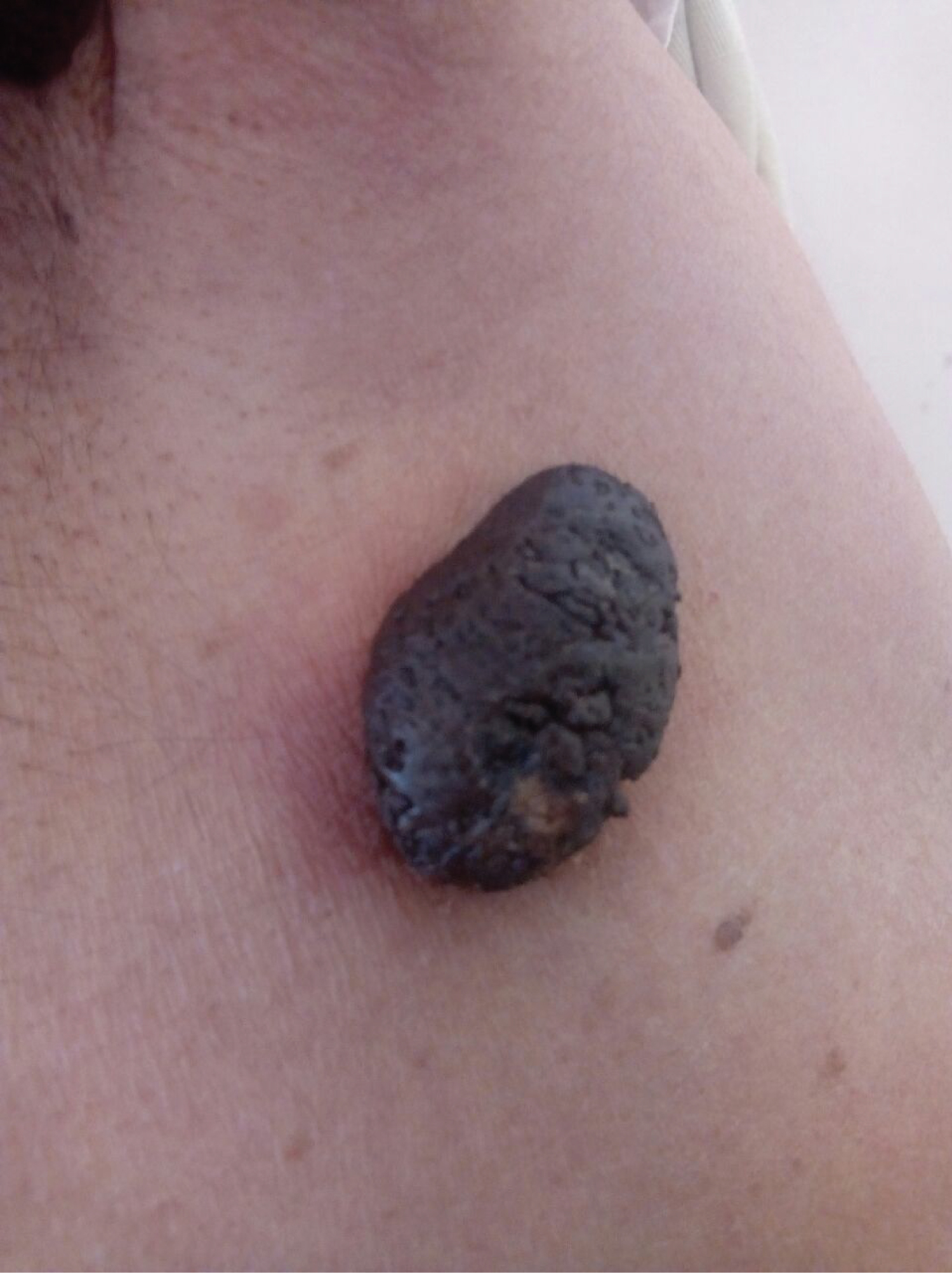
Figure 3: Dented hyperpigmented tumours and very limited cerebriforms.