A 51-year-old man with hypertension presented to the emergency department with sudden loss of consciousness (LOC). Two hours before presentation, his wife witnessed his abrupt collapse and called the emergency medical service. Vital signs were BP = 175/90, PR = 76, RR = 12, T = 36.9 ℃, GCS = 6/15 (E1,V1,M4), and his bedside glucometery was 138 mg/dL. While his facial and extremities movements were symmetric, there were abnormal pupil reflex, corneal reflex, gag reflex and bilateral plantar reflex. After intubation, he underwent non-contrast CT (NCCT) imaging of brain (Figure 1). Laboratory studies were insignificant.
To confirm basilar artery occlusion (BAO), he underwent brain and neck CT angiography (Figure 2 and Figure 3) and brain MRI (Figure 4). He admitted for endovascular basilar artery thrombectomy and had an uneventful follow up after 2 months.
BAO is a rare condition, more common in elderly population and usually caused by atherosclerosis or cardiac emboli [1]. The basilar artery supplies the pons and the midbrain. Depending on the site and rate of occlusion, different manifestations appear (such as vertigo in solo or sudden LOC). Several neuroimaging modalities maybe used. NCCT may reveal ischemic area hypo density or basilar artery hyper-density. Adding contrast to CT could improve the accuracy of study. Some MRI modalities (e.g., diffusion weighted imaging) are also diagnostic [1]. There are several ways to recanalize the artery with possible superiority of endovascular thrombectomy in comparison of intra-arterial/venous fibrinolytics [2,3].
The author would like to acknowledge Hadi Mirfazaelian M.D. for his editorial comments.
None.
None declared.
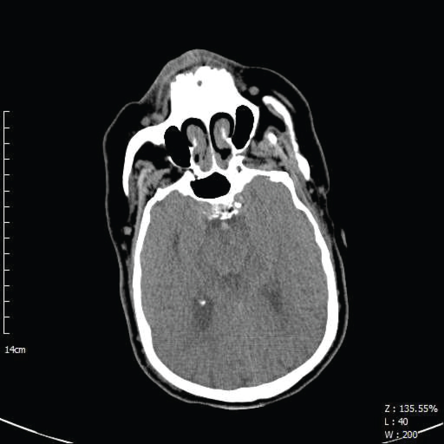
Figure 1: Brain CT without contrast. Hyperdensity in basilar artery indicating possible thrombosis (arrow).
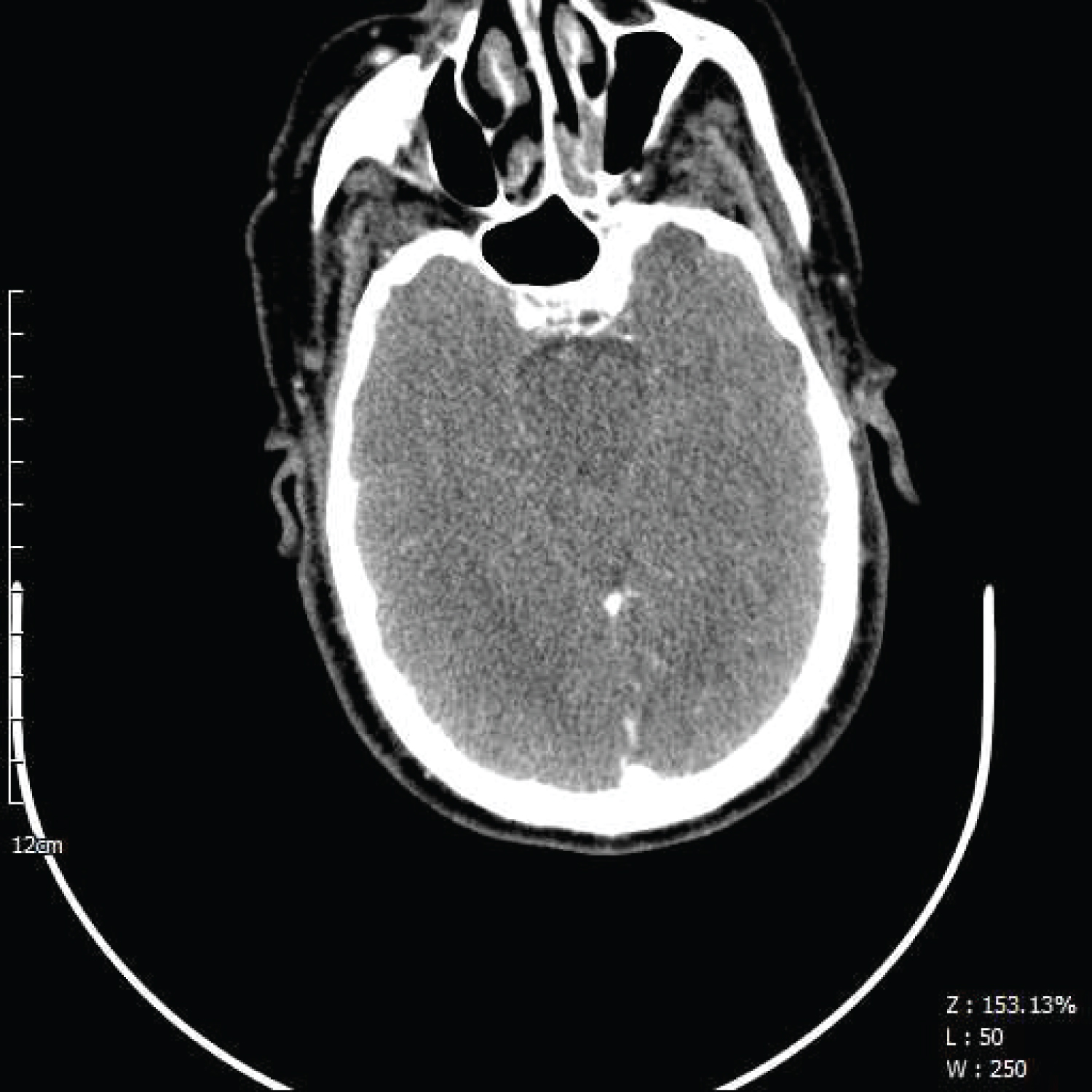
Figure 2: Brain CT with IV contrast. A) Black arrow indicates contrast in basilar artery. B) White arrow indicates contrast stoppedby basilar arteryocclusion.
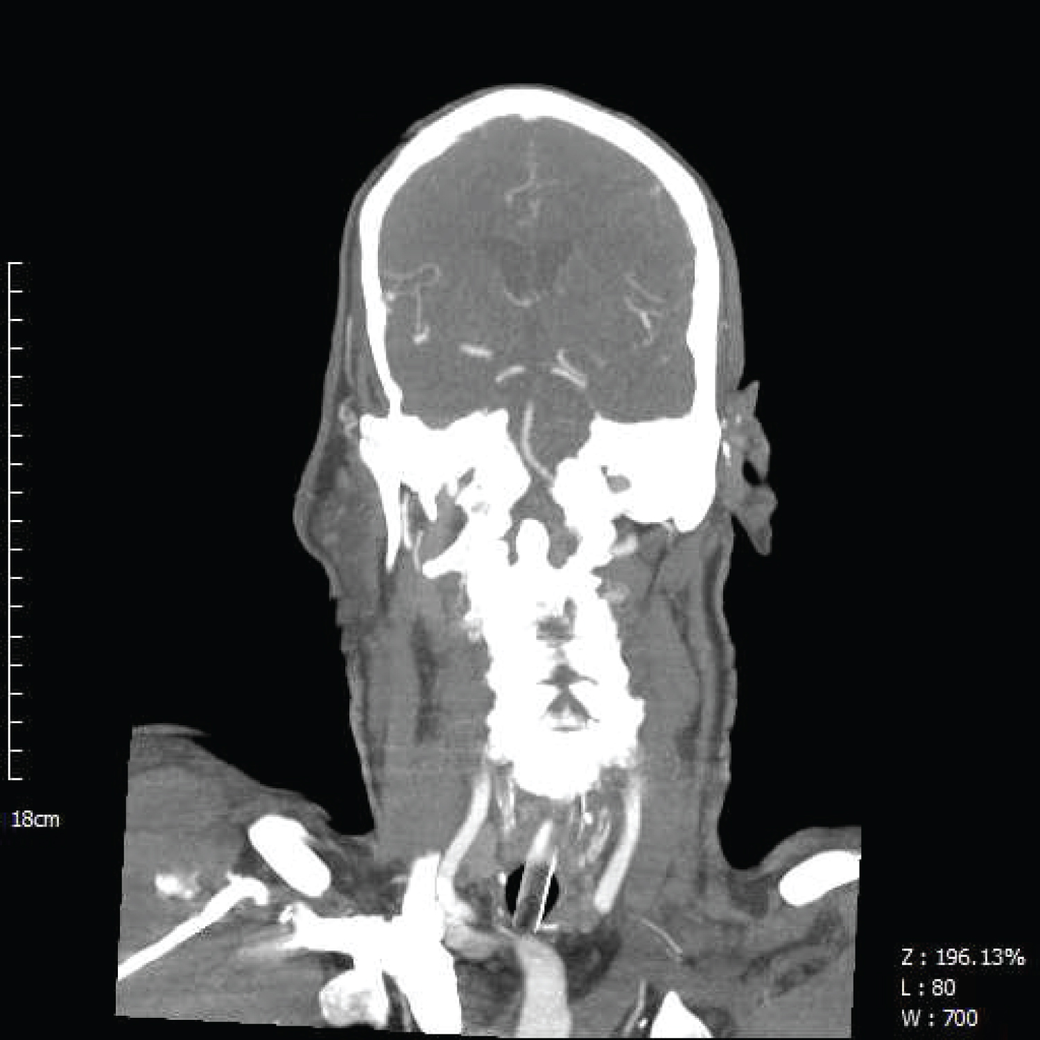
Figure 3: Arrow indicates contrast ceasing in basilar artery in coronal view.
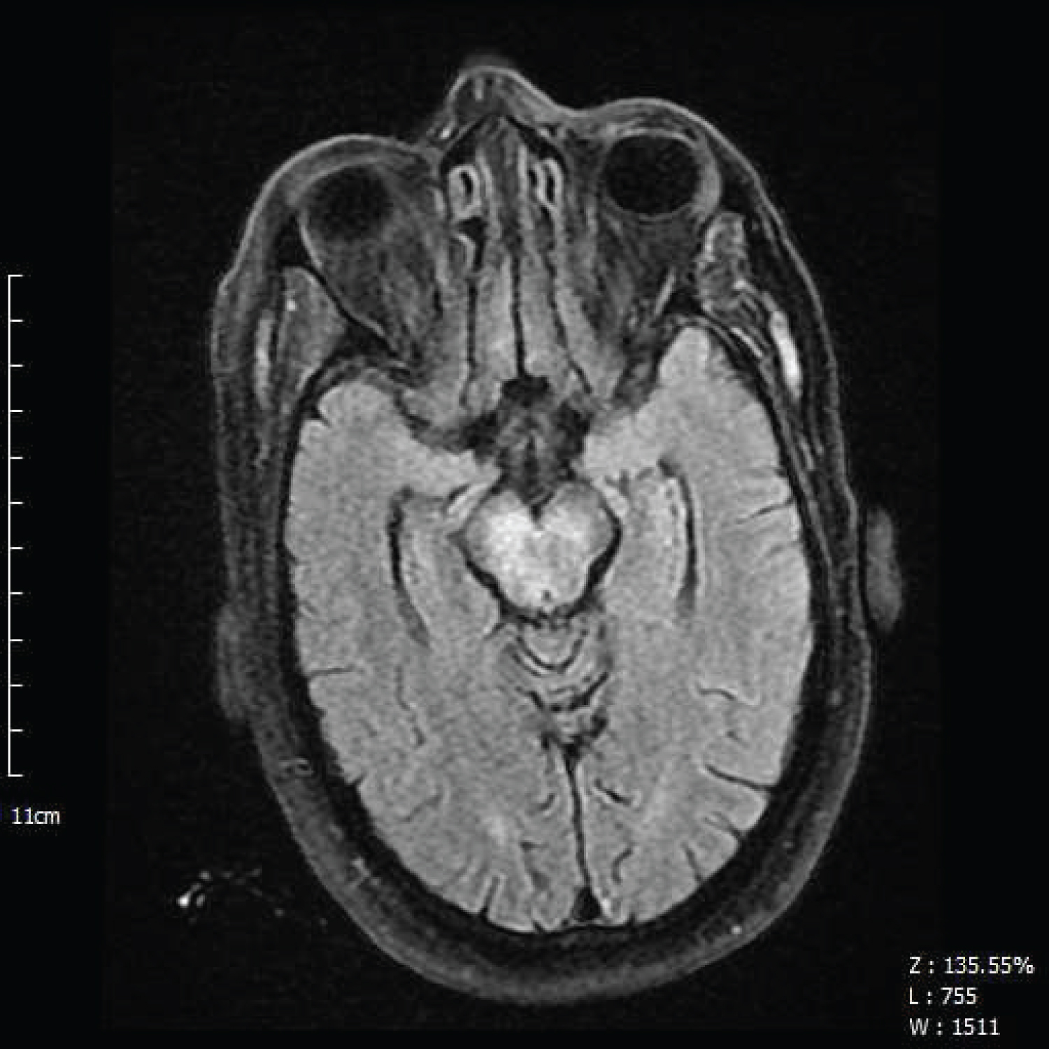
Figure 4: Diffusion weighted imaging modality of brain MRI shows restriction in mid-brain (arrow).