26-year-old lady admitted with shortness of breath and chest discomfort for last one and half months. She had no history of chest pain. She was not suffering from any major illness neither she was taking any medications on regular basis. On initial examination there was pallor and bipedal oedema. SpO2 was 94% in room air. Cardiovascular examinations reveal short systolic murmur in the pulmonary area with no radiation and continuous murmurs in left axillary area of same intensity. Chest examinations revealed normal vesicular breath sound in both lung fields. Electrocardiogram (ECG) revealed sinus tachycardia with heart rate of 116/min, regular in rhythm. Chest Xray was advised which revealed no significant abnormalities. Echocardiogram was advised which revealed normal Left Ventricular systolic function with normal chambers dimension and function. There was no significant valvular abnormality. Main pulmonary artery was normal in calibre. Left and Right Pulmonary artery was found to be stenosed (Figure 1 and Figure 2). It was long segment stenosis which was clearly visible in echocardiogram images and Video 1. Doppler examinations revealed pressure gradient of 22 mm of Hg and 31 mm of Hg in left and right pulmonary artery branch respectively. Complete Blood Count and other relevant laboratory investigations were also sent during admission and it revealed that total leukocyte count was 2 lakhs/cmm with numerous atypical blood cell (myelocytes, metamyelocyte, promyelocyte) including blast cell. She was diagnosed to have been suffering from Chronic Myeloid Leukaemia (CML). She was referred to Haematology department for further management. She was counselled properly for various approaches to BPAS (balloon plasty, stenting, surgical repair) and was advised to attend cardiology clinic after remission of CML for further treatment of BPAS.
There is no sponsor or fund support of any kind.
There are no conflicts of interest of any kind.
Sudeb Mukherjee: Conception and design, Acquisition of data, Analysis of data. Writing draft, final revision; Suhana Datta: Drafting the article, concept, design and revision.
We acknowledge contribution from the patient for giving consent for publication of this report.
The corresponding author is the guarantor of submission.
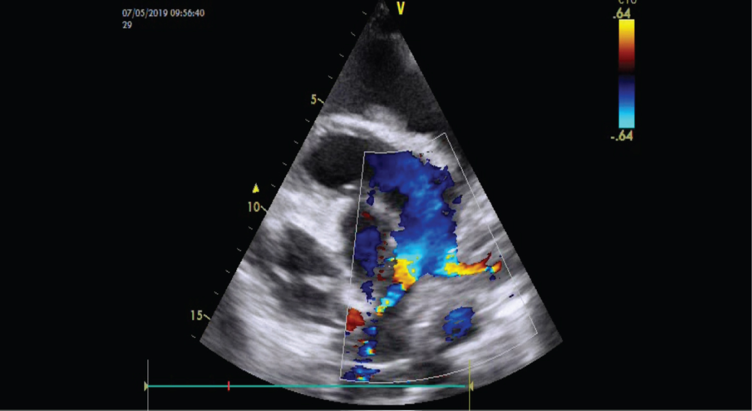
Figure 1: Showing the long stenosed segment of both pulmonary artery branches.
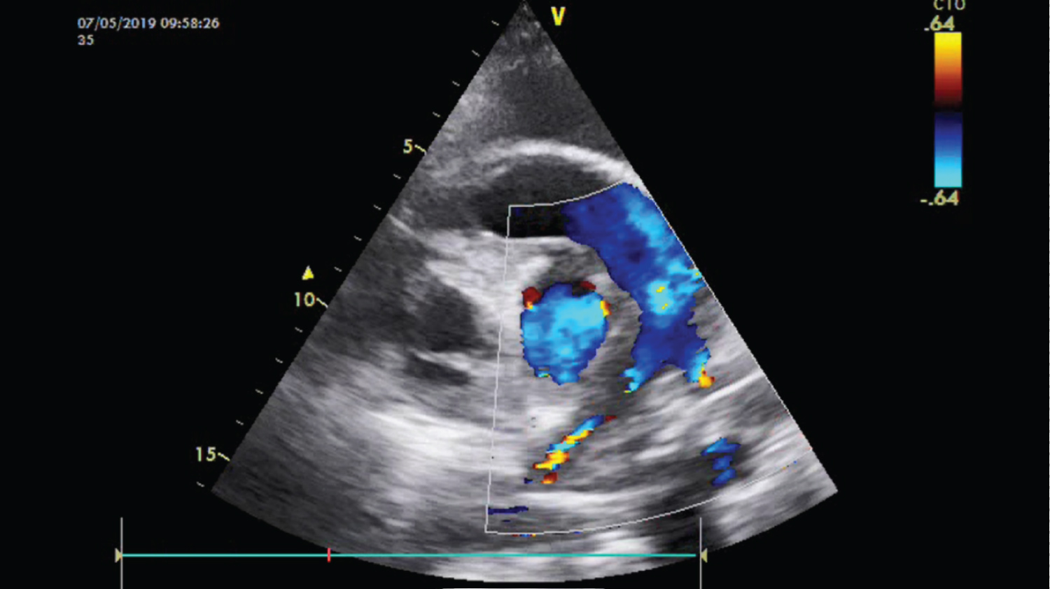
Figure 2: Showing normal pulmonary trunk and stenosed both branches.