A 45-year-old female with no known past medical history presented to the trauma unit as an unrestrained passenger in a motor vehicle accident. On arrival, her GCS was 8 and she was intubated. Focused Assessment with Sonography for Trauma (FAST) exam was negative, but initial chest imaging (Figure 1A and Figure 1B) revealed right-sided fractures of the first through seventh ribs, a fracture of the left first rib with bilateral consolidations, and a small right-sided hemopneumothorax. Further imaging showed hepatic vascular injury, Grade 2 splenic injury, Grade 5 renal injury, and a non-displaced fracture of the right L5 transverse process.
X-Ray eight hours later showed subcutaneous emphysema of the right chest wall (Figure 2A), with corresponding computed tomography (CT) showing worsening of the right-sided pneumothorax, as well as an intercostal pleurocutaneous fistula along the anterior superior chest wall (Figure 2B). A right-sided chest tube was then placed, and daily X-rays were performed to assess for resolution of the fistula (Figure 3). Within 12 hours improvement was seen, and within 36 hours the subcutaneous emphysema was nearly resolved (Figure 4).
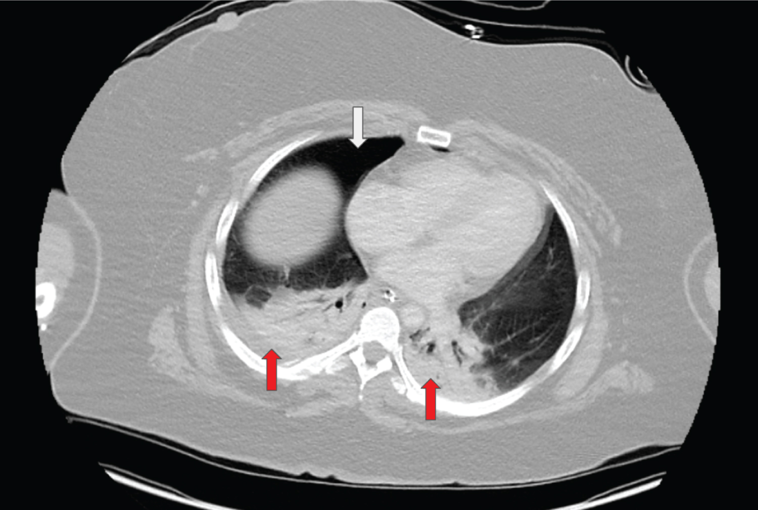
Figure 1a: Axial CT with contrast shows right-sided pneumothorax (white arrow) and bilateral layering consolidations (red arrows).
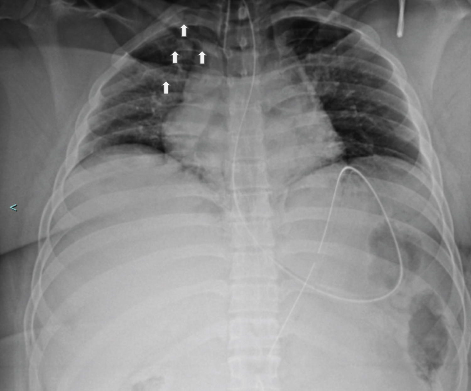
Figure 1b: Portable chest X-ray shows fractures of the right second through fourth ribs (white arrows). Corresponding CT (Figure 1A) confirmed fractures of the right first through seventh ribs, left first rib, and right L5 transverse process.
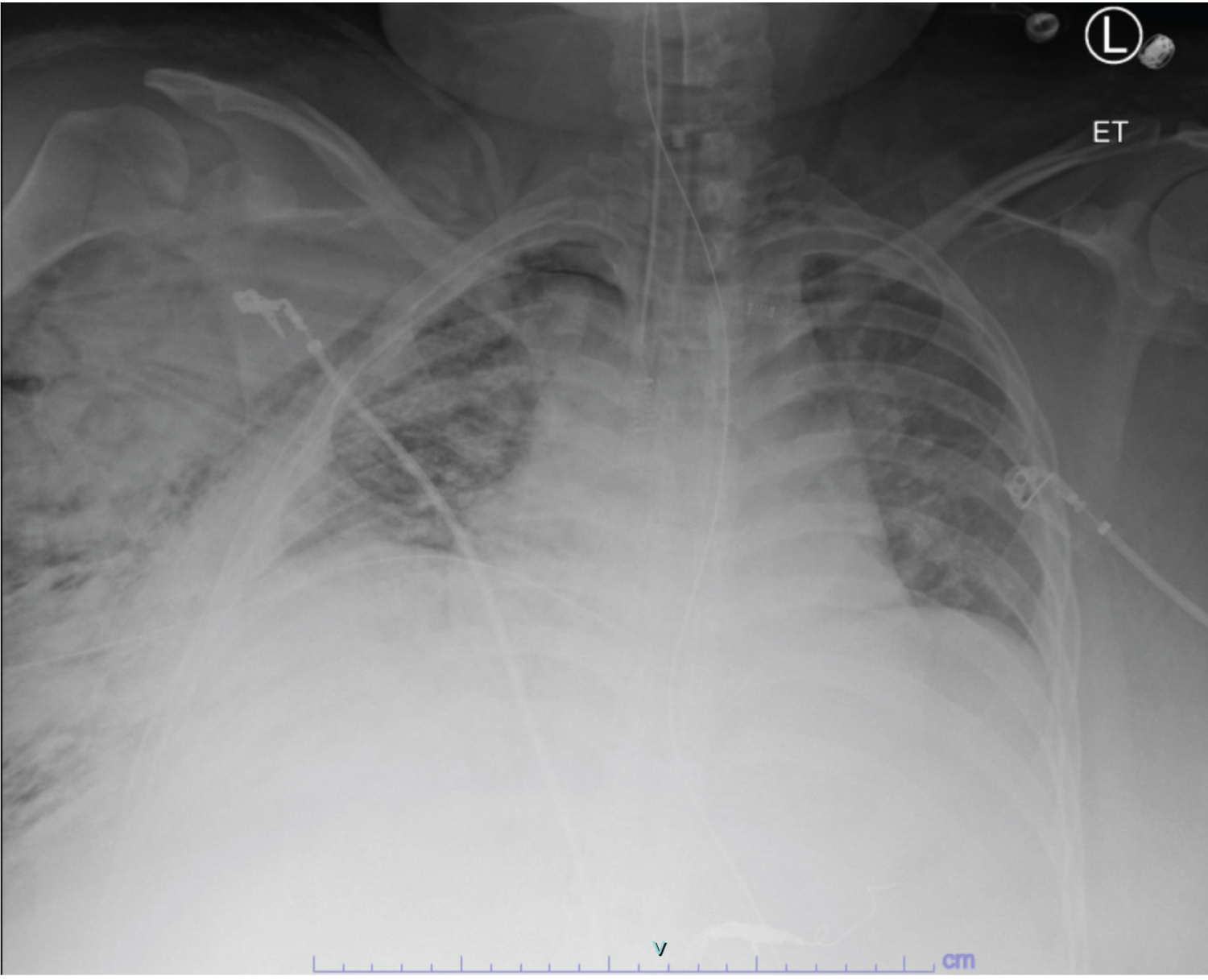
Figure 2a: Portable Chest X-ray taken immediately after chest tube placement shows the extent of the right-sided subcutaneous emphysema.
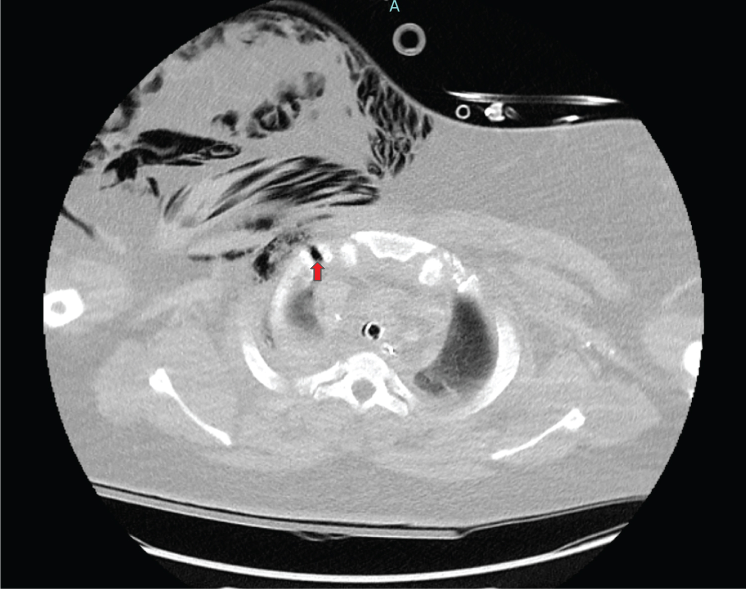
Figure 2b: Axial CT with contrast shows the right-sided pleurocutaneous fistula (red arrow) with resulting subcutaneous emphysema of the right chest.
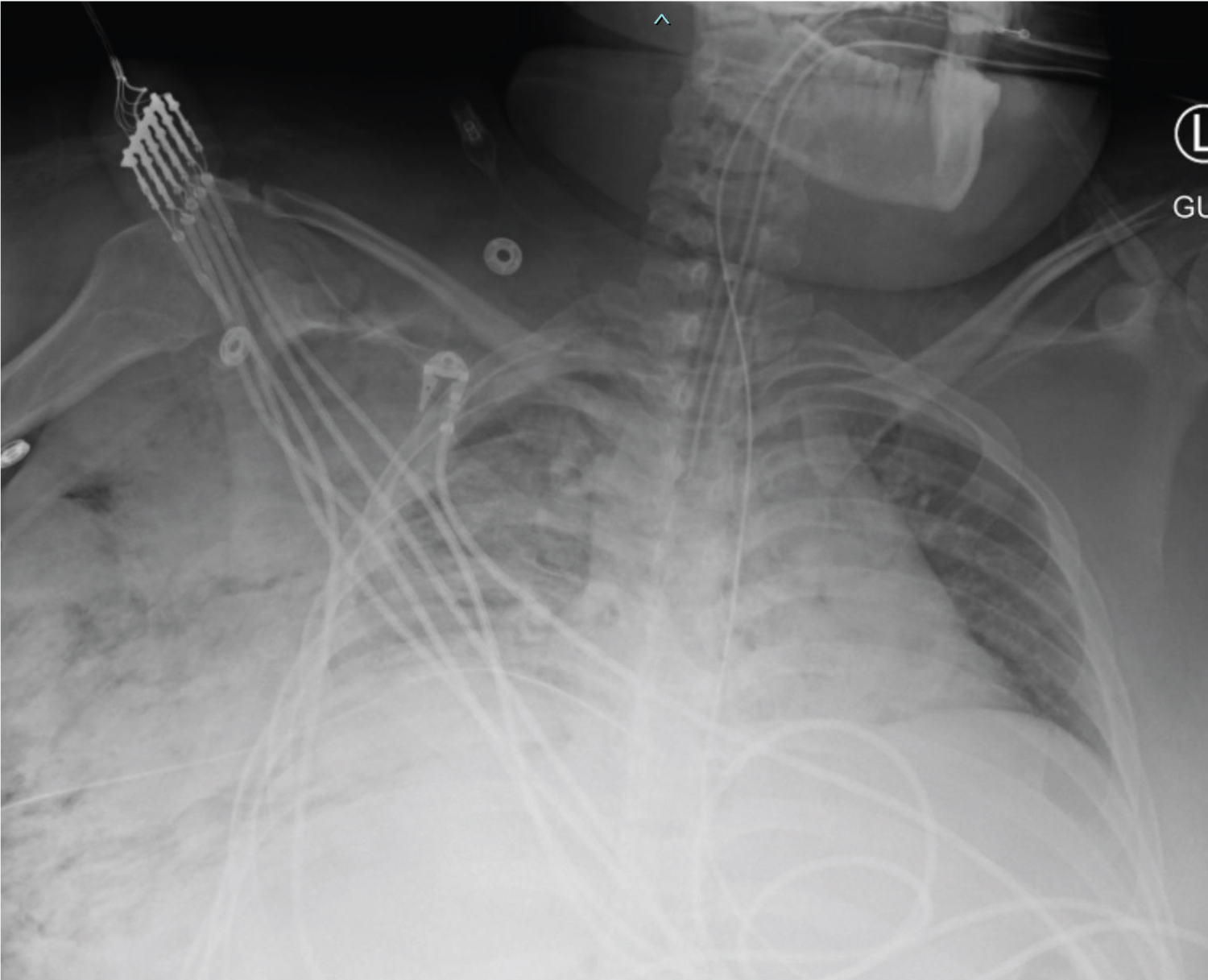
Figure 3: Portable chest X-ray taken 12 hours after chest tube placement shows improvement of right-sided subcutaneous emphysema.
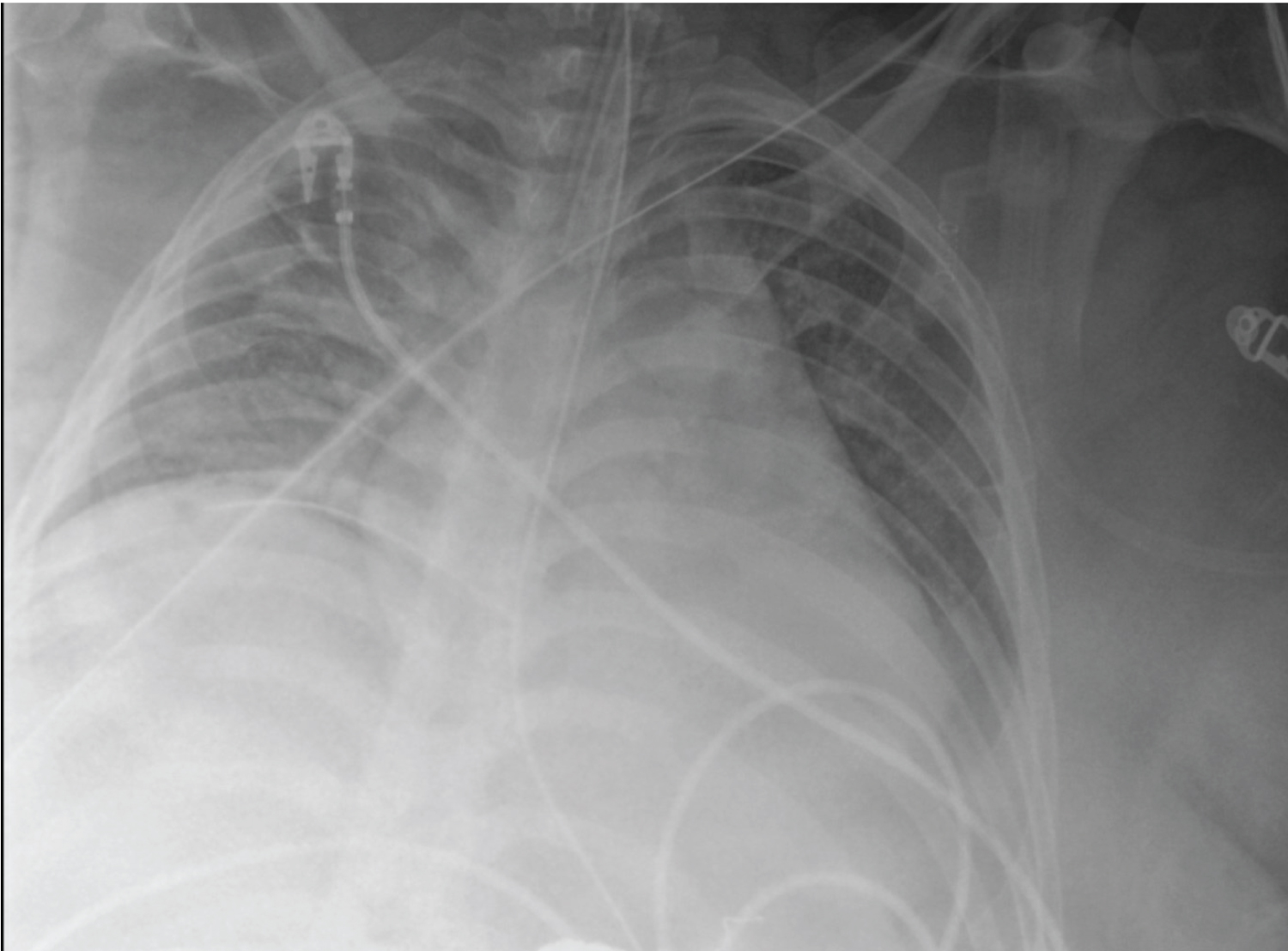
Figure 4: Portable Chest X-ray 36 hours after chest tube placement shows near total resolution of the right-sided subcutaneous emphysema.