The clinical characteristics and management strategies for pneumothorax are highly variable.
The purpose of the present study was to clarify the clinical characteristics and to determine the risk factors for recurrence and the surgical management of pneumothorax.
The clinical characteristics, as well as the risk factors for recurrence and surgical treatment, were retrospectively analyzed in 125 patients with pneumothorax that was managed in our hospital.
The incidence of pneumothorax was higher in men and current or former smokers. Patients with secondary pneumothorax were significantly older and required more frequent chest drainage than those with primary pneumothorax. Collapse rate was an independent predictor of the need for chest drainage. Chest drainage, recurrence, and the presence of bulla ≥ 2 cm were the independent predictors of the need for surgical treatment.
Patients with secondary pneumothorax and higher collapse rate frequently required chest drainage. Surgical treatment should be considered in patients who require chest drainage, those with recurrent episodes, or those with bulla ≥ 2 cm.
Pneumothorax, Bullous disease, Chest tube drainage
Most cases of pneumothorax comprise primary spontaneous pneumothorax (PSP), which usually occurs in young, tall men and results from rupture of subpleural blebs or bullae [1]. Secondary spontaneous pneumothorax (SSP), on the other hand, is associated with an underlying lung disease, such as emphysema or asthma; acute or chronic infections; lung cancer; and congenital disease, including cystic fibrosis, catamenial pneumothorax, and lymphangioleiomyomatosis.
Patients with small pneumothorax are managed by bed rest, oxygen supplementation, or manual aspiration [2-4]; whereas patients with larger or recurrent episodes of pneumothorax may require more aggressive therapies, such as chest tube drainage or surgery [5-9]. The purpose of the present study was to clarify the clinical characteristics and to determine the risk factors for recurrence and the surgical management of pneumothorax.
The medical records of 140 patients who were treated for pneumothorax at our hospital from April 2013 to September 2014 were retrospectively reviewed. Figure 1 shows the study design and exclusion criteria. The study protocol was approved (No.15-96) by the Ohashi Hospital Committee of Toho University Ohashi Medical Center.
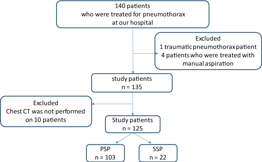 Figure 1: Study design and exclusion criteria.
PSP: Primary spontaneous pneumothorax; SSP: Secondary spontaneous pneumothorax; CT: Computed tomography. View Figure 1
PSP was defined based on medical history, chest radiography, or computed tomography (CT). We retrospectively collected the following variables: detailed history, number of pneumothorax episodes and treatment modalities, age, sex, smoking habits, laterality, collapse rate, need for hospitalization, and treatment period. Since manual needle aspiration was performed on only five symptomatic patients with small pneumothorax, we compared patients based on the presence or absence of chest drainage. The patients who underwent chest tube drainage were further compared based on the application of continuous negative pressure suction. The surgical management for all patients was wedge resection.
The pneumothorax collapse rate was calculated as a percentage, based on the method defined by Kircher (Figure 2), at the time of diagnosis of pneumothorax. Using the posteroanterior chest radiograph, the atelectatic lung area was subtracted from the area of the entire hemithorax; the result was then divided by the area of the entire hemithorax.
 Figure 2: Chest radiograph showing the calculation of the percentage of pneumothorax based on the method defined by Kircher.
Collapse rate = (1-ab/AB) × 100.
View Figure 2
At the time of lung expansion, chest CT was performed on 125 patients who were divided and compared according to the presence or absence of bleb or bullae. Patients with bleb or bullae were further subdivided and compared based on the number (single vs. multiple) and size (≥ 2 cm vs. < 2 cm) of the bleb or bullae.
Descriptive data were expressed as mean ± standard deviation (SD). Differences between groups were assessed by the Student's t-test or Mann-Whitney U-test, as appropriate. Variables with a P value < 0.05 in the univariate analysis were entered into a multivariate analysis using stepwise logistic regression to determine the predictors of the need for chest drainage and surgical treatment. All analyses were performed by SPSS Statistics (IBM; Tokyo, Japan). P values < 0.05 were considered significant.
Table 1 shows the clinical characteristics of the 125 patients. The incidence of pneumothorax was higher in men and current or former smokers. Approximately 74% required hospital admission. Comparison of the clinical findings based on treatment is shown in Table 2. Patients without chest drainage and those with chest drainage had significant differences in smoking habits, collapse rate, laterality, and surgical treatment but were comparable in terms of recurrence rate and treatment period. Among the patients with chest drainage, addition of continuous negative pressure suction significantly affected the duration of treatment and the need for surgical treatment.
Table 1: Characteristics of the study population. View Table 1
Table 2: Comparison of the clinical findings by treatment (n = 125). View Table 2
Table 3 shows the comparison of clinical findings based on the cause of pneumothorax. Patients with SSP were significantly older and required chest drainage more frequently than those with PSP. Majority of the cases were PSP; the underlying causes of SSP were emphysema (n = 7), cystic fibrosis (n = 4), pneumonitis (n = 10), and lung cancer (n = 1).
Table 3: Comparison of the clinical findings according to the cause of pneumothorax (n = 125). View Table 3
A comparison of the clinical findings based on the presence and number of blebs and/or bullae is shown in Table 4. Both groups had significant differences in terms of recurrence, collapse rate, and requirement for chest drainage and surgical treatment. Recurrence rate was significantly higher in patients with multiple bullae than in those with single bulla. On multivariate analysis, the independent predictor for the need for chest drainage was collapse rate [odds ratio (OR) 1.1516, 95% confidence interval (95% CI) 1.0855-1.2184; p < 0.001] (Table 5); the independent predictors for the need for surgical treatment were chest drainage (OR 3.4685, 95% CI 1.3299-9.0463; p = 0.0110), recurrence (OR 3.6552, 95% CI 1.0756-12.4211; p = 0.0378); and presence of bulla ≥ 2 cm (OR 0.2601, 95% CI 0.0856-0.7910; p = 0.0176) (Table 6).
Table 4: Comparison of the clinical findings based on the presence of cystic lesion (n = 125). View Table 4
Table 5: Multivariate analysis of the predictors of chest drainage (n = 125). View Table 5
Table 6: Multivariate analysis of the predictors of surgical treatment (n = 125). View Table 6
In this study, PSP was the most common cause of pneumothorax and occurred more frequently in young men with smoking habits, as previously reported. Many factors are involved in the pathogenesis of PSP. The rate of recurrence after the first episode of PSP was reported to be higher in smokers than in non-smokers [5,10-12]. The pathogenesis of PSP is believed to differ between smokers and non-smokers. Cheng Y reported that pathologic findings of respiratory bronchiolitis were seen in all smoking patients and in only 49% of non-smokers [13]. Bronchial abnormalities and distal airway inflammation or obstruction in smokers could lead to rupture of the visceral pleura and passage of air into the pleural space [5,10]. In this study, the recurrence rate of pneumothorax was higher in smokers than in non-smokers.
Comparison of the clinical findings based on the management of PSP patients demonstrated a significantly more frequent need for surgical treatment in patients who underwent chest tube drainage than in those without chest drainage. For PSP patients without chest drainage, surgical treatment was performed to prevent recurrence. Since almost all SSP patients were treated with chest tube drainage, we suggest that SSP cases undergo chest drainage with continuous aspiration and surgical treatment at an earlier stage after the diagnosis.
Although the pathophysiology of PSP recurrence in never-smokers remains unclear, the presence of bleb or bullae in about 81% of such cases was reported to be associated with PSP recurrence [14,15]. Kinoshita, et al. reported that recurrence rate was 40.7% in patients with bleb or bullae and only 11.1% in patients without bleb or bullae [16]. In this study, recurrence rate was also significantly higher in patients with multiple blebs or bullae than in those with single bleb or bulla.
In this study, the need for surgical treatment was significantly frequent for patients with PSP with bleb or bullae, SSP, treatment with chest tube drainage, and application of continuous negative pressure suction. However, the number of blebs or bullae was not a predictor of PSP prognosis. In patients with PSP, collapse rate was significantly associated with the need for chest tube drainage, whereas recurrence, presence of bleb or bullae ≥ 2 cm, and requirement for chest drainage might be the clinically useful predictors of the need for surgical treatment.
The present study had several limitations, including its retrospective design and inclusion of recurrent cases. Therefore, we could not evaluate recurrence accurately. Another limitation was the small population of SSP. In order to validate our findings and identify the predictors of recurrence and need for surgical treatment of pneumothorax, we are planning to perform a prospective study on patients with initial pneumothorax episode and to increase the number of subjects.
In conclusion, patients with SSP and higher collapse rate frequently required chest drainage. Surgical treatment should be considered for patients requiring chest drainage, those with recurrent episodes, and those with bullae > 2 cm.