A 51-year-old man with advanced ALS and home mechanical ventilator dependence presented with fever, tachycardia, hypotension and leukocytosis. Chest radiograph showed a large left pleural effusion (Figure 1) and an unenhanced chest CT confirmed a loculated effusion with gas bubbles (Figure 2) and large air-fluid level (Figure 3). A small-bore (14 F) thoracostomy tube was placed with an initial return of large quantity of malodorous gas then followed by purulent fluid which showed polymicrobial organisms on gram stain and culture including Streptococcus viridans, Staphylococcus aureus, Klebsiella pneumonia, and Bacteroides fragilis. His initial blood cultures grew Streptococcus but subsequent cultures were sterile. This combination of polymicrobial organisms represents an oral source from aspiration. Only after drainage of the empyema assisted by pleural infusions of alteplase and dornase-alfa and open insertion of a second (20 F) thoracostomy tube was the suspected lung abscess visible on repeat chest CT (Figure 4). He recovered uneventfully and returned home to complete 4 weeks total course of intravenous antibiotics.
 Figure 1: Initial upright portable chest radiograph demonstrating large left pleural effusion, likely loculated.
View Figure 1
Figure 1: Initial upright portable chest radiograph demonstrating large left pleural effusion, likely loculated.
View Figure 1
 Figure 2: Chest computed tomography without intravenous contrast demonstrating loculated fluid collections in pleural space, here in major fissure with multiple gas bubbles.
View Figure 2
Figure 2: Chest computed tomography without intravenous contrast demonstrating loculated fluid collections in pleural space, here in major fissure with multiple gas bubbles.
View Figure 2
 Figure 3: Chest computed tomography without intravenous contrast demonstrating large air-fluid level within pleura.
View Figure 3
Figure 3: Chest computed tomography without intravenous contrast demonstrating large air-fluid level within pleura.
View Figure 3
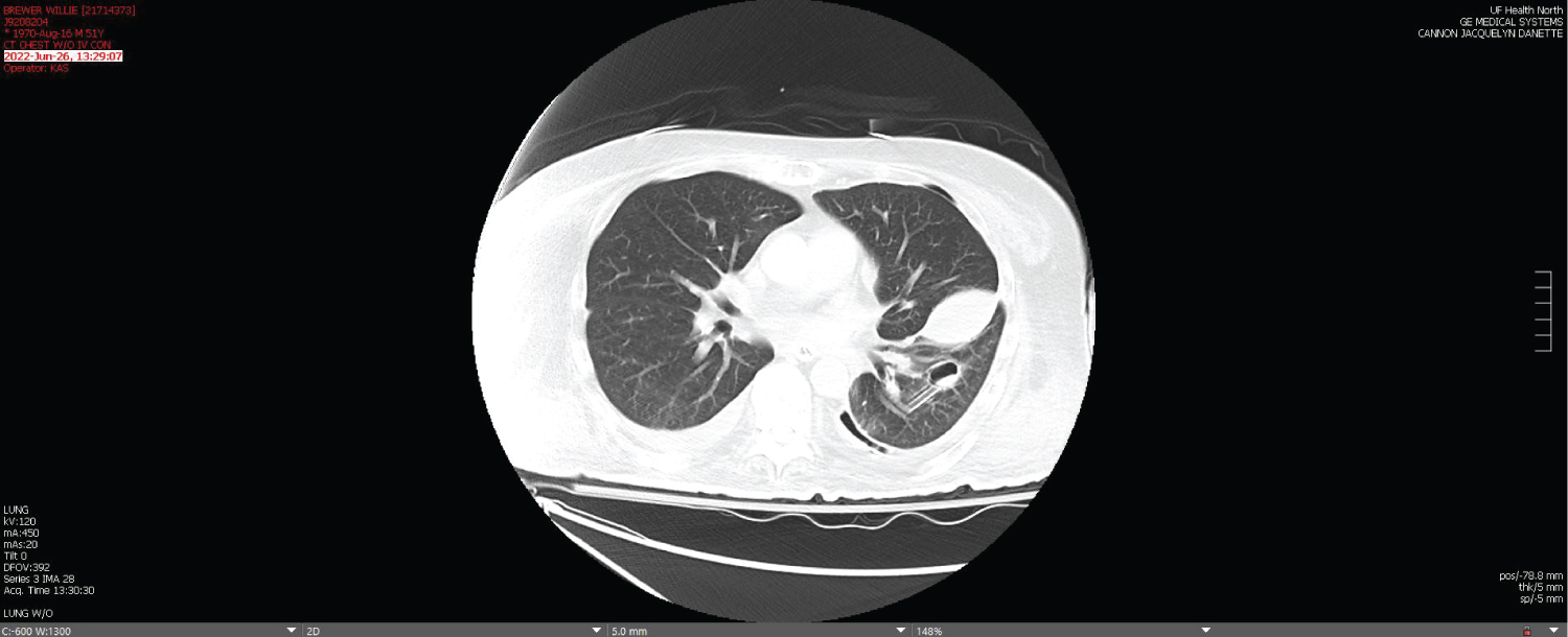 Figure 4: Repeat chest computed tomography without intravenous contrast 5 days later after placement of two thoracostomy tubes, demonstrating parenchymal lung abscess.
View Figure 4
Figure 4: Repeat chest computed tomography without intravenous contrast 5 days later after placement of two thoracostomy tubes, demonstrating parenchymal lung abscess.
View Figure 4