Cutaneous larva migrans (CLM) is a zoonotic infestation caused by penetration and migration of filariform larvae into the epidermal layer of skin derived from dogs and cats, namely Ancylostoma braziliense and Ancylostoma caninum. Infective filariform larvae penetrate the surface of the skin, and migrate beneath the epidermis by leaving prominent linear or serpiginous lesions called 'creeping eruption'. One case was reported of CLM with the main complaint of being very itchy and serpiginosa lesion with hyperemic papules. Treatment with pyrantel pamoate and mebendazole is not effective in this case. Patients were given alternative therapies using 5% permethrin cream for 3 days. On the sixth day after 5% permethrin therapy, the lesion underwent total resolution.
Cutaneuous larva migrans, Permethrin, Mebendazole
Cutaneous larva migrans (CLM) is one of the most common skin disorders in the tropics. Cutaneous larval migrans (CLM) is a typical clinical skin infection caused by active penetration of nematode larvae and epidermal migration of it, usually Ancylostoma braziliense. The most frequent location is in the lower extremities, especially in the legs. Although this disease can heal itself, further treatment is often needed in many cases [1,2].
The etiology of cutaneous larva migrans (CLM) is a zoonotic infestation caused by penetration and migration of filariform larvae (commonly a type of hookworm) into the epidermal layer of the skin through contact with the feces of infected animals, usually dogs and cats [3]. Clinically, cutaneous larval migrans (CLM) are characterized by tortuous erythematous pruritic lesions or serpiginosa with slightly raised or prominent pathways. This disease was first introduced by Lee, a British doctor, in 1874. The term "cutaneous migratory larvae" was coined by Crocker in 1893, and in 1929 the etiology of this disease was known as Ancylostoma larvae, so the terminology cutaneous larva migrans (CLM) known as Hookworm-related cutaneous larva migrans (HrCLM) [4].
During these years, the terms HrCLM and creeping eruption are considered to have the same meaning, whereas HrCLM itself describes a disease syndrome while creeping eruption is a clinical picture as a linear reddish lesion (serpiginosa), prominent and migrate in irregular patterns [3].
Cutaneous larva migrans (CLM) are actually self-limited which often heal themselves in 2 to 8 weeks, however, pruritus or itching can be very severe in the course of the disease. The recommended treatment options according to the literature are a single oral dose of albendazole 400 mg in adults, or 400 mg for 3 to 5 days (or 10-15 mg/kg, with a maximum dose of 800 mg daily for pediatric cases) to increase its effectiveness, or a single dose of ivermectin 12 mg for adults (or 150 μg/kg in pediatric cases), or topical application of thiabendazole 10%, three times a day for at least 15 days [1].
Another therapy that is commonly used in Indonesia is pyrantel pamoate at a dose of 10 mg/kg BB which is available in tablet or syrup form. Pirantel pamoate is a common therapy used to eradicate most types of worms. Pirantel pamoat and its derivatives work by causing depolarization and increasing the frequency of impulses in the worm muscles that cause the worms to die. Side effects of pyrantel pamoate are rarely reported and are only mild symptoms such as headaches and nausea [5].
The last few decades have been reported several types of larvae and worms that began to be resistant to anti-helmentic group's benzimidazole (BZ), especially sheep in 1983. In line with the development of treatment and use of antimicrobials that are not controlled, there are also resistance to other drug groups such as levamizole (Lev) and macrolitic lactones (Ivermectin). The Bogor Institute of Agriculture also reports that at present there have been multiple resistance of worms to the anti-helmentic drugs benzimidazole, levamizole and macrolitic lactones. Although there are no data that report the magnitude of resistance occurring in Indonesia, a report in Australia says that 80% of sheep farms have been declared resistant to benzimidazole and levamisole [6].
Until this journal was written, there were no real data regarding the reported incidence of treatment resistance in cases of cutaneous larva migrans (CLM). This literature report aims to report the possibility of starting cases of antihelmintic resistance case in humans.
A boy aged 1.5 years with complaints of itching and redness on the right and left buttocks since 10 days ago (Figure 1). The rash first appeared as a red pimple which the mother thought was an insect bite. After a few days, the red nodule extends to form a long curve, winding, protruding, and elongated. The skin disorder makes the child difficult to sleep and looks constantly scratching the buttocks. History of habits found children like to play with cats around the house and like to sit on the ground.
On physical examination found compos mentis awareness with good general condition. On the surface of the right and left buttocks of the skin there is a picture of the winding lesions (serpiginous papules) with hyperemic lesions around the skin (Figure 1). The results of history and physical examination can confirm the conclusion as a cutaneous larvae migrans. Patients were given 125 mg pyrantel pamoate therapy and concoction cream in the form of a 10 gram mometasone cream mixture with 500 mg mebendazole (Figure 1). The patient did not re-control the doctor and returned 1 month later with complaints of more extensive lesions and felt more itching, especially in the last few days (Figure 2). Considering that the child is 1.5-years-old, with thin and smooth skin, and lesions in the area around the anus, it is not possible to do therapy such as Chloretyl spray which has been carried out in CLM cases, so it is considered topical permethrin cream 5% which has been used as a scabicid can be done, with the hope that it can also for larvacid in CLM. Before we give therapy, we have explained to the patient's mother all the above considerations and the patient's mother agreed with the note always communicate with us. Topical permethrin is applied only to the lesion 2 times daily. Finally, we decided to give permethrin cream 5% which is applied twice a day to areas with skin disorders. The patient came back for re-examination 3 days later with complaints that the symptoms had disappeared and only the remnants of the lesions had dried up (Figure 3). The patient is scheduled for final control 3 days later with the lesion having total resolution with the symptoms disappearing and leaving a blackish spot known as hyperpigmentation postinflammatory (Figure 4). The patient returned to control 1 week after the last treatment and confirmed that the infection was completely resolved (Figure 5).
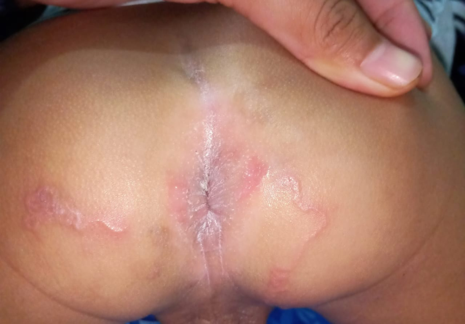 Figure 1: Serpiginosa lesions when patients first come to the clinic.
View Figure 1
Figure 1: Serpiginosa lesions when patients first come to the clinic.
View Figure 1
 Figure 2: Serpiginosa lesions, 1 month later with patients not re-control for treatment and serpiginosa lesions getting worse.
View Figure 2
Figure 2: Serpiginosa lesions, 1 month later with patients not re-control for treatment and serpiginosa lesions getting worse.
View Figure 2
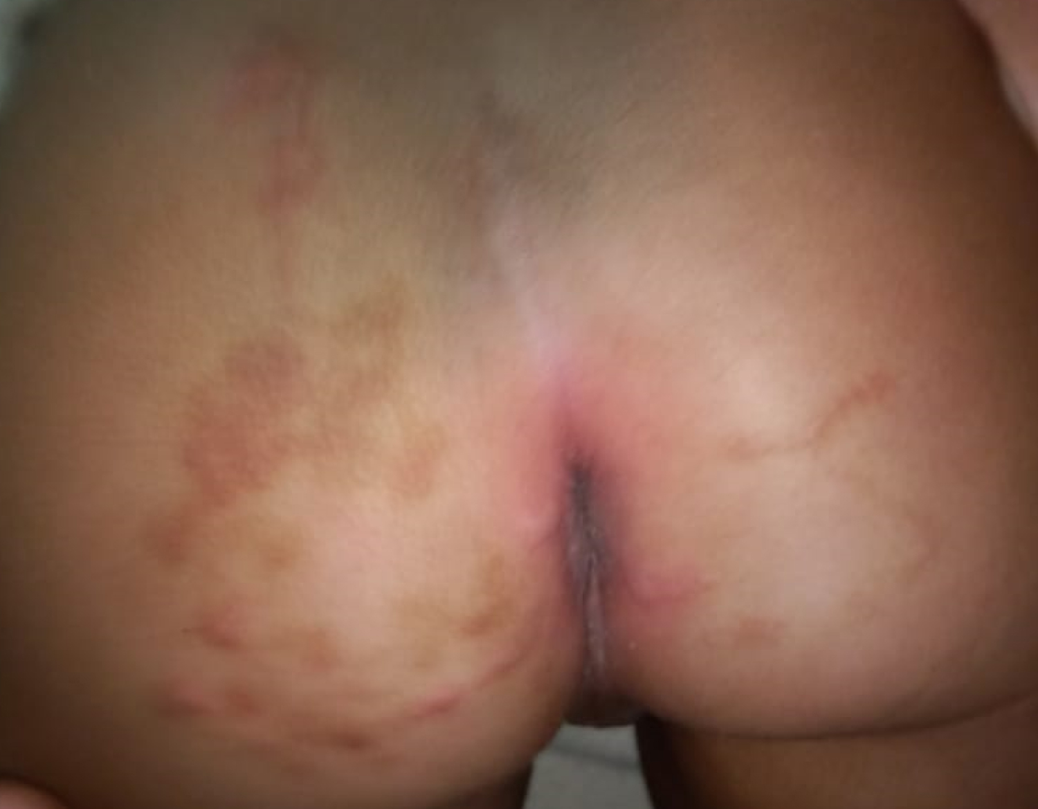 Figure 3: Lesions that start to heal after 3 days of treatment with permethrin cream 5%.
View Figure 3
Figure 3: Lesions that start to heal after 3 days of treatment with permethrin cream 5%.
View Figure 3
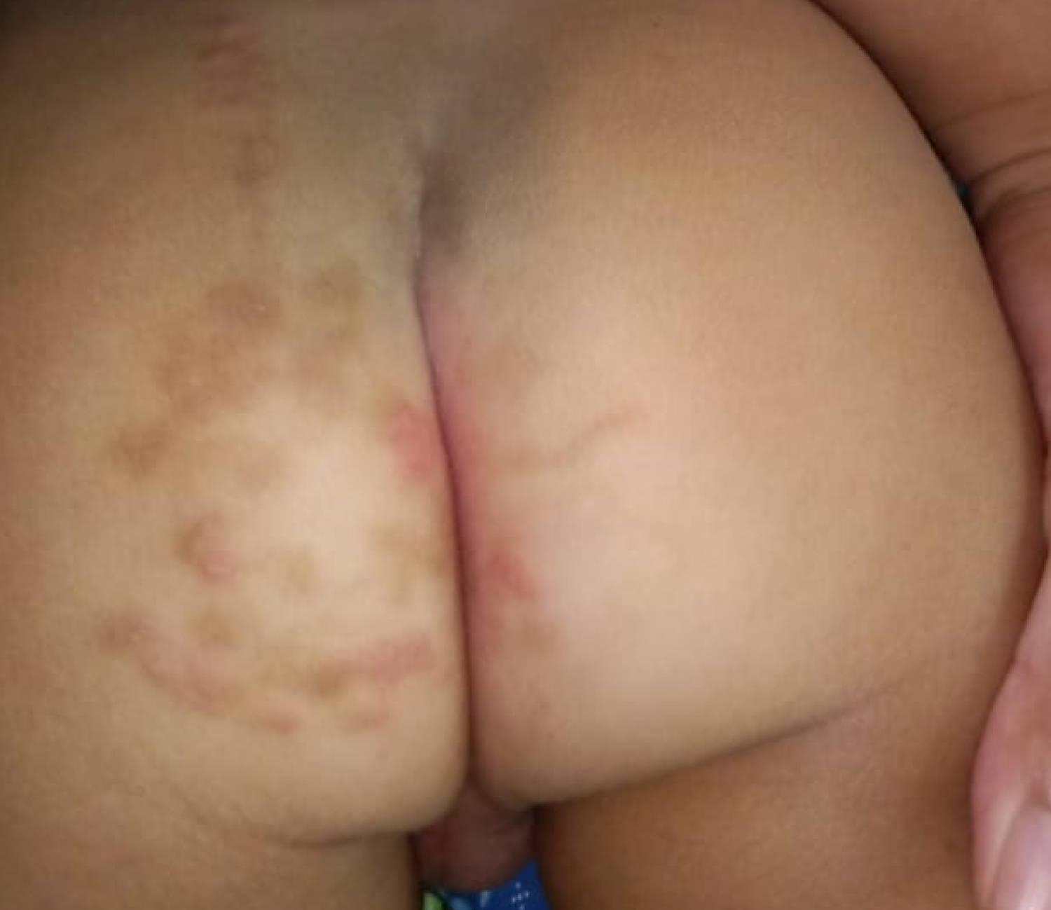 Figure 4: Total resolution by leaving hyperpigmentation post inflammatory lesions after the use of a permethrin cream 5% for 6 days.
View Figure 4
Figure 4: Total resolution by leaving hyperpigmentation post inflammatory lesions after the use of a permethrin cream 5% for 6 days.
View Figure 4
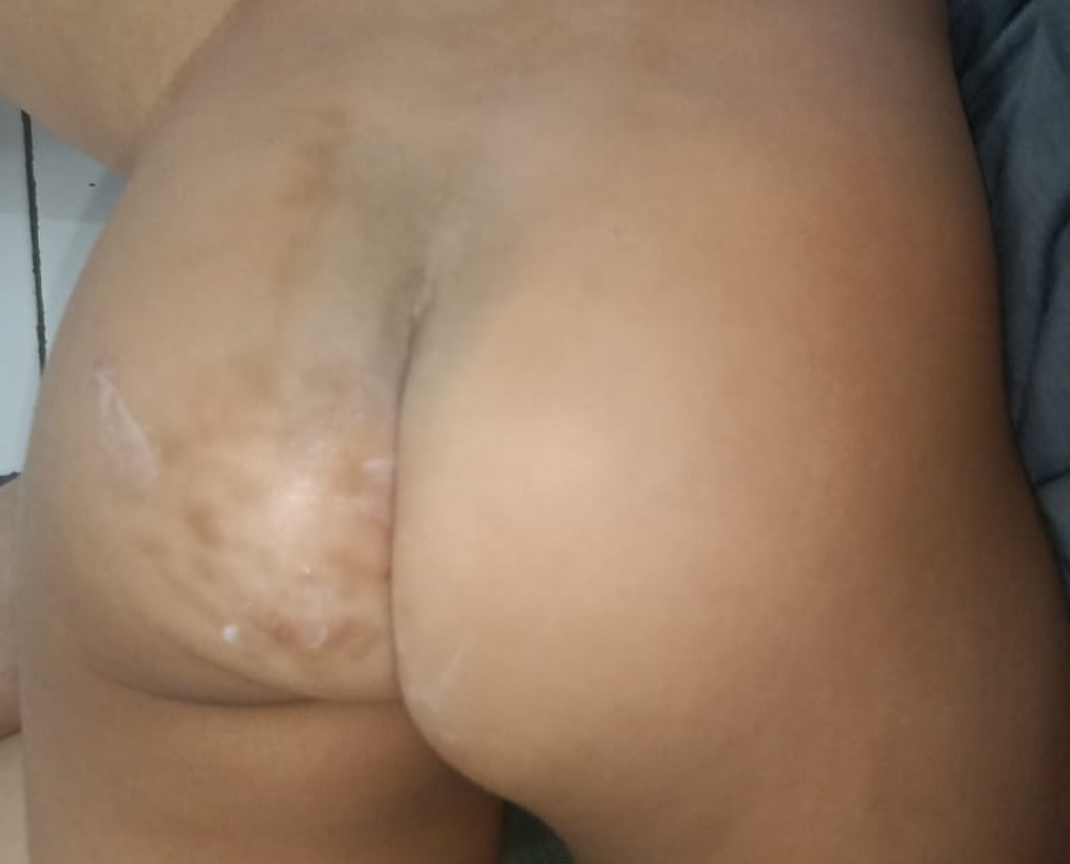 Figure 5: The infection was totally resolved.
View Figure 5
Figure 5: The infection was totally resolved.
View Figure 5
Cutaneous larva migrans (CLM), also known as creeping eruption, is a pruritic serpiginous hyperemic lesion caused by migratory larvae of hookworm through the epidermis. The most common parasites found are Ancylostoma braziliense (common in dogs and cats) and Ancylostoma caninum (common in dogs). Other larvae that cause CLM are Uncinaria stenocephala (dog hookworm), Bunostomum phlebotomum (cattle hookworm), and Ancylostoma ceylonicum (dog and cat hookworm). Ancylostoma caninum and Uncinaria stenocephala have been found in fox [1,4].
The life cycle of cutaneous larva migrans (CLM) is an adult hookworm infesting the intestines of certain host animals. Parasitic eggs are excreted with excrement and contaminate the soil or sand around the sewage. In the optimal environmental conditions, embryonated eggs hatch in the surface layer of the soil within two days into rhabditiform larvae. Rhabditiform larvae live by eating bacteria that are in the soil and/or feces. These rhabditiform larvae mature and shed their skin twice in 5 to 10 days to become infective filariform larvae. Filariform larvae can survive for several weeks to several months in optimal conditions. Ancylostoma braziliense larvae, the most common causative larvae with an average size of about 6.5 mm and have a diameter of 0.5 mm [1,4].
Humans are accidental hosts as infection sites. Infection in humans begins when the filariform larvae come in direct contact and penetrate the stratum corneum. Larvae usually live in shallow layers of sand/soil, within a few inches where eggs are stored. Beach or open field is a common reservoir for filariform larvae. Infection in humans usually occurs when humans walk barefoot or with open-type shoes or lying without clothes on sand/soil, especially sandy beaches that have been contaminated by infected dog and cat feces. Larvae can also be found in sand, and loose soil on construction sites, gardens, fields, or under houses. Simultaneous infestation or combination between hookworm species is common [1,4].
The pathophysiology of larval penetration is through proteases and hyaluronidases released by filariform larvae so that filariform larvae can penetrate skin cracks, hair follicles, sweat glands and even intact skin by digesting keratin in the epidermal layer. After penetrating the skin, filariform larvae release their cuticles. Until then, the larvae had no functioning mouth parts. After the cuticle is released, the larvae start migrating for about 7 days. During the process of larval penetration, collagenase deficiency occurs and the larvae are unable to invade the dermis but also cannot reach the blood vessel or lymphatic vessels to reach the intestine and complete their life cycle, as in the right animal host (dog or cat). The process of collagenase deficiency causes the larvae to remain confined to the epidermis. The larvae crawl aimlessly in the epidermis in the serpiginous route at speeds of 2 mm to 2 cm per day. The speed of migration varies depending on larval species, but generally does not exceed 1 cm a day. Larvae usually die in subcutaneous tissue within 2 to 8 weeks without being able to complete their life cycle in the human body. In other words, humans are a dead-end host for larvae. Migration to internal organs is very rarely reported but can occur in rare cases [1,4].
Stinging or tingling can be experienced within 30 minutes after the larvae penetrate to the skin followed by the appearance of reddish-brown itchy papules or nonspecific eruptions within a few hours at the site of penetration. The incubation period is around 5 to 15 days, with a range of a few minutes to 165 days. In rare cases, CLM can present with bilateral lesions, in severe cases, a patient can have hundreds of lesions. Shih, et al. reported a patient with CLM who had an eruption lesion similar to popular urticaria after a trip to Thailand. Therefore it is important to know the patient's history of exposure to contaminated sand or soil. Cutaneous larvae migrans (CLM) usually appear with a single dermatological symptom, but in rare cases can be associated with Loffler's syndrome which is characterized by pulmonary infiltrates and peripheral eosinophilia [1,4].
According to the literature, CLM's first-line therapy is ivermectin (150-200 μg/kg body weight) single dose or albendazole (400-800 mg/day) single dose orally for three days with cure rates ranging from 94 to 100 percent. Other safe and effective alternative treatments are topical tiabendazole and topical albendazole topically applied twice a day for 10 days (this drug is not available in all countries). Another method of treatment is invasive methods such as cryotherapy/frozen surgery using liquid nitrogen and ethyl chloride which are no longer recommended [1].
Case reports in the last 2 years, find several alternative options in the treatment of CLM. Case report from Andreas, et al. who used krin ivermetin 1% once a day in male patients aged 20 years with the outcome of symptoms had disappeared within 3 days and only appeared post-inflammatory hyperpigmentation on 14th day [7]. The second case report from Fischer, et al. also uses ivermectin 1% cream twice a day in women aged 34 years with total remission results after 2 weeks of use [8]. The third case report is from Francesca, et al. who gave ivermectin 1% cream twice daily for 3 days with a total resolution of serpiginous lesions and skin lesions that healed completely on the fifth day [9].
Beyond the success of new treatments with ivermectin cream. Contradictory results come from the case report Veraldi, et al. who treated 14 patients (12 adult patients and 2 pediatric patients) with ivermectin 1% cream twice a day for 2 weeks using occlusion tape. The results of this study concluded that ivermectin 1% cream was not effective in the treatment of CLM and found only 1 case of children who experienced total remission [10].
From the case report above and compare it with tracking case reports that there is a possibility of finding CLM cases that will be difficult to cure with general treatment or in this case treatment with ivermectin, albendazole and their derivatives. This difficult thing may be caused by various factors is the possibility of the mechanism of drug resistance in infectious larvae. This hypothesis was proposed because there have begun to appear cases of anti-helmentic resistance of the benzimidazole (BZ) group and other drug classes such as levamizole (Lev) and macrolitic lactones (Ivermectin) since 1983 [3].
In this case report also found ineffective treatment with general regimens namely pyrantel pamoate and mebendazole. Considering that the child is 1.5-years-old, with thin and smooth skin, and lesions in the area around the anus, it is not possible to do therapy such as Chloretyl spray which has been carried out in CLM cases in Indonesia for decades, Another alternative choice in the treatment of CLM according to the literature is oral and topical ivermectin which is very difficult to obtain, especially in Indonesia. Therefore we need another alternative treatment that is better to overcome this. One alternative is to use is permethrin.
Permethrin is a pyrethroid derivative or synthetic used by topical scabicides and pediculicides, ovicidal, with a cure rate of 97-99% for head lice, better tolerated than lindane, available in topical form. Pyrethroids are members of a major class of neurotoxic insecticides. Pyrethroid is a synthetic analog of the natural insecticide ester of chrysanthemum acid (pyretrin I) and pyretric acid (pyrethrin II), which was originally found in Chrysanthemum cinerafolis flowers. The alcohol portion of pyrethrin has three natural variations giving rise to pyrethrin I and II series, jasmolin I and II, and cinerin I and II [11,12].
Pyrethrin and pyrethroids can affect the peripheral and central nervous system of insects. Pyrethrin and pyrethroid initially stimulate nerve cells to produce repetitive release and eventually cause paralysis. The effect of pyrethroids affecting only a few sodium channels needs to be influenced by pyrethroids to produce repeated release. Modifications made by pyetoridids cause sodium channels to continue to open and deactivate so that excessive sodium reabsorption occurs and causes excessive excitation of nerve cells with the end result of damage to peripheral and central nerve damage to the target (insects or worms) [12].
The above phase is the initial damage caused by pyrethrin and pyrethroids on insects or worms which are actually only disabling (sublethal). The amplitude of the sodium current continues without decreasing until the hyperexcitability level exceeds the cell's capacity to maintain sodium pump activity. Higher lipophilicity provides a better level of paralysis because pyrethroids can penetrate to the target faster [12].
Pyrethroids type I (eg., Permethrin) are generally good paralytic agents because of their ability to induce repeated axon firing, loss of consciousness, coordination and hyperactivity in the target body (insects or worms) followed by more serious paralysis [12].
Type II compounds, characterized by deltamethrin, have a cyano group in the a-benzylic position (carbon-a of 3-phenoxybenzyl alcohol) and cause a pronounced seizure phase that results in better killing because the depolarization of axons and nerve terminals cannot be restored. The different physiological effects are explained by the fact that the duration of sodium current modified by Type I compounds only lasts tens or hundreds of milliseconds, while the duration of Type II compounds lasts for a few seconds or longer. But type II compounds are not uncommon and are not suitable for treatment in humans [12].
This case report is a stepping stone to uncovering a new paradigm of the possibility that topical permethrin can be used as an alternative treatment for cutaneous larvae migrans (CLM) that cannot be cured with regular treatment.
Cutaneous larva migrans (CLM) is a zoonotic infestation caused by penetration and migration of filariform larvae into the epidermal layer of skin derived from dogs and cats, namely Ancylostoma braziliense and Ancylostoma caninum. One case was reported of CLM in a 1.5-year-old child, based on clinical symptoms of itching with serpiginosa papule erythema. Treatment with pyrantel pamoat syrup 125 mg single dose and topical mebendazole for 3 weeks is not effective in this case. Patients were given alternative therapies using permethrin 5% cream twice a day which was applied only to the lesion area. On the third day after therapy the lesions improved and the patient's parents felt very satisfied, the lesions experienced spontaneous resolution after 3 days of application, and all lesions completely disappeared after the 10 days with Hiperpigmentation post-Inflammation.