Foot infections are the most frequent cause for hospitalization and the immediate forerunner to lower-extremity amputation in Diabetes [1]. Infection usually starts in ulcerated soft tissues but can spread contiguously to underlying bone [2] leading to osteomyelitis, which can affect any bone but most frequently the forefoot (90%), followed by the midfoot (5%) and the hind foot (5%). Forefoot have a better prognosis than midfoot and hind foot osteomyelitis [3]. Despite the variety of available treatment options, including surgical procedures and antimicrobial therapy, bone infections are still a challenge to the professionals. In this case report we demonstrated the application of a synthetic, biodegradable and biocompatible form of calcium sulphate as a drug delivery system to treat a diabetic foot ulcer complicated by osteomyelitis.
A 62-year-old Female patient with Type 2 diabetes (Hb A1 C > 7) of 12 years duration with controlled hypertension and was referred to our Hycare Wound Care Centre for immediate assessment due to a limb threatening foot infection. The diabetologist had counselled the patient with regards to the likelihood of amputation. Local examination revealed tenderness with increased local temperature, swelling and erythema, with complaints of pyrexia, pain on the left fore foot and restricted movement of the left leg. Neurological testing demonstrated profound peripheral neuropathy with no ability to appreciate a 10 g monofilament or vibration sensation within the foot. Radiograph demonstrated the presence of osteomyelitis at the hallux (Figure 1) in the left fore foot. Patient underwent surgery by means of an incision and drainage procedure with local antibiotic administration to augment systemic antibiotics. HPS calcium sulfate powder was mixed with 1 g of vancomycin hydrochloride and 80 mg of gentamicin sulfate in the form of pellets. Vancomycin and gentamicin were selected as the antibiotics based on in vitro sensitivities of isolates obtained from diabetic foot infections. Sequestrate were excised, the bone was fenestrated and packed with the pellets, and primary closure was carried out. The beads were packed into the wound and around the remaining bone to ensure thorough coverage within the foot. The skin edges were apposed with single non-absorbable interrupted sutures and dressed appropriately. The ulcer was defined as fully healed when epithelization was complete, and the intervention was considered successful when there was no reoccurrence for 12 months.
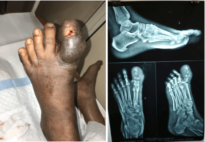 Figure 1: Foot x-ray showing osteomyelitis at the hallux.
View Figure 1
Figure 1: Foot x-ray showing osteomyelitis at the hallux.
View Figure 1
Osteomyelitis is challenging to treat due to multidrug resistance of common pathogens and poor penetration of antibiotics into bone [4]. Hemihydrate form of Calcium Sulfate is produced using a synthetic process resulting in pure grade with no traces of toxic impurities. It has the advantage of delivering a wider spectrum of antibiotic combinations into the affected joint.
Osteomyelitis is challenging to treat due to both multidrug resistance microbes and to poor penetration of antibiotics into the bone. Inadequate release of antibiotics to the site of infected bone is a frequent problem associated with systemic antibiotic therapy and sometimes even with local drug delivery system. Purified synthetic calcium sulphate is absorbable, can be readily mixed with antibiotics and positioned at the site of infection. This technique provides advantages over other local antibiotic delivery systems as it is biodegradable, predicable elution characteristics, osteoconductive and finally it can fill the dead space.
Post-Operative Radiograph evaluation was performed (Figure 2A and Figure 2B) in the patient after 2 weeks and later at 4 weeks to find out the bony integration of the calcium sulphate impregnated with antibiotic beads. Patient achieved healing with a median time span of 4 weeks and no recurrence within 12 months after intervention (Figure 2C). The need for postoperative antibiotics was decided as per clinical assessment and here the patient was prescribed with Ceftriaxone (500 mg)/day for a period of 5 days. Foot X-ray of the patient after 14 weeks is presented in Figure 3A and Figure 3B.
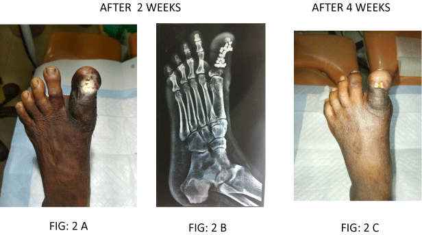 Figure 2: Post-operative x-ray showing partially absorbed calcium sulfate beads.
View Figure 2
Figure 2: Post-operative x-ray showing partially absorbed calcium sulfate beads.
View Figure 2
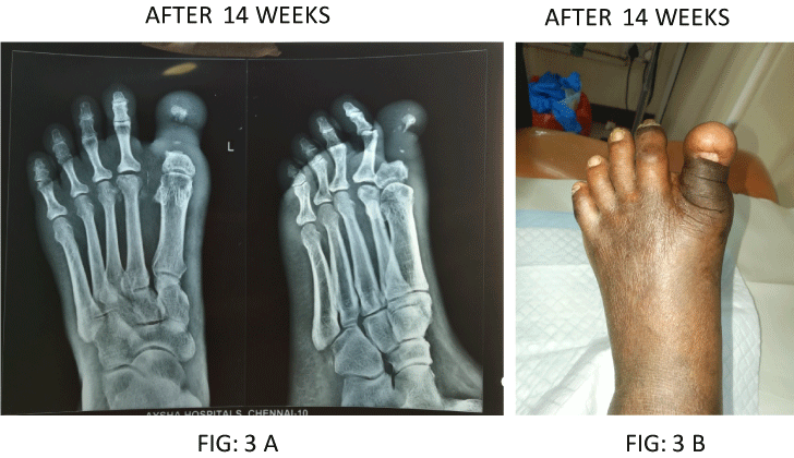 Figure 3: Post-operative x-ray completely absorbed calcium sulfate beads.
View Figure 3
Figure 3: Post-operative x-ray completely absorbed calcium sulfate beads.
View Figure 3
This procedure is found to be safe and effective for the treatment of forefoot diabetic osteomyelitis. No adverse reaction was noted in the patient and now with confidence our centre offers this treatment to the patients with midfoot and calcaneal osteomyelitis.
Our ultimate aim is to eradicate infection, heal the ulceration and reduce the need for intravenous antibiotics in the treatment of osteomyelitis. Hence this protocol was adopted to achieve an alternative route of administration of antibiotics in the management of Diabetic foot ulcers.