Uranyl acetate (UA) is one of the heavy metals used to stain tissue in sample preparation for transmission electron microscopy (TEM). It gives excellent contrast to ultrastructure morphology. Unfortunately, UA is very toxic and has proven to be a carcinogen and radioactive reagent. Meanwhile, UA-Zero is a UA replacement with improved staining property has shown better for staining due to its safety profile and environmental-friendly properties. Therefore, en-bloc staining with UA-Zero provides better quality in sharpness, brightness, and contrast of electron micrograph images for kidney and other samples.
Uranyl acetate, UA-Zero, Uranyl acetate replacement, Transmission electron microscopy
TEM delivers valuable information in morphological science, including histology and histopathology [1]. TEM is a microscopy-based technique that visualizes the particulars of the ultrastructural morphology of the cells [2] when tissue reacts to a high-energy electron beam. It produces dark regions when the electron transmits or bright in areas where the electrons pass through the tissue. The grey part will be present when both fields interface. Thus, it yields TEM micrographs as dark, bright, and grey fields in the images [3].
This contrast shall be presented between the mass per unit area of the tissue when it reacts with heavy metal. UA is a heavy metal used and plays an essential role in TEM. It can bind easily with the overall components of the tissue after staining. This UA-Zero gives good-quality images in the aspect of contrast as visualized with TEM [4]. Besides, the uranium atom has a large atomic number and radius, providing a solid electron beam scattering effect to the micrograph [5] as in UA-Zero. However, UA needs to be handled carefully due to its toxicity which is a potentially carcinogenic element. The use of UA, which is a radioactive item, needs approval by the authorities and needs to abide by rigorous rules. Users are required to handle UA carefully while storing, shipping, or waste disposal [6].
Due to the UA's toxicity, carcinogenicity, and radioactive, UA-Zero was used to replace UA. UA-Zero is a non-radioactive and uranium-free staining solution. It is safe to use, transport, and dispose of because it contains no radioactive materials [7]. UA-Zero can be a direct replacement for UA without any changes to the present protocol, and no additional processing is required to provide the highly contrasted imaging.
In this study, we would like to examine the outcome of the electron micrograph from TEM after the replacement by using UA-Zero while maintaining or enhancing the quality of the electron's sharpness, contrast, and brightness micrograph images compared to UA staining from kidney samples [8].
Freshly dissected mouse kidney tissues from mice sourced from the Laboratory Animal Resource Unit, Institute for Medical Research, National Institutes of Health (NIH) fixed in 2% phosphate-buffered glutaraldehyde. The tissues were then post-fixed with 1% osmium tetroxide and en-block-stained with UA Zero and 1% UA. The tissues were then dehydrated with 30%, 50%, 70%, and 100% (3X) acetone, embedded in Agar 100 epoxy resin, and sectioned to a 90 nm thickness. The residual of Reynalds' unbound stain was removed by rinsing the sample grid with distilled water after 10 minutes. The stained ultra-thin sections were examined with TEM (FEI Techni G2) at 100 kW.
The TEM images showed good quality sharpness, contrast and brightness in the stained kidney sections for UA-Zero samples compared to UA-stained (Figure 1 & Figure 2). The other ultra-structures of the mice kidney tissues, namely nuclear and mitochondria were clearly seen (Figure 3 & Figure 4) compared to UA stained (Figure 5, Figure 6, Figure 7 and Figure 8). There were no significant charging effects on kidney sections by using UA-Zero. This method of replacement of UA to UA-Zero is seen to produce high quality ultrastructural images while it is less carcinogenic and radioactive to its technicians.
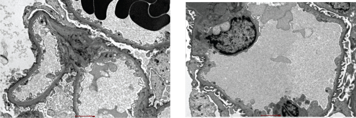 Figure 1 and 2: Mice kidney sections were stained using UA-Zero protocol. Foot processes and glomerular basement membrane can be viewed clearly with good quality contrast and sharpness. (Magnification at 1100X and scale bar is 2 um).
View Figure 1 and 2
Figure 1 and 2: Mice kidney sections were stained using UA-Zero protocol. Foot processes and glomerular basement membrane can be viewed clearly with good quality contrast and sharpness. (Magnification at 1100X and scale bar is 2 um).
View Figure 1 and 2
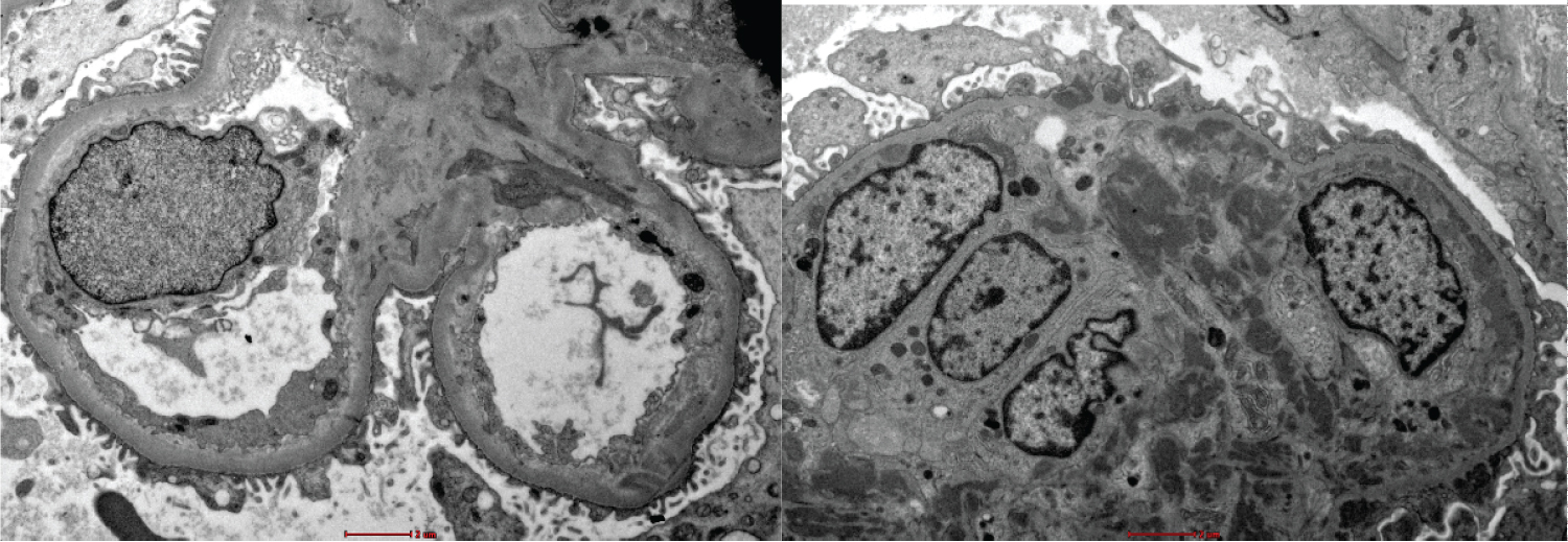 Figure 3 and 4: Mice kidney sections were stained using UA-Zero protocol. Nucleus and mitochondria can be viewed clearly with its good contrast and sharpness. (Magnification at 1100X and scale bar is 2 um).
View Figure 3 and 4
Figure 3 and 4: Mice kidney sections were stained using UA-Zero protocol. Nucleus and mitochondria can be viewed clearly with its good contrast and sharpness. (Magnification at 1100X and scale bar is 2 um).
View Figure 3 and 4
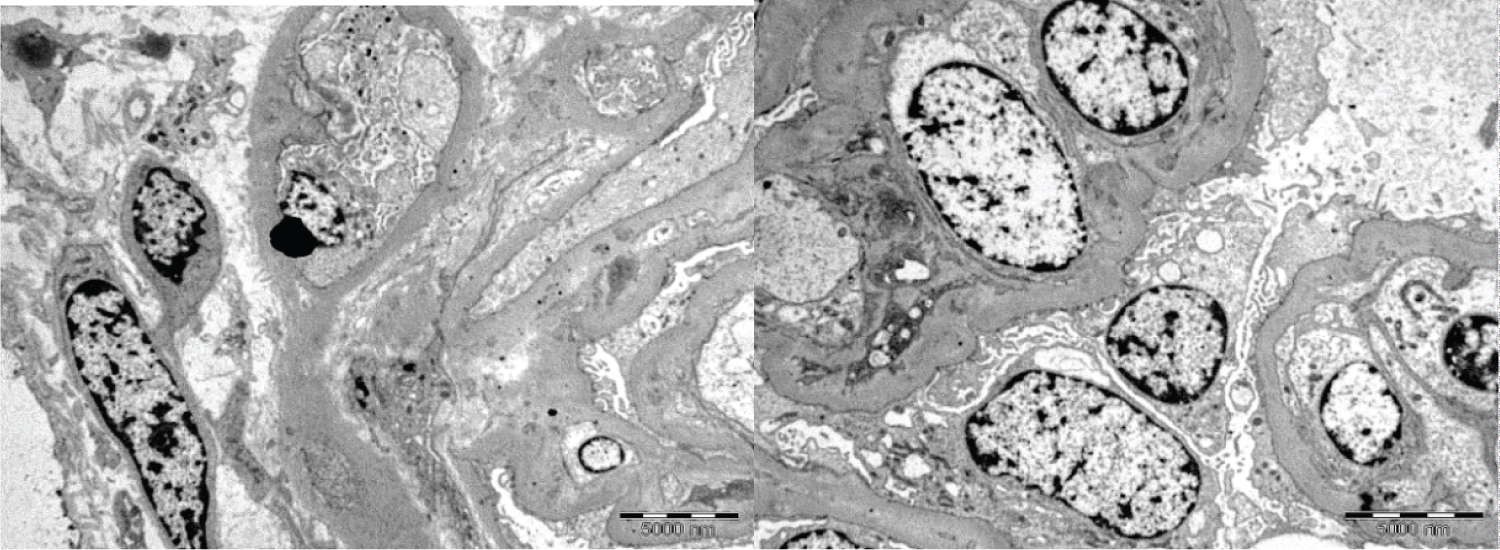 Figure 5 and 6: Comparing with mice kidney sections were stained using UA protocol. The contrast among nucleus and mitochondria less clear when compare with stained using UA-Zero as above. (Magnification at 1100X and scale bar is 5 um).
View Figure 5 and 6
Figure 5 and 6: Comparing with mice kidney sections were stained using UA protocol. The contrast among nucleus and mitochondria less clear when compare with stained using UA-Zero as above. (Magnification at 1100X and scale bar is 5 um).
View Figure 5 and 6
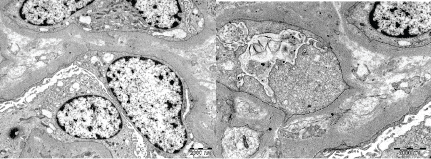 Figure 7 and 8: Another group of mice kidney sections were stained using UA protocol. Nucleus and membrane less sharp and contrast lesser compare with those stained with UA-Zero. (Magnification at 1100X and scale bar is 2 um).
View Figure 7 and 8
Figure 7 and 8: Another group of mice kidney sections were stained using UA protocol. Nucleus and membrane less sharp and contrast lesser compare with those stained with UA-Zero. (Magnification at 1100X and scale bar is 2 um).
View Figure 7 and 8
A study by Modla, et al. (2010) [9] showed that UA staining could increase the contrast and brightness of TEM images. However, in our research, we added the UA-Zero during en-bloc staining to increase the penetration of UA-Zero into the tissue and membrane to give the optimum contrast and brightness before sectioning. It has to produce higher-quality ultrastructural images than using UA staining.
The modified procedure is a better alternative for a safer contrast stain. While being more confident of the staining outcome as it does not compromise image quality, especially in diagnostic studies [8]. Compared with previous year's cases with UA during staining, it showed less contrast and less sharpness in the ultrastructural image (Figure 5, Figure 6, Figure 7 and Figure 8). With this comparison, it was clear that staining during en-bloc was very helpful for its performance. Using UA-Zero can also provide better images in contrast and brightness.
A few steps must be considered for TEM ultrastructure to be of acceptable quality during the contrasting protocol. The reliability of ultrastructural images on the tissue sections should be maintained. The UA-zero should create the contrast that allows viewing the organelles of significance [10]. Thus, the UA-Zero produced a high-quality ultrastructural image in this current study.
Furthermore, other staining reagents are safe for use, including oolong tea extract (OTE) and neodymium for TEM staining, which are not toxic and not radioactive [11,12]. However, we have not studied these staining reagents and cannot make any direct comparison. It should be an area that we could learn in the future.
Using the UA-Zero can reduce UA's toxicity and carcinogenicity to users and the environment. At the same time, using UA-Zero will maintain excellent ultrastructural images on the electron micrograph's sharpness, contrast, and brightness. After comparing it with the previous year's image, we can see an improvement in the image quality. This method UA-Zero is recommended to replace conventional UA in the future for all TEM samples. At the same time, it is less carcinogenic to its technicians and researchers.
We want to thank the Director-General of Health, Malaysia, for his permission to publish this paper. We also like to extend our gratitude to the Director of the Institute for Medical Research, Malaysia, for supporting this study.
The authors declare there is no conflict of interest in conducting this study.