Limbus vertebra is marginal interosseous herniation of the nucleus pulposus. It is generally considered to be an incidental finding without any symptoms unless posterior herniation causes nerve compression. However, recent findings have suggested limbus vertebra may be associated with a specific gene and intervertebral disk degeneration. This article reports a case of a 40-year-old male patient with limbus vertebra, intervertebral disk degeneration, and low back pain, followed by a comprehensive literature review.
Limbus vertebra, Low back pain, Intervertebral disk degeneration, COL11A1 polymorphism
Limbus vertebra was first described by Schmorl in 1927 [1]. Schmorl postulated limbus vertebra is formed from an intrabody herniation of disc material during childhood or adolescence [2]. On plain films, it appears as a triangular osseous fragment adjacent to the corner of a vertebral body. It was frequently mistaken for an infection, fracture, or tumor initially. However, pathologic studies of these fragments revealed the presence of "disc material" [3]. Then, in 1976, Ghelman and Freiberger provided the pathophysiologic evidence via discography, which demonstrated injection of contrast into the nucleus pulposus indeed extends around the limbus vertebra [4].
The most common presentation of limbus vertebra is at the anterosuperior margin of a single vertebral body in the lumbar spine. Postero inferior corner and other regional involvement can also be seen albeit much less in frequency [4]. Generally, anterior limbus vertebra (ALV) is thought to be asymptomatic, whereas posterior limbus vertebra (PLV) can mimic intervertebral disc herniation symptoms due to nerve compression [5,6]. However, recent data seem to suggest TT genotype of COL11A1 polymorphism being a risk factor for limbus vertebra [7] and the association of ALV with intervertebral disk degeneration (IDD) [8]. This case reports a patient who presented to the clinic with low back pain with ALV and IDD.
40-year-old male patient with chronic low back pain presented with an acute exacerbation of his low back pain for two weeks. He denied any recent trauma at the site. He noticed low back pain for the first time in his mid-30's immediately after shoveling the snow. He had been experiencing intermittent flare-ups of his low back pain since that time, which occurred at a frequency of approximately once a year. It was usually preceded by lifting a heavy object. Each episode typically lasted for about a week and resolved spontaneously without requiring any treatment.
Upon further questioning, the patient stated he had been athletic and physically active all his life. He played three sports in high school, and he continues to run, play softball, and lift weights regularly into his adulthood. He also endorsed having participated in an endurance event where he ran 5 miles in mud while clearing 13 obstacle courses about a week before the onset of his recent low back pain. The pain was located over midline lumbar spine. The pain was 5 out of 10 on a pain scale. The pain was achy in quality. It was aggravated by activity and alleviated by rest. He denied any weakness or radiation of the pain into lower extremities. He also did not report any associated constitutional symptoms, inflammatory low back pain symptoms, saddle anesthesia, or fecal and urinary incontinence.
His physical examination findings were unremarkable except for spasm of the paravertebral musculature and decreased range of motion in the lumbar spine secondary to pain. His hip examination was unremarkable. The motor strength, straight leg test, sensation, and reflexes in the lower extremities were also normal.
Plain radiographic evaluation (Figure 1) of the lumbar spine demonstrated normal vertebral body height and alignment in the lumbar spine. However, he was found to have mild-to-moderate degenerative disc disease at L4-5 and a triangular osseous fragment adjacent to the anterosuperior corner of L5 vertebral body consistent with ALV. Due to the lack of any worrisome neurological, inflammatory, or "alarm" symptoms and signs, the decision was made not to pursue MRI evaluation. Instead, conservative treatments were rendered, including relative rest, modified activities as tolerated, and physical therapy for six weeks for range of motion and strengthening exercises for his core and paraspinal muscles. His low back pain subsequently resolved after about two months without any recurrence thus far.
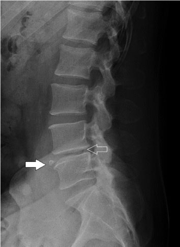 Figure 1: Mild to moderate degenerative disc disease at L4-5 (open arrow). A limbus vertebra was also noted at the anterosuperior aspect of the L5 vertebral body (closed arrow). View Figure 1
Limbus vertebra is considered to be a consequence of remote injury to an immature spine. The vertebral ring apophysis ossifies during childhood and adolescence until it eventually fuses with the vertebral body through skeletal maturation in early adulthood [9]. During this vulnerable period, chronic stress, trauma, or congenital abnormality could cause an intrabody marginal herniation of the nucleus pulposus between the ring apophysis and the adjacent vertebral body, resulting in limbus vertebra [4,10]. The detached fragment represents a portion of the ring apophysis that failed to fuse with the rest of the vertebra, which ossified separately [4,11].
Limbus vertebra is believed to be frequently localized at the anterosuperior margin of the vertebral body secondary to upper vertebral bodies being smaller compared to the adjacent lower ones [4]. During loading of the back in flexion, the anterior portion of the disc is likely forced into the superior end plate of the larger vertebra below [12]. Pathophysiology of limbus vertebra is also postulated to be similar in Schmorl's nodes and Scheuermann's disease where the nuclear material extrudes more centrally and at multiple levels in the lower thoracic spine, respectably [13,14]. As such, it is not uncommon for limbus vertebra, Schmorl's nodes, and Scheuermann's disease to coexist in the same patient.
The typical roentgenographic appearance of limbus vertebra is a small, corticated, triangular, osseous density with sclerosis of the surface of the bone defect adjacent to the corner of a vertebral body in adults [4]. As such, plain radiographic examination of the spine is usually adequate to make the diagnosis [15]. However, in children and adolescents, only an irregular destructive-appearing process may be present on the vertebral margin rendering the diagnosis more ambiguous [4]. In cases where roentgenographic appearance may not be sufficient, especially for PLV where the lesions at the L5 and S1 levels are often obscured by pelvic structures, CT and MRI have been shown to be helpful in confirming the diagnosis of limbus vertebra [6,15]. This would be especially important for patients with PLV who may require surgery due to failed conservative treatment from nerve compression. The surgery for PLV may require techniques and equipment that are not routinely used for an intervertebral disc herniation [6].
Unlike PLV, there has been a long controversy about the clinical significance of ALV. Many considered ALV to be an incidental finding in asymptomatic patients [5,16]. However, MRI studies performed on adolescents with ALV revealed a high frequency of associated IDD similar to Scheuermann's disease [13]. Furthermore, Henales, et al. [17] published 13 pediatric patients with ALV who presented with symptomatic low back pain in 1993. Most recently, Koyama, et al. [8] reported a high prevalence of low back pain, limbus vertebra, and IDD in 104 Japanese collegiate gymnasts and significant association of ALV with IDD in these collegiate gymnasts. In addition, Koyama, et al. [7] also found COL11A1 genotype and sporting experience being risk factors for limbus vertebra, and the risk of limbus vertebra decreasing with age. Acosta, et al. [18] have also suggested sporting experience as being an important risk factor for limbus vertebra, and Baranto, et al. [19] reported that the frequency of apophyseal changes indeed did not increase over time in a 15-year follow up of top athletes by MRI studies, which further lent support to the theory that limbus vertebra occurs during childhood and adolescence.
Regarding the patient in the case study, ALV likely occurred during his adolescence due to his vigorous sporting experience. Then, the accumulation of repeated bending force applied onto his extruded disc from his regular athletic activities probably contributed to the development of progressive IDD and chronic low back pain. This case supports the association of ALV with IDD and low back pain. As such, ALV should be included in the differential diagnosis of patients presenting with low back pain. Also, further studies need to be carried out to elucidate other potential genetic risk factors for limbus vertebra besides COL11A1 polymorphism.
• Limbus vertebra is marginal interosseous herniation of the nucleus pulposus due to congenital abnormality, chronic stress and trauma to an immature spine during childhood and adolescence. The pathophysiology of limbus vertebra is likely to be similar and related to Schmorl's nodes and Scheuermann's disease.
• Limbus vertebra can be easily diagnosed via plain films in adults by visualizing a small, corticated, triangular, osseous density with sclerosis of the surface of the bone defect adjacent to the corner of a vertebral body. However, such appearance may not be so obvious in children and adolescents, which may necessitate ordering CT or MRI to confirm the diagnosis.
• Anterior limbus vertebra (ALV) is the most common presentation of limbus vertebra, which has been previously thought to be asymptomatic. However, recent data suggest a strong relationship of ALV with sporting experience, TT genotype of COL11A1 polymorphism, intervertebral disk degeneration and low back pain.
• Posterior limbus vertebra is far less frequently seen compared to ALV, but may cause symptomatic nerve compression that may require surgery if conservative treatment fails.
• Limbus vertebra needs to be considered in the patients presenting with low back pain, and further studies need to be performed to elucidate other genetic risk factors that may be associated with limbus vertebra besides COL11A1 polymorphism.
There aren't any potential conflict of interest.
There is no government, commercial, private foundation, pharmaceutical or industry support.
The patient has provided written informed consent for his case details and images to be published.