The development of small or moderate size left ventricular thrombus (LVT) is a well-known complication in various cardiac conditions with the highest rate observed in acute anterior myocardial infarction and congestive heart failure (CHF) as a result of severe left ventricular (LV) systolic dysfunction, but a huge LVT that almost protrudes to the aortic valve is an exceptionally rare phenomenon.
This is a case of a 38-year-old male with a history of chronic methamphetamine use presented with cardiomyopathy and massive LV thrombosis. The thrombosis measured 4.4 cm × 2.1 cm which almost protrudes to the aortic valve. The patient was started on Anti coagulants and was closely monitored at the ICU. The patient died on the 4th day with multi organ failure.
There is an increasing trend that methamphetamines and related substances are now among the most abused drugs worldwide. Clinicians should maintain a high degree of suspicion when assessing all patients with a history of methamphetamine abuse, especially chronic users and those who may have heart failure symptoms with no prior history of coronary artery disease, hypertension, valvular disease, and congenital heart disease. Physicians should also be aware of the cardiac complications of methamphetamine and other related substances such as formation of left ventricular thrombus.
Left ventricular thrombus is defined as an echo-dense mass, contiguous but distinct from the endocardium, located in an area of a synergy that was seen in both systole and diastole in at least two echocardiographic views [1]. The combination of blood stasis, endothelial injury and hypercoagulability, often referred to as Virchow's triad, is a prerequisite for in vivo thrombus formation [2]. Anterior location of the infarction, left ventricular ejection fraction of < 35% and apical dyskinesia or aneurysm were the major variables related to thrombus formation [1].
Cardiomyopathies are either confined to the heart or are part of a generalized systemic disorder, both often leading to cardiovascular death or progressive heart failure-related disability. Other diseases that cause heart muscle dysfunction are excluded, such as coronary artery disease, hypertension or abnormalities of the heart valves. Toxic cardiomyopathy is an example of this, often which the cardiotoxicity is associated with very high levels of exposure or acute overdoses, in which acute electrocardiographic and hemodynamic abnormalities may reflect both direct drug effect and systemic toxicity. Cocaine, amphetamines and related catecholaminergic stimulants can produce chronic cardiomyopathy as well as acute ischemia and tachyarrhythmia [3]. The aim of this report is to present a rare case of large left ventricular thrombus in a patient with chronic history of methamphetamine use who presented with symptoms of heart failure.
D.C., 38-year-old Male, Filipino, born on October 10, 1980 in Batangas, residing at Manila city, admitted for the first time at Tondo Medical Center, on October 8, 2019 with a chief complaint of difficulty of breathing.
Two months prior to admission, patient had episodes of difficulty of breathing upon excursion associated with non productive cough. Patient did not seek consult and no medications were taken.
One month prior to admission, still with aforementioned symptoms but now with associated 3 pillow-orthopnea and bipedal edema. Patient still did not seek consult and no medications were taken. During the interim patient had progressive symptoms until the dyspnea is felt even at rest.
One day prior to admission, still with above symptoms but now with severe difficulty of breathing. Patient was brought to a private physician where the patient was advised to transfer to a larger hospital with an ICU setting because the patient was assessed to be hypotensive. Patient was then transferred to our institution and was subsequently admitted.
Patient denies any history of hypertension, diabetes mellitus, cardiac problem, asthma and previous PTB treatment. Previous hospitalization was due to pneumonia last 2012. Patient had no history of any heredofamilial diseases. Patient worked as a garbage collector since 2015. Patient is a 30-pack-year smoker, alcoholic beverage drinker and a chronic methamphetamine user since 2012.
On admission, patient was stretcher bourne, stuporous, weak looking, can speak in phrases, medium build, medium skin tone, edematous, with a height of 163 cm, weight of 60 kg, Body Mass Index of 22.6 kg/m2, in cardiorespiratory distress with the following vital signs: Blood pressure: 60/40, Heart Rate: 117, Respiratory Rate: 28, Temperature: 36.7 ℃, O2 saturation: 95% on room air. Patient had anicteric sclera, pale palpebral conjunctiva, no tonsillopharyngeal congestion and no cervical lymphadenopathy. Chest had symmetrical chest expansion, no lagging, with subcoastal retractions and crackles heard on both lung fields, mid to base, negative for bronchophony, egophony and whispered pectoriloquy. Precordium was dynamic, heart has fast rate, regular rhythm, with no murmur noted. Neck veins were distended. Abdomen was flabby, normoactive bowel sounds noted, soft and non-tender. Extremities were grossly normal, with grade II bipedal edema. Neurologic exam revealed GCS 15 (E4V5M6), with no noted motor and sensory deficit. Cranial nerves were intact. While admitted, patient was noted with hypotension attributed to cardiogenic shock secondary to toxic cardiomyopathy. Laboratory results revealed elevated BUN and creatinine levels secondary to prerenal azotemia. Chest X-ray revealed Cardiomegaly with congestive changes and pneumonia both lower lobes. 2-Dimensional Echocardiogram revealed a dilated left ventricular dimension with increased LV mass index and normal relative wall thickness with hypokinesia of almost all left ventricular segments except basal inferolateral left ventricular free wall. Left ventricular ejection fraction was 22% and there was A large echogenic left ventricular mass is noted (4.4 cm × 2.1 cm) which identified as a large left ventricular thrombus (Figure 1).
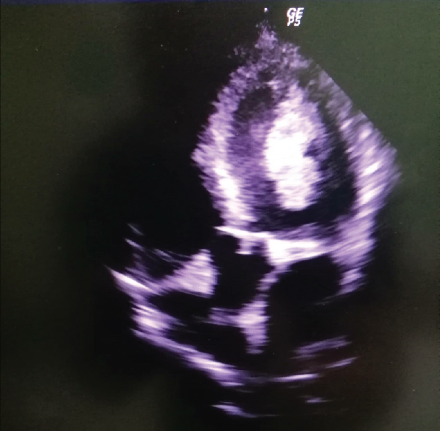 Figure 1: Large left ventricular thrombus.
View Figure 1
Figure 1: Large left ventricular thrombus.
View Figure 1
The patient became profoundly hypotensive which was likely due to cardiogenic shock despite being on maximum vasopressors. Pneumonia was managed with broad spectrum antibiotics. Patient was also started with vitamin K antagonist for the large thrombus. Interventional radiology and vascular surgery were consulted but the patient�s condition worsened developing acute kidney injury with severe metabolic acidosis. The patient could not tolerate continuos renal replacement therapy. The patient had 1 episode of ventricular fibrillation and expired after 4 days of being hospitalized.
We are presented with a case of a 38-year-old male who is a chronic methamphetamine user for 7 years who developed difficulty of breathing, orthopnea and bipedal edema. Patient was also noted to be hypotensive and is attributed to cardiogenic shock. Upon further analysis, it was found out on his 2d Echo that the patient has Eccentric left ventricular hypertrophy with multi-segmental wall motion abnormality suggestive of coronary artery disease with depressed global systolic function with grade III diastolic dysfunction, ejection fraction of 22%, left ventricular size of 6.3 cm, dilated left atrium, large left ventricular thrombus measuring 4.4 cm × 2.1 cm which is a rare case for a middle aged patient with no other co-morbidities.
In vivo thrombus formation is attributed to combinations of blood stasis, endothelial injury and hypercoagulability, often referred to as Virchow's triad [1]. Risk factors for thrombus formation include large infarct size (EF < 30%), severe apical akinesia or dyskinesis, formation of ventricular aneurysms, and anterior myocardial infarction [4]. Prevalence of left ventricular thrombus formation in general population is low. In a retrospective study by Lee JM on Left ventricular thrombus and subsequent thromboembolism, incidence of left ventricular thrombus was 7 per 10,000 patients [5]. Eighty percent of these cases were related to ischemia, while the rest were due to dilated cardiomyopathy and stress-induced cardiomyopathy.
Toxic cardiomyopathy is usually defined as a myocardial disorder in which the heart muscle is structurally and functionally abnormal, in the absence of coronary artery disease, hypertension, valvular disease and congenital heart disease sufficient to cause the observed myocardial abnormality. One of the common causes of toxic cardiomyopathies is the stimulant drugs (cocaine, methamphetamines) [6].
There is a growing body of evidence that methamphetamine abuse is associated with cardiomyopathy. Evidence points toward contributions from drug-induced vasospasm and ischemia, direct toxicity of methamphetamine, as well as deleterious effects of excess catecholamines on cardiomyocytes [7]. Methamphetamines exert its sympathomimetic effects by stimulating the central nervous system through excessive release of excitatory neurotransmitters such as norepinephrine, epinephrine, dopamine and serotonin while simultaneously blocking the reuptake at the sympathetic synaptic receptors which eventually leads to several clinical effects such as inducing euphoria, intensifying emotions, altering self esteem, and increasing alertness, aggression, and sexual appetite.
The Philippines ranked as the country with the second highest abuse rate for methamphetamine or �shabu� in East Asia, according to the latest United Nations World Drug Report [8]. In a study conducted at Philippine General Hospital by Duya J, et al. [9] on the clinical, electrocardiographic and echocardio graphic profile of adult Filipino patients with methamphetamine induced cardiomyopathy, they reported that in 22 patients seen from January to October 2016, the most common complaints prompting ER consult were dyspnea (77%) and paroxysmal nocturnal dyspnea (68%). About 77% were in NYHA functional class II-III and 30% were in cardiogenic shock. Echocardiographic data revealed 73% had left ventricular dilatation, with mean ejectionfraction of 29% by teicholz and 13% by Simpsons.
Diagnostic modalities to detect left ventricular thrombus include echocardiography. Two dimensional Transthoracic Echocardiography is the technique used most often for assessing the presence, shape and size of a left ventricular mural thrombus. It has 85-90% specificity and 95% sensitivity in detecting left ventricular thrombus. Computed tomography scan provides about the same specificity and sensitivity with echocardiography but is not routinely used because it requires intravenous injection of radiographic contrast material and exposes the patient to ionising radiation. Cardiac magnetic resonance imaging with contrast (delayed enhancement) has significantly better accuracy than Transthoracic Echocardiography. It allows for a relatively rapid assessment of thrombus presence, size, and location and is nowadays considered the gold standard [2,10,11].
Pharmacologic management of left ventricular thrombus is mainly focused on anticoagulation therapy. Anticoagulation is considered in patients with such large anterior wall motion abnormalities, if they are at low risk of bleeding, to prevent the development of thrombi. Consensus is that mural thrombi, once diagnosed, require oral anticoagulant therapy with vitamin K antagonists for up to 6 months [12]. Intravenous thrombolysis has also been used for treatment of documented LV thrombus. In a report of 16 patients with LV thrombus on echocardiography, urokinase was infused intravenously at a rate of 60,000 U/h for 2-8 days in combination with intravenous heparin (200 units/kg × 12 h). LV thrombi were successfully lysed in 10 of 16 patients. None of the patients suffered from clinical embolism, and therapy had to be discontinued in only one patient due to haematuria. It was concluded that fibrinolytic agents are capable of lysing ventricular thrombi but that the risks of this therapy are too high. [1] In a study by Jaidka A, et al. on the treatment of left ventricular thrombus using warfarin versus oral anticoagulants following anterior myocardial infarction that triple therapy using Aspirin 81 mg/tab daily plus clopidogrel 75 mg/tab daily plus warfarin versus direct oral anticoagulants is proved to show similar rates of thrombus resolution at three to six months.
There is an increasing trend that methamphetamines and related substances are now among the most abused drugs worldwide. Clinicians should maintain a high degree of suspicion when assessing all patients with a history of methamphetamine abuse, especially chronic users and those who may have heart failure symptoms with no prior history of coronary artery disease, hypertension, valvular disease, and congenital heart disease. Physicians should also be aware of the cardiac complications of methamphetamine and other related substances such as formation of left ventricular thrombus.