
Myiasis refers to infestation of human tissue by larvae of flies of the order Diptera [1]. Clinical classification is determined by the anatomical site involved, with recognised forms including cutaneous, ophthalmic, nasopharyngeal, enteric, auricular, oral and urogenital myiasis. Cutaneous myiasis occurs worldwide but is more frequently reported in tropical and subtropical regions, where environmental conditions favour fly breeding. Nevertheless, wound myiasis may also occur in temperate climates when susceptible wounds are exposed to ovipositing flies. Other risk factors include exposed or open wounds, frailty, poor hygiene, low socioeconomic conditions, psychiatric illness, alcoholism, diabetes mellitus, peripheral vascular disease and malignancy [2]. Wound myiasis most frequently occurs in neglected ulcers; however, infestation of acute traumatic wounds is rare, particularly when involving the head and neck.
There are few population-based estimates of human myiasis incidence. National registry data from Ecuador suggest an annual incidence of up to 23 cases per 100,000 population in some regions, with substantially lower rates reported in non-tropical areas [3]. Narrative reviews from Europe indicate that wound myiasis accounts for approximately one-third of reported cutaneous myiasis cases among hospital presentations [2]. Despite this, rigorous epidemiological studies specifically quantifying the incidence of traumatic wound myiasis are lacking.
Although wound myiasis is uncommon in Australian hospital practice, clinicians should recognise that it may occur in both tropical and temperate environments when open wounds are exposed to flies. There is also limited guidance regarding the practical management of traumatic wound myiasis, including the role of systemic antiparasitic therapy. This report describes an unusual case of acute traumatic forehead wound myiasis in a non-tropical setting and discusses key diagnostic and management considerations.
A woman in her late 90s presented to the Emergency Department of Canberra Hospital, Australian Capital Territory, Australia, after being found on the ground in her garden following an unwitnessed fall. The injury occurred in Canberra, Australian Capital Territory, during the Australian autumn. The wound remained exposed for an estimated 5-7 hours before hospital assessment. The patient was able to recall the circumstances of the fall and denied any loss of consciousness. She reported tripping while walking in the garden and falling forwards, striking her head and sustaining a frontal scalp injury. There were no prodromal symptoms such as chest pain, dyspnoea or dizziness. According to her son, her cognition was at baseline. She had a history of recurrent falls.
Her medical history included type 2 diabetes mellitus, chronic kidney disease, hypertension and hypercholesterolaemia. She lived with her son, was independent in personal activities of daily living, and mobilised with a single-point stick. There was no recent intercurrent illness or change in oral intake, and no history of recent travel to tropical or subtropical regions.
On examination, the patient was haemodynamically stable but appeared clinically dehydrated. An approximately 8 cm central transverse forehead laceration extending down to the muscle layer was present (Figure 1, Figure 2 and Figure 3), heavily contaminated with soil, with visible larval infestation within a deep wound pocket. There was associated periorbital bruising and swelling. Multiple smaller superficial skin tears and bruises were noted on the extremities. Neurological examination demonstrated intact cranial nerve function, normal speech and movement of all four limbs, with no focal neurological deficit. No other clinically significant traumatic injuries were identified.
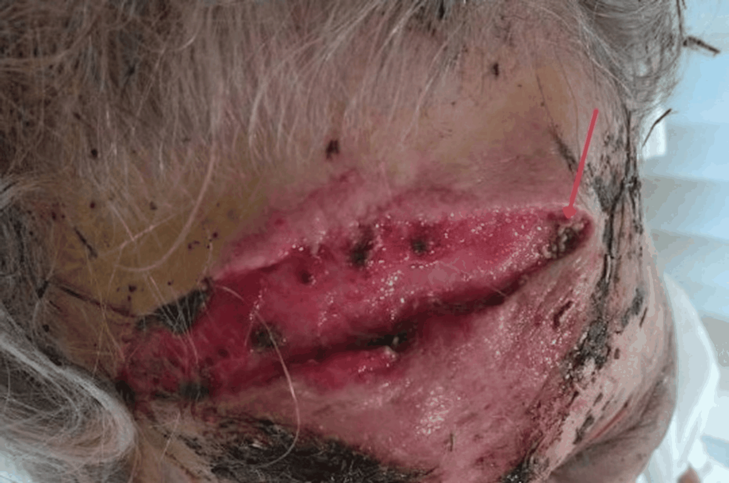 Figure 1: Clinical photograph of a transverse forehead laceration with visible larval infestation (red arrow) prior to definitive surgical debridement.
View Figure 1
Figure 1: Clinical photograph of a transverse forehead laceration with visible larval infestation (red arrow) prior to definitive surgical debridement.
View Figure 1
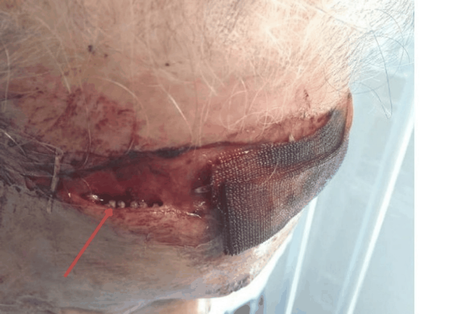 Figure 2: Clinical photograph showing visible larvae within a deep wound pocket (red arrow).
View Figure 2
Figure 2: Clinical photograph showing visible larvae within a deep wound pocket (red arrow).
View Figure 2
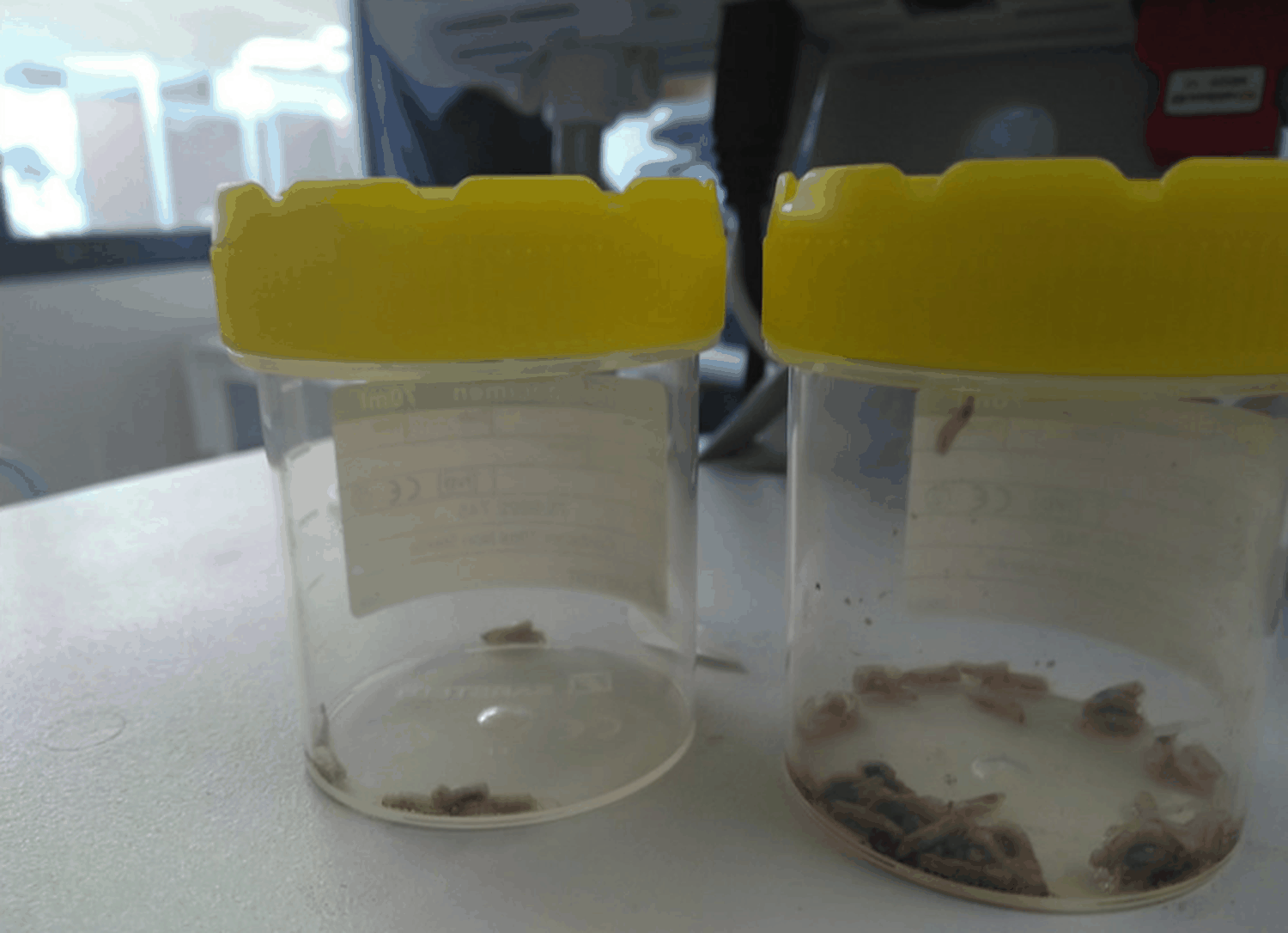 Figure 3: Wound-derived larvae collected in a specimen container following debridement. Species identification was not performed.
View Figure 3
Figure 3: Wound-derived larvae collected in a specimen container following debridement. Species identification was not performed.
View Figure 3
Initial blood investigations demonstrated a haemoglobin concentration of 130 g/L and a platelet count of 200 × 10 9 /L. The white cell count was elevated at 17 × 10 9 /L, with a neutrophil predominance of 15.81 × 10 9 /L, lymphopenia at 0.34 × 10 9 /L, and an eosinophil count of 0 × 10 9 /L. C-reactive protein was 3.5 mg/L. Renal function testing showed an acute deterioration from baseline, with a urea concentration of 15.3 mmol/L and a creatinine level of 150 µmol/L (baseline creatinine 97 µmol/L; baseline urea 14.8 mmol/L), corresponding to an estimated glomerular filtration rate of 25 mL/min/1.73 m 2 (baseline 43 mL/min/1.73 m 2 ), consistent with stage 1 acute kidney injury. Serum sodium was 143 mmol/L and potassium was 4.7 mmol/L. Creatine kinase was markedly elevated at 3368 U/L suggestive of rhabdomyolysis, likely related to prolonged immobilisation following the fall. Computed tomography of the brain (Figure 4) demonstrated a frontal scalp soft tissue defect with associated haemorrhage, without evidence of acute displaced calvarial fracture or intracranial haemorrhage.
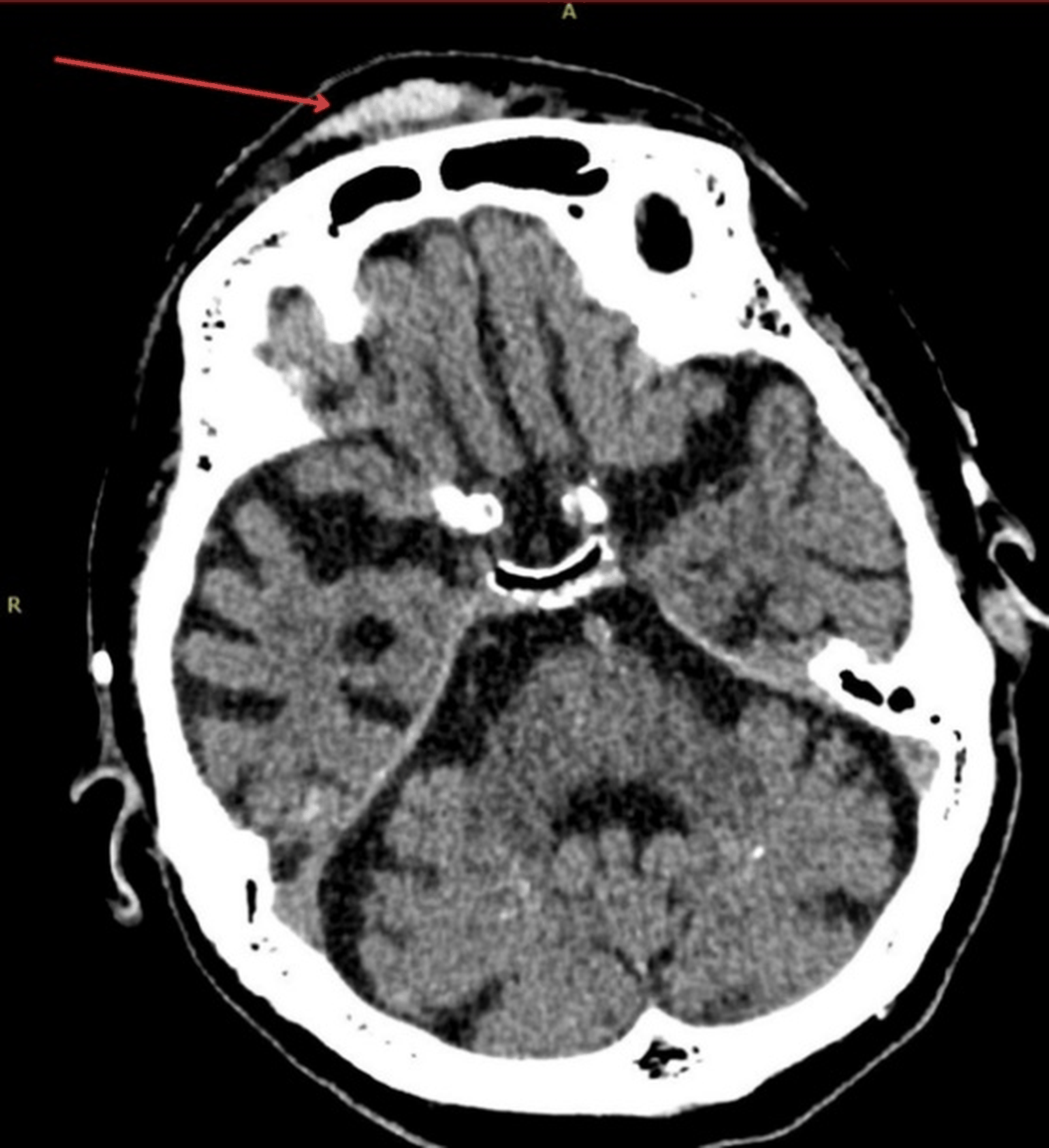 Figure 4: Axial non-contrast computed tomography of the brain showing a frontal scalp soft tissue defect (red arrow).
View Figure 4
Figure 4: Axial non-contrast computed tomography of the brain showing a frontal scalp soft tissue defect (red arrow).
View Figure 4
The wound was fully exposed and irrigated with copious normal saline. Mechanical removal of larvae was attempted at the bedside using forceps; however, a large number of larvae were identified within a deep pocket of the laceration, and complete extraction was not achievable in this setting. Supportive care was provided, including intravenous fluids and empirical intravenous cefazolin. Given the extent and depth of infestation, the patient was referred for operative management and subsequently underwent formal surgical debridement and wound washout in a sterile operating theatre environment under the care of the plastic surgery team. Intraoperative findings confirmed residual larvae within the wound cavity, which were completely removed, and devitalised tissue was debrided. Postoperatively, a short course of oral cephalexin was prescribed.
Renal function improved with intravenous fluid therapy and returned to baseline levels. The patient remained clinically stable and was discharged from hospital approximately one week later. At review one week following surgery, the forehead wound demonstrated satisfactory healing, with no evidence of recurrent infestation or secondary infection. The patient reported resolution of symptoms and was able to resume her usual activities, with ongoing community wound care support arranged.
The most plausible mechanism of infestation in this case is oviposition by adult flies onto the exposed traumatic wound during the prolonged period before rescue. The patient remained outdoors following her fall, with an open laceration heavily contaminated by soil and organic material. Moist, blood-containing wounds are well recognised to attract gravid flies, which may deposit eggs directly onto exposed tissue. Depending on the fly species and ambient environmental conditions, eggs may hatch within several hours, allowing larvae to become established before hospital presentation.
Although wound myiasis is more frequently reported in warmer climates, sporadic cases continue to occur in temperate countries, particularly in patients with neglected wounds, frailty, or delayed access to medical care. Diagnosis relies on direct wound inspection and may be missed if wounds are inadequately examined. A limitation of this case is that the larvae were not submitted for formal entomological identification. Consequently, the causative fly species could not be confirmed, and it cannot be determined whether infestation resulted from an obligate or facultative myiasis-producing species. The diagnosis therefore relied on direct clinical observation of larvae within the wound.
Management of wound myiasis centres on complete mechanical removal of larvae, thorough irrigation and debridement [4]. Occlusive dressings (such as petroleum jelly under an occlusive film) have been reported to facilitate extraction by inducing local hypoxia and encouraging larval migration to the surface [5-7].
Although oral or topical ivermectin has been described as an adjunctive therapy in selected cases of myiasis, particularly in extensive, cavitary, or inaccessible infestations, there is no high-quality evidence supporting its routine use following definitive surgical management [8]. Available data are largely limited to case reports and small case series, often involving malignant or deeply invasive disease, where complete mechanical removal is not feasible [9-13].
Given the absence of robust data on recurrence following operative debridement of wound myiasis, future studies comparing reinfestation rates in patients managed with and without adjunctive antiparasitic therapy would be valuable in informing evidence-based practice.
This report has several limitations. First, entomological identification of the larvae was not performed, preventing species confirmation and limiting epidemiological interpretation. Second, although the patient was estimated to have remained outdoors for several hours following the fall, the exact timing of fly oviposition and larval development cannot be established. Third, as a single case report, this study cannot determine whether adjunctive antiparasitic therapy provides additional benefit following complete operative debridement. Future cases incorporating species identification and longer-term follow-up may help clarify the epidemiology and optimal management of traumatic wound myiasis.
This case illustrates that traumatic wound myiasis may occur in temperate regions of Australia following prolonged exposure of an open wound outdoors. Prompt recognition, meticulous wound examination and complete mechanical removal remain the cornerstones of management. Species identification may provide valuable epidemiological information and should be considered in future cases whenever feasible. Management should prioritise complete mechanical removal of larvae with adequate irrigation and debridement, with escalation to operative management when bedside extraction is incomplete. Evidence supporting the routine use of adjunctive antiparasitic therapy remains limited, and treatment decisions should therefore be individualised according to the extent of infestation and the feasibility of achieving definitive mechanical clearance.
The authors received no specific funding for this work from the National Institutes of Health (NIH), the Wellcome Trust, or any other funding agency in the public, commercial, or not-for-profit sectors.
The authors contributed equally to this work. Both authors were involved in the conception of the report, manuscript preparation, critical revision of the manuscript, and approval of the final version for publication