A septate uterus is the most common congenital anomaly of the uterus. Pregnancy in a septate uterus may progress asymptomatically to term. However, complications may arise and threaten the obstetrical or even vital prognosis of the pregnant woman. The authors report a rare case of uterine rupture in the second trimester of pregnancy in a septated uterus.
The septate uterus is a congenital uterine malformation characterised by the presence of a septum dividing the uterine cavity [1]. It affects 2% of women of childbearing age, with poor fertility results [2]. This malformed uterine environment can also be a source of serious obstetric complications such as uterine rupture. Rupture of a pregnant septate uterus is a rare occurrence and constitutes a catastrophic obstetric emergency with unpleasant maternal and foetal consequences. Rupture in the 2 nd trimester of pregnancy is even rarer. We report a case of spontaneous uterine rupture in a non-scarring septated uterus in the second trimester of pregnancy. The interest of this observation is to show the obstetrical risk in women with these uterine malformations.
Mrs B.A aged 25 years, a shopkeeper, was referred to us from her local health centre for severe anaemia decompensated during pregnancy.
On history taking, she had been suffering from dizziness for 2 weeks, which had prompted a consultation at her local health centre, where she had received repeated blood transfusions without success. She also noted the onset of acute abdominal pain, which had been present since the day before her admission, with no evidence of metrorrhagia.
The patient was gravida 4, para 3, and the mother of three live children delivered at term by vaginal delivery. The pregnancy had not benefited from any antenatal consultation. Gestational age, estimated on the basis of the date of the last menstrual period, was 17 weeks of amenorrhoea. The medical and surgical history was unremarkable.
On admission, she presented with an altered general condition, pale conjunctivae and signs of shock requiring resuscitation measures. Abdominal examination revealed an enlarged abdomen and peritoneal irritation syndrome.
Gynaecological examination revealed a single closed cervix and a slightly enlarged uterus. Culdocentesis yielded blackish, incoagulable blood. The immediate urine beta HCG test was positive. The biological work-up carried out two hours before admission showed a haemoglobin level of 5.5 g/dl and platelets of 105,000/ul. Her blood group was rhesus B positive (B+). No radiological work-up could be performed. The diagnostic hypotheses put forward were: haemoperitoneum of aetiology to be explored: ruptured cyst in a pregnancy of 17 weeks of amenorrhoea? Ruptured ectopic pregnancy? Spontaneous rupture of a solid intra-abdominal organ in a pregnancy of 17 weeks of amenorrhoea? An exploratory laparotomy under general anaesthetic was indicated as a matter of urgency. At coeliotomy, we found a large haemoperitoneum of around 3 litres, which we aspirated. Exploration revealed a corpoeal partitioned uterus dividing the uterine body into two asymmetric cavities, the larger of which was on the left. The right cavity was the one carrying the product of conception. There was a uterine rupture at the fundic level extending to the right horn, allowing the foetus to escape in the intra-abdominal cavity (Figure 1).
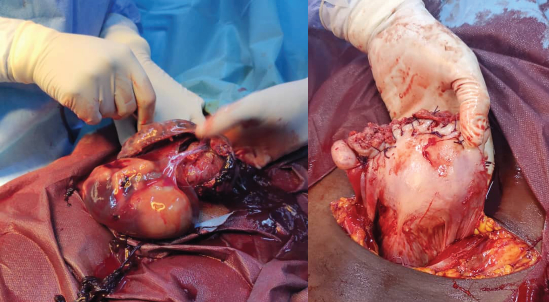 Figure 1: Intraoperative diagnosis of ruptured septate uterus.
View Figure 1
Figure 1: Intraoperative diagnosis of ruptured septate uterus.
View Figure 1
The edges were haemorrhagic. We proceeded with closure of the uterine breach followed by total right salpingectomy with good haemostasis. The right ovary and contralateral adnexa looked healthy. Prior to skin closure, we observed cardiac arrest, which led to successful resuscitation measures. Four hours later, her anaemia decompensated, leading to death in the context of a lack of labile product.
Partitioned uterus is the most common uterine anomaly [3-5]. It is caused by incomplete resorption of Muller's duct during embryogenesis. This malformation accounts for approximately 35% of all uterine malformations [4]. The uterine septum may be partial or total. Partial septated uterus involves a partial division of the uterus, whereas total septated uterus involves a complete division of the uterus from the uterine fundus to the external os of the cervix [1]. In our patient, the uterus was corporally divided.
The septated uterus is most often asymptomatic and the diagnosis is made incidentally during an X-ray examination or a surgical procedure performed for another purpose [6,7].
Treatment outside pregnancy is based on hysteroscopic resection of the septum [1]. The uterine malformation was unrecognised in our patient.
Pregnant women with a septated uterus may asymptotically carry their pregnancy to term and give live birth [3,6,7]. This was true for our patient who had already carried three (03) pregnancies to term. However, a septate uterus carries a higher risk of recurrent miscarriage, prematurity and infertility. This could be explained by poor blood supply to the septum and the narrowness of the compartment where the egg is implanted. Other more serious complications, such as uterine rupture, can occur. This was the case with our patient. The occurrence of spontaneous uterine rupture in the 2 nd trimester of pregnancy in our patient, who had asymptomatically carried three pregnancies to term, could be explained by the implantation of the egg in the small compartment. Uterine rupture remains a frequent and dramatic situation in developing countries with limited health resources such as ours [8]. Delays in consultation, evacuation and the lack of labile blood products could be factors contributing to the failure of our patient's management.