
Successful embryo implantation is a highly coordinated biological process that depends on precise temporal and molecular interactions between the embryo and the maternal endometrium. Endometrial receptivity is established during a limited window of implantation and is regulated by complex networks involving hormonal signaling, immune modulation, angiogenesis, cytokine balance, and gene expression dynamics. Progesterone-driven decidualization of stromal cells, coupled with tightly regulated estrogen signaling, creates a permissive environment for embryo attachment and invasion. Immune cells, particularly decidual natural killer cells and macrophages, play a central role in maintaining immune tolerance while simultaneously supporting vascular remodeling and tissue homeostasis. Cytokines, chemokines, and growth factors further fine-tune this environment by regulating trophoblast invasion, angiogenesis, and cellular communication at the maternal–fetal interface. Disruption of these molecular and cellular processes contributes to implantation failure, recurrent implantation failure, and adverse outcomes in assisted reproductive technologies (ART). Emerging evidence highlights the influence of oxidative stress, metabolic health, epigenetic regulation, and controlled ovarian stimulation on endometrial function and receptivity. Advances in molecular diagnostics, including transcriptomic profiling and personalized embryo transfer strategies, have improved the understanding and clinical management of implantation disorders. This review integrates current knowledge on the hormonal, immune, vascular, and molecular mechanisms governing endometrial receptivity and implantation, with a focus on their implications for infertility and ART outcomes, providing insights into potential biomarkers and therapeutic targets for improving reproductive success.
Endometrial receptivity, Embryo implantation, Epigenetic regulation, Gene expression, Cytokine signalling, Angiogenesis, Decidualization
A successful embryo implantation depends on the precise temporal and spatial coordination between a developmentally competent embryo and a receptive endometrium, making implantation a highly regulated and complex biological event [1]. Despite substantial progress in assisted reproductive technologies (ART), implantation failure remains a major obstacle to achieving successful pregnancy outcomes [2]. Notably, even after the transfer of morphologically high-quality embryos, a considerable proportion of couples undergoing in vitro fertilization (IVF) experience repeated implantation failure, highlighting both the biological complexity of implantation and the limitations of current clinical evaluation strategies [3]. This persistent challenge emphasizes the need to move beyond conventional morphological and hormonal assessments toward a deeper exploration of the molecular and cellular determinants of endometrial receptivity [4].
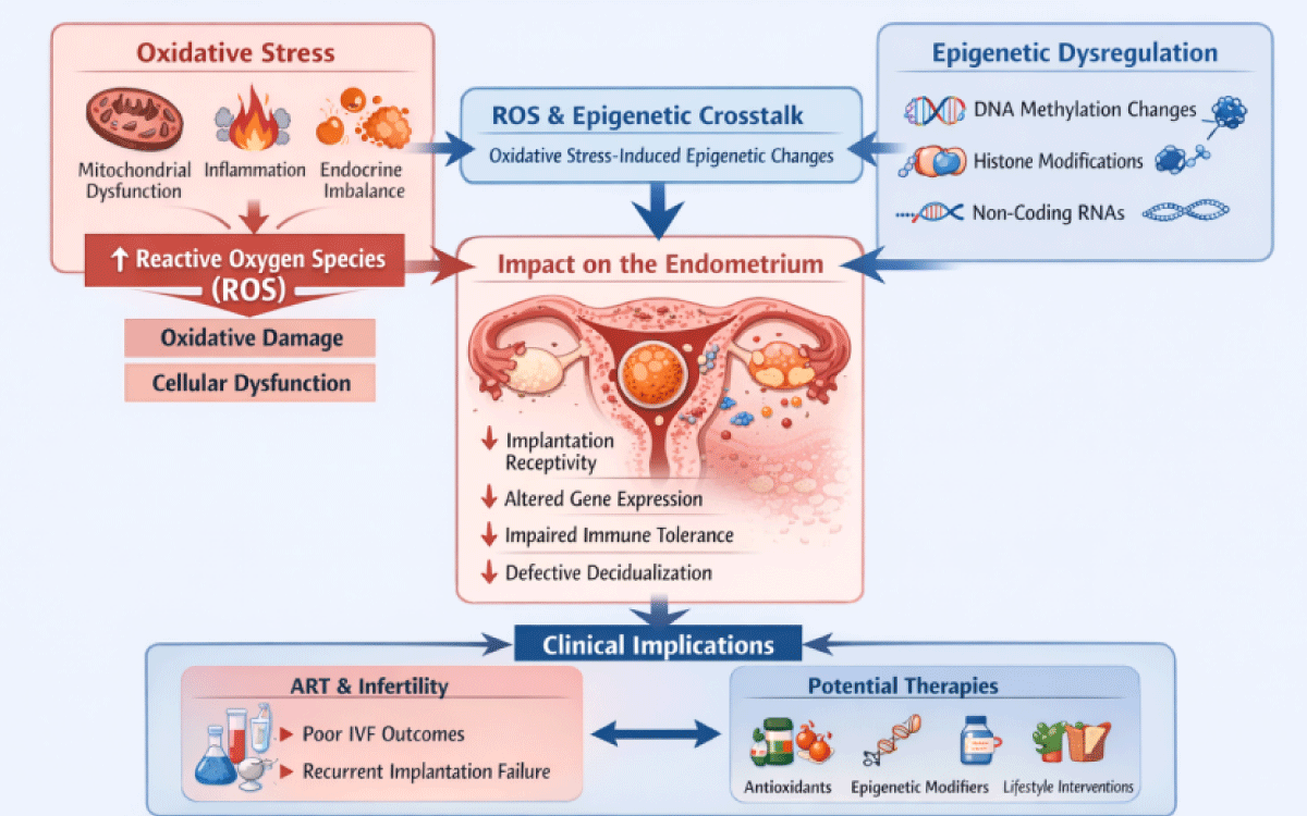 Figure 1: Molecular interplay between oxidative stress and epigenetic dysregulation in implantation failure.
View Figure 1
Several clinical factors, including hormonal imbalances, uterine structural abnormalities, inflammatory conditions, immune dysregulation, and embryo aneuploidy, have traditionally been associated with implantation failure [5]. Endocrine disturbances such as progesterone resistance, thyroid dysfunction, and altered gonadotropin signaling are known to disrupt the endometrial window of implantation [6]. In addition, impaired embryo-endometrium communication has been linked to chronic inflammation, abnormal cytokine expression, defective decidualization, and altered uterine perfusion. Although advances in imaging techniques, hormonal surveillance, and embryo grading systems have improved diagnostic accuracy, these approaches fail to explain implantation failure in a substantial subset of patients who present with morphologically optimal embryos, apparently normal endocrine profiles, and structurally normal uteri [7].
In response to this clinical paradox, research attention has increasingly shifted toward molecular alterations within the endometrium that may compromise receptivity in the absence of overt clinical abnormalities. Emerging evidence suggests that implantation failure may result from dysregulated gene expression, aberrant intracellular signaling, and impaired adaptive responses within the endometrial microenvironment rather than from gross structural defects [8]. Among the mechanisms governing endometrial adaptability throughout the menstrual cycle, epigenetic regulation has gained considerable interest [9]. Epigenetic processes-including DNA methylation, histone modifications, and non-coding RNA-mediated regulation-enable tightly controlled yet dynamic modulation of gene expression necessary for preparing the endometrium for implantation [10].
Endometrial receptivity is now recognized as being largely governed by epigenetic regulation [11]. Through reversible epigenetic modifications, the endometrium can respond efficiently to cyclical hormonal fluctuations without alterations to the underlying DNA sequence [12]. However, these epigenetic mechanisms are highly sensitive to metabolic and environmental influences, particularly oxidative stress [13]. Increasing evidence links reproductive dysfunction to oxidative stress, defined as an imbalance between reactive oxygen species (ROS) production and antioxidant defense mechanisms [14].
This review explores the hypothesis that oxidative stress contributes to implantation failure by functioning as an upstream driver of epigenetic instability within the endometrium [15]. By integrating molecular, cellular, and clinical evidence, we aim to provide a comprehensive overview of how redox imbalance and epigenetic alterations converge to impair implantation processes [16]. Such insights may help inform future clinical strategies aimed at improving implantation outcomes [17,18].
This narrative review was conducted to synthesize and critically evaluate existing evidence on the relationship between human implantation failure and oxidative stress-induced epigenetic instability. A comprehensive literature search was performed using electronic databases including PubMed, Scopus, and Web of Science. Relevant studies published predominantly within the past ten years were identified using combinations of keywords such as implantation failure, endometrial receptivity, oxidative stress, reactive oxygen species, epigenetics, DNA methylation, histone modification, and assisted reproductive technologies.
Priority was given to original research articles, clinical studies, and relevant review papers focusing on human reproductive biology. In addition, in vitro studies and experimental animal models providing mechanistic insights applicable to human endometrial function were considered. Publications that did not address implantation or endometrial biology, as well as those not available in English, were excluded from the analysis.
To ensure comprehensive coverage, the reference lists of selected articles were manually screened to identify additional relevant studies. The included literature was analyzed thematically, integrating molecular, cellular, and clinical findings to provide an overarching perspective on oxidative stress-epigenetic interactions in implantation failure. As this work was designed as a narrative review, no formal quality assessment or meta-analysis was undertaken.
Reactive oxygen species (ROS), including superoxide anions, hydrogen peroxide, and hydroxyl radicals, are generated as natural by products of normal cellular metabolism [19]. At physiological concentrations, ROS play essential roles in reproductive tissues by supporting angiogenesis, intracellular signaling, and tissue remodeling [20]. Within the endometrium, tightly regulated ROS production facilitates the vascular adaptations necessary for implantation, stromal cell differentiation, and cyclic tissue renewal [21]. However, when ROS generation becomes excessive or prolonged, endogenous antioxidant defense mechanisms may be overwhelmed, resulting in oxidative stress [22].
The endometrium is particularly vulnerable to oxidative stress due to its high proliferative activity, recurrent cycles of hypoxia and reoxygenation, and hormonally driven metabolic demands [23]. Multiple factors contribute to increased ROS production within the endometrial environment, including endocrine disturbances, metabolic dysfunction, inflammatory cell infiltration, mitochondrial impairment, and exposure to environmental stressors [24]. Clinical studies have demonstrated elevated markers of oxidative stress in the endometrial tissue, uterine fluid, and systemic circulation of infertile women. These alterations-characterized by increased lipid peroxidation, reduced antioxidant enzyme activity, and disrupted redox signalling-have been associated with unfavorable implantation outcomes [25].
Excessive oxidative stress can adversely affect key processes required for successful implantation, including endometrial cell viability, stromal decidualization, cytokine secretion, and maintenance of vascular integrity [26]. Importantly, the consequences of oxidative stress extend beyond transient cellular injury; sustained redox imbalance may induce persistent molecular alterations by disrupting regulatory pathways that govern endometrial function. As a result, increasing attention has been directed toward understanding how oxidative stress influences epigenetic regulation within the endometrium, potentially contributing to long-term impairment of receptivity [27].
Epigenetic mechanisms enable the endometrium to respond rapidly and reversibly to hormonal signals by modulating gene expression without altering the underlying DNA sequence [28]. Key epigenetic regulators-including DNA methylation, histone modifications, and non-coding RNAs-collectively influence chromatin organization and transcriptional activity, ensuring precise temporal control of genes essential for implantation [29]. DNA methylation patterns within the endometrium are closely linked to progesterone responsiveness and undergo dynamic changes across the menstrual cycle [30]. Aberrant methylation of genes involved in cell adhesion, immune tolerance, and decidualization has been identified in women experiencing implantation failure [31]. In parallel, histone modifications regulate transcriptional activation or repression by altering chromatin accessibility at implantation-specific genomic loci [32].
Non-coding RNAs, particularly microRNAs, play a critical role in post-transcriptional gene regulation during the window of implantation. Altered microRNA expression profiles have been associated with impaired endometrial receptivity, disrupted immune signaling, and defective embryo–endometrium communication. Collectively, these observations underscore epigenetic regulation as a fundamental determinant of implantation competence. However, epigenetic homeostasis is highly sensitive to redox balance, and oxidative stress can disrupt these regulatory processes, thereby compromising endometrial receptivity.
Beyond facilitating cyclical gene regulation, epigenetic mechanisms allow the endometrium to integrate hormonal, immune, and metabolic cues within a dynamically remodeling tissue environment. This integrative capacity is essential for coordinating stromal decidualization, epithelial differentiation, vascular remodeling, and immune tolerance during the implantation period [33].
Importantly, epigenetic alterations induced by oxidative stress may persist beyond the initial exposure, leading to sustained changes in endometrial gene expression across successive menstrual cycles [34]. Such persistence may help explain repeated implantation failure in women who display normalized hormonal profiles yet continue to exhibit impaired endometrial receptivity [28,35]. Moreover, recurrent inflammatory or metabolic insults may cumulatively diminish the endometrium’s responsiveness to embryonic signals by progressively reducing epigenetic flexibility. Together, these findings support the concept that implantation failure arises from an integrated response to redox imbalance within the endometrial microenvironment rather than from isolated epigenetic defects [36].
Oxidative stress can influence the epigenetic machinery through both direct and indirect mechanisms. Reactive oxygen species (ROS) are capable of disrupting DNA methylation patterns by modifying DNA bases, altering the availability of methyl donors, and interfering with the activity of DNA methyltransferases. In addition, oxidative stress has been shown to affect the acetylation and methylation states of enzymes responsible for histone modification, thereby influencing chromatin structure and gene regulation [37].
ROS also regulate the expression of non-coding RNAs involved in implantation-related pathways. Oxidative stress-induced alterations in microRNA expression may persist beyond the initial insult, giving rise to an epigenetic “memory” that affects subsequent menstrual cycles [37]. Beyond direct effects on epigenetic enzymes, oxidative stress can modify intracellular signaling pathways that govern chromatin dynamics and transcriptional responses [38]. Redox-sensitive signaling cascades influence the recruitment of epigenetic regulators to implantation-associated gene promoters, thereby reshaping transcriptional programs essential for endometrial receptivity [39]. Persistent oxidative stress may therefore lock the endometrium into a maladaptive epigenetic state, limiting its ability to respond appropriately to cyclical hormonal signals [40].
Notably, oxidative stress-induced epigenetic alterations may extend beyond a single menstrual cycle, resulting in temporally persistent effects. Even in cycles characterized by optimal hormonal support or improved embryo quality, this form of epigenetic memory may contribute to repeated implantation failure [41]. This mechanism provides a molecular explanation for the limited success of therapeutic approaches that focus solely on endocrine correction or embryo selection without addressing the underlying endometrial molecular environment [42]. Collectively, these observations suggest the existence of a self-perpetuating cycle in which oxidative stress and epigenetic dysregulation reinforce one another, progressively impairing endometrial function and reducing implantation potential. Breaking this cycle may require integrated therapeutic strategies that simultaneously restore redox balance and reestablish epigenetic plasticity within the endometrium [43].
Oxidative stress-driven epigenetic alterations can affect numerous biochemical pathways critical for successful implantation [44]. Genes involved in extracellular matrix remodeling and cell adhesion are particularly vulnerable. Proper expression of adhesion molecules and matrix-modifying enzymes is essential for stable embryo attachment and controlled trophoblast invasion [45]. Epigenetic silencing or inappropriate activation of these genes may disrupt epithelial–stromal interactions, leading to defective embryo anchoring or abnormal invasion dynamics, even in the absence of overt anatomical abnormalities [41].
Immune tolerance pathways within the endometrium are also highly sensitive to oxidative-epigenetic perturbations [46]. Successful implantation requires a finely tuned immune environment that supports acceptance of the semi-allogeneic embryo while maintaining local immune defense [47]. Oxidative stress–induced epigenetic reprogramming of genes involved in cytokine signaling, antigen presentation, and immune cell recruitment may shift the endometrial milieu toward a pro-inflammatory state [48]. Such immune imbalance can promote excessive immune activation, reduce regulatory immune cell function, and ultimately compromise embryonic survival during the peri-implantation period [49].
Angiogenic signaling represents another key target of oxidative-epigenetic dysregulation. Adequate vascular remodeling during early implantation is essential for nutrient exchange, oxygen delivery, and endometrial expansion [50]. Epigenetic modifications affecting angiogenic mediators may impair endothelial function and vascular stability, resulting in insufficient perfusion at the implantation site [51]. This vascular insufficiency may further exacerbate local hypoxia and oxidative stress, creating a vicious cycle that progressively undermines endometrial receptivity [52].
In addition to these mechanisms, oxidative stress has a profound impact on stromal cell decidualization, a progesterone-dependent differentiation process that is critical for implantation and early pregnancy maintenance. Epigenetic disruption of transcriptional networks governing progesterone responsiveness may delay or impair decidual transformation, even when circulating hormone levels are adequate [53,54].
Despite continued advancements in embryo selection techniques and laboratory optimization, implantation rates have not improved proportionally within assisted reproductive technology (ART) programs [55]. This discrepancy highlights a fundamental limitation of current ART paradigms, which remain largely embryo-centric while underestimating the importance of endometrial molecular competence [56]. The growing recognition of oxidative stress–mediated epigenetic dysregulation provides a compelling framework to explain persistently poor ART outcomes, particularly in patients with repeated implantation failure [57].
Despite continued advances in embryo selection strategies and laboratory optimization, implantation rates have not increased proportionally within assisted reproductive technology (ART) programs [58]. This discrepancy highlights a fundamental limitation of prevailing ART paradigms, which remain largely embryo-focused while underestimating the importance of endometrial molecular competence [59]. Increasing evidence linking oxidative stress-mediated epigenetic dysregulation to impaired receptivity offers insight into persistently suboptimal ART outcomes, particularly among patients with repeated implantation failure [60].
Controlled ovarian stimulation, a central component of ART, induces supraphysiological hormonal environments that can substantially alter both local and systemic redox homeostasis [61]. Elevated estrogen concentrations, altered luteal phase dynamics, and inflammation associated with stimulation protocols may enhance reactive oxygen species production within the endometrium. Although these effects are often transient, repeated ART cycles may lead to cumulative oxidative stress, increasing the likelihood of stable epigenetic alterations in endometrial cells [62,63]. Such changes may disrupt the finely regulated transcriptional programs required for receptivity, even when embryo quality is optimal.
Exogenous hormonal supplementation is routinely employed in ART to correct presumed luteal phase insufficiency [64]. However, oxidative stress–induced epigenetic alterations may impair progesterone receptor signaling or downstream transcriptional responsiveness, thereby diminishing the effectiveness of hormonal support [65]. This mechanism may explain implantation failure in some patients despite adequate serum progesterone levels and appropriately timed embryo transfers [66,67]. In these cases, the endometrium may be hormonally exposed yet functionally unresponsive due to epigenetically altered gene expression profiles [68,69].
The declining success of successive ART attempts can also be interpreted within this oxidative–epigenetic framework. Recurrent procedural interventions, hormonal manipulation, and psychological stress may contribute cumulatively to sustained oxidative burden, progressively reducing epigenetic flexibility within the endometrium [70]. Over time, this may lead to a refractory endometrial state that becomes increasingly resistant to conventional therapeutic approaches [71]. Recognition of this possibility underscores the importance of minimizing unnecessary cycle repetition and exploring adjunctive strategies aimed at restoring endometrial molecular integrity.
From a translational perspective, targeting oxidative stress and epigenetic dysregulation presents new opportunities to enhance ART outcomes. Interventions aimed at restoring redox balance-including lifestyle modification, metabolic optimization, and therapies designed to reduce systemic inflammation-may indirectly improve endometrial receptivity [15,14].
Antioxidant supplementation has been explored as a strategy to mitigate oxidative stress in reproductive medicine; however, reported outcomes have been variable and often inconsistent [72-74]. While some studies demonstrate modest improvements in implantation or pregnancy rates, others report no significant benefit. Factors contributing to this variability likely include heterogeneity in patient populations, differences in baseline oxidative status, variation in antioxidant formulations and dosages, and, critically, the timing of intervention relative to the implantation window [74,75]. Moreover, antioxidant therapies may have limited clinical efficacy if they fail to reverse established epigenetic alterations that persist within the endometrium [76].
Future therapeutic strategies may therefore require coordinated approaches that simultaneously restore redox homeostasis and reestablish epigenetic plasticity [77]. Lifestyle-based interventions-such as dietary modification, weight management, stress reduction, and physical activity-may play an important supportive role by reducing systemic inflammation and oxidative load. Optimization of metabolic and endocrine health, particularly in women with insulin resistance or thyroid dysfunction, may further enhance the endometrial environment and improve responsiveness to implantation signals [78].
Although still largely experimental in reproductive medicine, pharmacological modulation of epigenetic pathways represents an emerging area of interest [79,80]. When applied in a controlled and tissue-specific manner, agents targeting DNA methylation, histone acetylation, or non-coding RNA expression may hold therapeutic potential for improving endometrial receptivity [81].
To clarify the temporal relationship between oxidative stress, epigenetic alterations, and implantation outcomes, well-designed longitudinal clinical studies are urgently needed. Prospective investigations assessing redox and epigenetic markers across multiple menstrual cycles, as well as before and after targeted interventions, would be instrumental in establishing causality and identifying patient subgroups most likely to benefit from specific therapies [82]. Ultimately, incorporation of molecular profiling into clinical practice may facilitate personalized treatment strategies aimed at improving implantation success and reproductive outcomes [83].
Despite growing recognition of the role of oxidative stress and epigenetic dysregulation in implantation failure, the current evidence base is constrained by several important limitations. A major concern is that much of the available data derives from cross-sectional or observational studies, which limits the ability to establish causal relationships [84]. Although associations have been reported between oxidative stress markers, epigenetic modifications, and poor implantation outcomes, it remains unclear whether these molecular changes represent primary drivers of implantation failure or secondary consequences of underlying endocrine, metabolic, or inflammatory conditions [85].
A significant gap in the literature is the lack of long-term human studies. Most investigations rely on single time-point assessments of oxidative stress or epigenetic profiles, which fail to capture the cyclical and dynamic nature of endometrial remodeling across menstrual cycles [86]. Consequently, the temporal interplay between redox imbalance, epigenetic reprogramming, and implantation competence remains poorly understood. Longitudinal study designs that monitor molecular changes before, during, and after the implantation window are required to better evaluate persistence, reversibility, and clinical relevance [87,88].
Ethical and practical challenges associated with obtaining human endometrial samples further constrain study design. Repeated biopsies within the same cycle or across multiple cycles are often not feasible, resulting in limited sample sizes and reduced statistical power [89]. In addition, variability in biopsy timing relative to the menstrual cycle introduces heterogeneity that complicates cross-study comparisons and data interpretation. Standardization of sampling protocols and reporting practices remains insufficient within the field [90].
Another limitation relates to the translational applicability of in vitro and animal models. Although these systems provide valuable mechanistic insights into oxidative–epigenetic interactions, their relevance to human reproduction is restricted by species-specific differences in reproductive physiology, implantation mechanisms, and endometrial architecture [91].
Methodological heterogeneity across studies-including variation in oxidative stress markers, epigenetic assays, analytical platforms, and outcome definitions-further complicates data synthesis [89,90]. The absence of standardized biomarkers for assessing oxidative stress and epigenetic stability in the endometrium hampers the development of clinically useful diagnostic tools and contributes to inconsistent findings in therapeutic trials, particularly those involving antioxidant interventions [92].
Future research should prioritize integrative and interdisciplinary approaches that combine genomic, clinical, and environmental data to provide a more comprehensive understanding of implantation failure [93]. Advancing this field will require rigorously designed longitudinal studies, improved methodological consistency, and careful patient stratification. Addressing these limitations will be essential for translating insights into oxidative stress-epigenetic interactions into effective, individualized therapies aimed at improving implantation outcomes.
Implantation failure remains a complex and multifactorial challenge in human reproduction that cannot be fully explained by uterine anatomy, hormonal status, or embryo quality alone. A substantial proportion of implantation failures persist despite advances in ART and improved embryo selection techniques, underscoring the critical role of endometrial functional competence in reproductive success. Increasing evidence suggests that subtle molecular disturbances within the endometrium, rather than overt clinical abnormalities, play a central role in impaired implantation.
Oxidative stress and epigenetic dysregulation have emerged as interrelated and biologically plausible mechanisms contributing to progressive deterioration of endometrial function. Prolonged or excessive oxidative stress can disrupt redox-sensitive signaling pathways and epigenetic machinery, leading to aberrant gene expression patterns that compromise receptivity.
These molecular alterations may affect multiple implantation-related processes, including stromal decidualization, immune tolerance, angiogenesis, and embryo-endometrium communication. Importantly, oxidative stress–induced epigenetic modifications may persist beyond the initial insult, providing a molecular explanation for repeated or unexplained implantation failure even in the presence of optimal hormonal support and morphologically competent embryos.
Identifying redox imbalance as a trigger for epigenetic reprogramming offers an integrative framework that incorporates endocrine, metabolic, inflammatory, and environmental influences on endometrial function. This perspective challenges traditional embryo-centric models of infertility and highlights the need for more comprehensive evaluation of endometrial molecular integrity.
From a clinical standpoint, incorporating oxidative stress and epigenetic markers into diagnostic and therapeutic strategies may improve patient stratification, identify individuals at increased risk of implantation failure, and guide the development of more targeted interventions. Future progress in this field will depend on well-designed longitudinal studies that clarify causal relationships, define clinically relevant biomarkers, and evaluate interventions aimed at restoring both redox balance and epigenetic plasticity. Ultimately, integrating molecular insights into routine clinical practice holds promise for improving implantation success, reducing the burden of repeated ART cycles, and advancing personalized reproductive medicine.