Fetiform teratoma (homunculus) is a term that has been given to a rare form of teratoma which resembles fetus [1]. There are very few cases that reported in this entity. Because of its risk of malignancy; fetiform teratoma should be distinguished from fetus in fetu and ectopic pregnancy [1]. Fetus in fetu is in fact a rare case of intra-abdominal mass; usually seen in infants [2]. It is a congenital anomaly secondary to abnormal embryogenesis in a diamniotic monochorionic pregnancy [3]. Teratoma is the presence of variety of tissues in an abnormal location of the body [4]. Even the presence of high differentiated tissue does not exclude the diagnosis of teratoma as it happens in the case of homunculus. Teratoma, unlike fetus in fetu- does not show vertebral axis or regional distribution of the organs. In some cases of homunculus cephalic region is more developed; in others lower extremies and caudal region are more developed. The case with more developed cephalic region is more common.
We report a case of mature cystic teratoma containing a fetiform teratoma (homunculus) and to date there are only less than 20 cases to be reported.
Thirty three-years-old patient presented with abdominal distention and intermittent abdominal pain. She gave vaginal birth 2 times; last one three years ago. She didn’t have history of any abdominal operation, family history, or comorbidities. Her menstrual periods were regular. Review of the systems were unremarkable.
In the pelvic examination, a 10 cm abdominal mass palpated which deviated to the left side. The pelvic mass had a soft consistency and free mobility. In the ultrasonography, 80*80*70 mm measured complex lesion that resembles dermoid cyst was visualised at the right ovary. The routine laboratory that as at the admission were at normal ranges. Tumor markers were negative.
Laparoscopic surgical excision was done. At the exploration; the left tuba, the left ovary and uterus were visualised normal. Right ovarian cystectomy was performed without any spillage into the abdomen. The excised material was a complete homonculus which resembles the lower half of a miniature human body with buttocks, a pair of legs with recognizable feet; and an umbilical cord at the umbilicus area as seen in the Figure 1a, Figure 1b and Figure 2. There was rudimenter upper extremities not equal in size. Terminal digits were not present. The skin was covered by lanugo hair. The final pathological result was 100 * 80 * 30 mm measured adnexial mass containing hair, dermal tissue, sebaceous tissue. It was also containing bone, cartilage, blood tissues and mucosa; the tissues derived from the three germlayers. It was a benign cysticteratoma lined by normal skin. A relatively well developed osteo cartillage skeleton was identified in the pathological evaluation. There wasn’t any problem at postop follow-up. The patient was discharged on the second post-operative day.
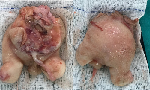 Figure 1: a) Caudal dominant fetiform structure covered with lanugo hair; b) Lower extremities, genital organ and umbilical cord are seen.
View Figure 1
Figure 1: a) Caudal dominant fetiform structure covered with lanugo hair; b) Lower extremities, genital organ and umbilical cord are seen.
View Figure 1
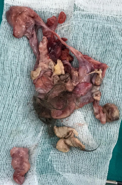 Figure 2: Cranial part and organ resembling structures of homunculus.
View Figure 2
Figure 2: Cranial part and organ resembling structures of homunculus.
View Figure 2
Mature cysticteratomas are common benign ovarian tumors especially seen in reproductive age. The majority of them composed disorganised embryonic germlayers: Ectoderm, mesoderm and endoderm. Rarely these tumors had a high degree of differentiation to a fetiform tissues: Homunculus. Although this adnexial masses are benign in nature; the totipotency of these tumors take great interest of the researchers. For differentiating homunculus from fetus in fetu; zygosity can be used. Homunculus as well as most ovarian teratomas are homozygos at loci. Fetus in fetu is just identical to the host him/herself and show heterozygocity. Further more homonculus can be differentiated from the ectopic pregnancy by blood beta hcg levels. As it is seen in our case, in the case of homonculus beta hcg is negative. Also in ectopic pregnancy presence of chorionic villusis documented at pathology report; where as in homonculus cases there is not any presence of chorionic villus.
The conflict about the histogenesis of the teratomas still continues. The most accepted theory is misplaced blastomere and the development of the germ cell. There is infact an abnormal development of the tissue; which sometimes resembles a fetiform tissues.
In this case the developmental pattern of lower part of the human body is shown rather than the cranial part. Its hows two lower extremites, two less developed upper extremities, a umbilical cord, and genital structures. The mass itself actually resembles an acranial fetus like structure. There was no internal organs but the blood vessels and complete nature of the bony tissues were present at the histopathological evulation of the mass.
In summary, within this case we report a rare case of homunculus in mature cysticteratoma.