Neutrophils are the most abundant leukocytes in humans and among the first cells to arrive on the site of inflammatory immune response. Due to their key role in inflammation, neutrophil functions such as locomotion, cytokine production, phagocytosis, respiratory burst and release of extracellular traps are extensively studied.
Develop a simple, fast and cheap method to isolate neutrophils from human blood.
A blood sample of 15 ml was obtained from healthy subjects. Subsequently, neutrophils where obtained using the modified density gradient separation method. Finally cell viability and morphology was analysed.
An average of 1.5 × 10⁷ cells/ml was obtained with a > 98% neutrophil purity and a viability greater than 95%.
Our protocol demonstrates that neutrophils can be isolated without investing a lot of money per procedure, which means this protocol will simplify the struggle of scientists to isolate neutrophils in terms of steps and expensive material.
Neutrophil, Protocol, Gelatin
Neutrophils are the most abundant leukocytes in humans and among the first cells to arrive on the site of inflammatory immune response. Due to their key role in inflammation, neutrophil functions such as locomotion, cytokine production, phagocytosis, respiratory burst and release of extracellular traps are extensively studied. In the presentstudy, we present a simplified neutrophil isolation protocol which has excellent reproducibility and low cost. Taken together, our findings point to the fact that this protocol can be used in any lab around the world, but is mainly focused in labs with low income that wish to isolate neutrophils for their studies.
Healthy subjects (HS) were recruited at the Family Medicine Deparment of the University Hospital "Dr. José E. González". All procedures were reviewed and approved by the Research and Ethics Committee of our institution with the number IN18-0005. All participants provided a written informed consent.
1. Extract 15 ml of peripheral venous blood using tubes with EDTA.
2. Place 7.5 ml of sedimentation fluid (3% Gelatin [Difco Gelatin 214340], 0.9% NaCl and 0.1% Dextrose at the bottom of a 50 ml conical tube. (1:2 ratio).
3. Place the 15 ml of venous blood over the sedimentation fluid and mix by inversion 3 times.
4. The tubes will we kept at room temperature for 15 min to allow the red blood cells to sediment.
5. After 15 min the leukocyte-rich plasma is obtained.
6. In a 15 ml conical tube 3 ml of Ficoll-Paque Plus (General Electric, GE17-1440-02) are added. Next, the leukocyte-rich plasma is added carefully on top of the Ficoll-Paque Plus to avoid mixing, two phases must be formed.
7. Centrifuge the tube at 1500 rpm for 20 min at 4 ℃. Acceleration (9) Deceleration (9).
8. A polymorpho nuclear and red blood cell pellet is obtained, the supernatant is eliminated and the pellet is resuspended in the red blood cell lysis solution (1 ml of PBS and 5 ml of distilled water).
9. The lysis solution is mixed by inversion for 45 seconds and then 2 ml of 3% NaCl are added.
10. Centrifuge the tube at 1500 rpm for 5 min at 4 ℃. Acceleration (9) Deceleration (9).
11. Eliminate the supernatant and gently resuspend the polymorphonuclear cell button in 1 ml of PBSG (PBS + Glucose 1 g/L).
After this, cell viability is analysed with trypan blue and morphology is evaluated with methylene blue stain 0.1% or safranin stain 0.5% (Figure 1).
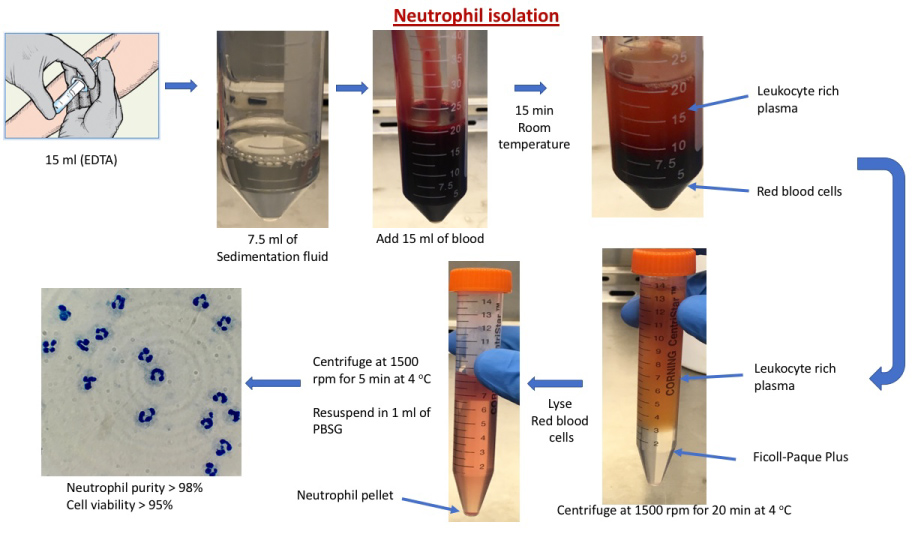 Figure 1: Flow diagram of the Neutrophil isolation protocol.
View Figure 1
Figure 1: Flow diagram of the Neutrophil isolation protocol.
View Figure 1
Since this is a protocol article no statistical analysis was done.
Using this neutrophil isolation protocol an average of 1.5 × 10⁷ cels/ml was obtained with a > 98% neutrophil purity and a viability greater than 95% (n = 15) (Figure 2 and Figure 3).
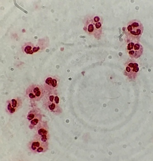 Figure 2: Neutrophils stained with safranin 0.5%.
View Figure 2
Figure 2: Neutrophils stained with safranin 0.5%.
View Figure 2
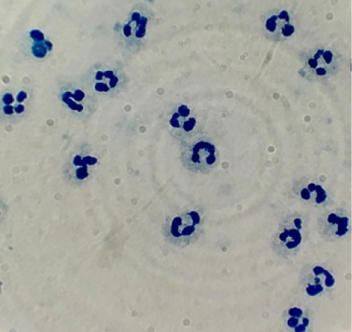 Figure 3: Neutrophils stained with methylene blue 0.1%.
View Figure 3
Figure 3: Neutrophils stained with methylene blue 0.1%.
View Figure 3
Different methodologies for the isolation of neutrophils were tested [1-6], but the best method we found to isolate neutrophils was the one described by GarcÍa-GarcÍa, et al. [7].
The density gradient separation method reported by GarcÍa-GarcÍa, et al. provided the best method to isolate human neutrophils from whole blood using Dextran and Ficoll-Paque Plus. This method is based on the mononuclear leukocyte separation method by Boyum (1968) which was modified for neutrophil separation by Ferrante and Thong (1980). The problem with this method is that Dextran is an expensive powder, making it difficult to do multiple isolation procedures without having to invest a lot of money. For example, 500 g of Dextran 200,000 costs around $ 1,200 dollars, in comparison, 500 g of Gelatin costs around $ 95 dollars.
Gelatin has a molecular weight of 100,000 to 120,000, this macromolecule helps aggregate red blood cells and thus is able to sediment them very fast. Red blood cellaggregation represents a balance of forces at the cell surfaces, and aggregation results when the macromolecular bridging force exceeds the electrostatic repulsive force and the mechanical shearing force. The larger a macromolecule is, the more effective it is at inducing red blood cell aggregation because of a weaker electrical repulsion due to the longer intercellular distance, and a stronger bridging force due to the larger adsorption area on the cell surface [8-10].
In terms of cell viability and number of neutrophils obtained per procedure Dextran and Gelatin obtain similar results, although gelatin takes only 15 min to sediment red blood cells while dextran takes 30-60 min, which makes our protocol suitable for labs with low income that wish to isolate neutrophils for their studies.
We know of no conflicts of interest associated with this publication, and there has been no significant financial support for this work that could have influenced its outcome.