Infertility affects 15% of couples worldwide. It is estimated that roughly half of the infertility cases are due to male factors. Male infertility could be caused by various reasons including failure in spermatogenesis, defects in sperm transportation or accessory gland function, genetic or environmental factors, and sexual disorders. Among these causes, spermatogenic defect is the primary one in male infertility. Spermatogenesis is a sophisticated multistep process involving three major phases: Mitotic, meiotic, and post-meiotic phases. Other cellular events such as apoptosis of spermatogenic cell, migration, and differentiation also play vital roles in the process of spermatogenesis. The dynamic balance between cell proliferation and apoptosis determines the number of cells in the seminiferous tubules of the testis. Compare between the effect of honey bee, bovine colostrum and G-CSF on homing of HSCs that are used for treatment of induced infertility in experimental animals.
Seventy male mice were randomly divided into seven groups, six of them injected with cyclophosphamide to be infertile, then first group treated with honey, second treated with bovine colostrum, third treated with MSCs, fourth treated with G-CSF, fifth treated with wheat germ, sixth saved as positive control group and seventh left as negative control group. The 2nd generation of stem cells were injected intraperitoneally. Different tissue and blood samples were taken at the end of study for tests.
Male fertility is improved by increasing sperm count and motility and level of sex hormones is elevated after the period of study.
Expression of SCP-3, GFRa1, and sca-1 in testis of azoospermatic male mice induced with cyclophosphamide after treatment with honey, bovine colostrum and umbilical cord derived mesenchymal stem cells.
Bovine colostrum, Cyclophosphamide, G-CSF, Honey, UCB-MSCs, Wheat germ oil
The regenerative-medicine in this decade could be a basis of many diseases treatment of the future, mainly the degenerative disease that cured through neither medical therapy nor surgery [1], especially testicle degeneration [2,3]. Testicular degeneration causes azoospermia. Azoospermia is a condition which is no spermatozoon produced by testicular seminiferous tubules, and makes infertility in the male and it means no offspring [2]. Testicular degeneration is the main cause of infertility in the male, the etiology varies such as genetic alteration, mechanical trauma, neoplastic changes and aging or senility. Current stem cell therapy usually is through stem cell transplantation which is cultured in vitro previously.
This is obviously very expensive. Therefore, it is needed a novel therapy based on mobilization and differentiation of own body-derived stem cells using natural bee honey. Rapidly mobilized stem cells in an adequate number towards defected tissue, in turn will differentiate to be certain cells (seminiferous tubules) from a certain tissue and will replace the damaged and apoptotic cells due to degenerative diseases, in this case, the differentiation becoming seminiferous tubules, sertoli and interstitial cells of Leydig. It is important to find out an innovation of therapy through auto-regeneration-induced of seminiferous tubule cells using beneficial of natural bee honey in reproducing of spermatozoa [4].
Regeneration of seminiferous tubule and subsequently sertoli cells will provide support, nutrients and other environmental factors for young spermatozoa and male behavior to allow spermatogenesis and the ability of conception to happen. Natural bee honey, a nutrient source from bee [5,6] has antibacterial and antioxidant potencies. Antioxidant is an important substance in protecting individual against free radicals. An adequate antioxidant consumption can reduce the prevalence of cancers, cardiovascular disease, cataract, digestive tract disorder and other degenerative diseases [7] and in present study, the testicular degeneration. Bee honey consumption will improve the digestive system, has a good result in diarrhea therapy and reproductive system disorder as well. As conceptual solution, it needs further study to explore the beneficial of natural bee honey to induce auto regeneration of testicular seminiferous tubule as a source of spermatozoa-producing tissue [4]. Also bovine colostrum is the pre milk fluid produced from mammary glands during the first 2 to 4 days after birth. It is a rich natural source of nutrients, antibodies, and growth factors for the newborn. It is augmenting mobilization of stem cells [8]. Mobilization of progenitor cells is also achieved with cytokines such as granulocyte colony-stimulating factor (G-CSF). G-CSF is preferred because of fewer side-effects and higher mobilization efficacy several investigators reported a dose-response effect to G-CSF up to 10 mg/kg. Despite the fact that a dose of 10 to 16 mg/kg G-CSF is accepted as standard use by most centres world-wide [9].
However, there is still controversy about histopathological findings for the detection of spermatogenesis level. Stem cells underwent proliferation and spontaneously differentiated into sperm whereas Sertoli cells attached and provided a somatic support. Transcripts specific for various stages of spermatogenesis were up-regulated by RT-PCR studies suggesting VSELs (Sca1) and SSCs (Gfra) proliferate (Pcna), undergo spermatogenesis (prohibitin), meiosis (Scp3) and differentiate into sperm (post-meiotic marker protamine) [6].
EMDM (sigma Aldrich, USA), RNA Extraction Kit (Sigma Aldrich, USA), RT-PCR (Sigma Aldrich, USA), Primers (Sigma Aldrich, USA), Honey (Faculty of Agriculture, Zagazig University, Egypt), Bovine colostrum (Faculty of Agriculture, Zagazig University, Egypt), G-CSF (Sigma Aldrich, USA), Wheat germ oil (Sigma Aldrich, USA), Cyclophosphamide (CP) was purchased from Sigma chemical Company (st. Louis, Mo, USA). Follicle Stimulating Hormone (FSH) ELISA kit (Lonza Bioproducts, Belgium), Testosterone ELISA Kit (Lonza Bioproducts, Belgium), Luteinizing Hormone (LH) ELISA kit (Lonza Bioproducts, Belgium).
For this study umbilical cord blood was used for extraction of mesenchymal stem cell (MSCs), and Male Swiss Albino mice from the Animal House Lab at Faculty of Science, Zagazig university. The animals were maintained on standard casein diet and water ad libitum at the Animal House Laboratory and housed in a temperature-controlled 25 ± 3.2 ℃ on light/dark cycle of 12/12 hours and artificially illuminated room, free from any source of chemical contamination. Seventy healthy male mice aging 6 months and weighed between 28-30 g were used. The seventy healthy male mice were fed and watered under controlled temperature (25 ℃-30 ℃). To ensure adequate adaptation, they were observed in this environment for 7 days prior to commencing treatment.
This study was done according to animal rights. All animal procedures were conducted according to guidelines provided by Zagazig University Institutional Animal Care and Use Committee under an approved protocol. The seventy male mice were randomly divided into seven groups and six groups of these male mice were injected with cyclophosphamide (50 mg/kg) daily for 7 days intraperitoneally to be infertile [10]. These groups are divided as follow:
Group 1: Ten infertile mice without treatment as positive control.
Group 2: Ten healthy mice were injected with saline intraperitoneally as negative control.
Group 3: Ten infertile mice were injected with umbilcal cord derived mesenchymal stem cells intraperitoneally.
Group 4: Ten infertile mice were injected with honey bee (1 g/kg) orally for two weeks daily.
Group 5: Ten infertile mice were injected with bovine colostrum (15 µl/g) orally for 10 days daily.
Group 6: Ten infertile mice were injected with G-CSF (250 µg/kg) intraperitoneally for 5 days daily.
Group 7: Ten infertile mice were injected with wheat germ (250 mg/kg) orally for 21 days daily.
To isolate MSC from cord blood, CB is collected into a sterile bag containing the anticoagulant citrate-phosphate-dextrose (CPD). The CB is then processed by density gradient centrifugation to obtain mononuclear cells (MNC). These are cultured until the outgrowth of fibroblastoid cell colonies appears. After reaching a subconfluent stage, cells are harvested, expanded, and characterized as cord blood mesenchymal stromal cells (CB-MSC) according to standard criteria: Plastic adherence, fibroblast morphology, CFU-f assay, proliferation potential, immune phenotype, and differentiation potential. Apparently, the frequency of MSC in CB is extremely low. Thus, not every CB unit will provide adequate MSC isolation yields. Different strategies have been proposed aiming to optimize the isolation success by selecting CB units of optimal quality: Decontaminate the external surfaces of the CB blood bag with 70% ethanol. Transfer 25 ml of CB from the collection bag to a 50-ml polypropylene tube. Add 25 ml of PBS/EDTA. When appropriate, remove a small aliquot for cell counting and for sterility testing. Add 10 ml of Ficoll-Paque Plus to a 50-ml polypropylene tube and carefully layer 25 ml of 1:1 diluted UCB on top. Centrifuge 30 min at 430 × g, low acceleration and no brake. Remove the plasma phase. Using a sterile Pasteur pipette, carefully transfer the buffy coat interface into a new 50-ml polypropylene tube, and fill the tube with PBS/EDTA. Centrifuge 10 min at 430 × g. 9. Wash with PBS/EDTA two to three times until the supernatant is clear. Suspend the pellet in 10 ml complete medium and remove a small aliquot for counting mononuclear cells (MNC). Seed the MNC at a density of 1 × 10⁶ cells/cm2 plastic surface into FBS-precoated 6-well plates. Discard the non-adherent cells after 1-3 days of incubation and add fresh MSCGM™ every 3-4 days. Monitor the culture for the presence of individual adherent fibroblastoid cells by phase-contrast microscopy for 14-30 days. Passage cells when they have reached 70% confluency in primary culture. If no colonies of fibroblastoid cells appear within 4-6 weeks, discard the culture [11].
Before culture, cells were tested to detect their viability by trypan blue test, the dye was diluted with PBS (0.4 trypan blue/PBS) and then 100 ul of the sample was added to equal volume of dye. Viable cell do not take blue Color according to De Bruijn, et al. [12].
The mononuclear cell suspension obtained was resuspended in complete culuture medium high glucose IMDM, 4.5 g/l glucose with L-glutamine (Lonza Bioproducts, Belgium) containing 10% FBS, 1% Pencillin, streptomycin-Amphoteracin mixture (Lonza Bioproducts, Belgium). Cells were cultured at concentration of 5*106 per 25-cm2 culture flask then they were incubated at 37 ℃ in 5% humidified Co2 incubator (Heraeus, Germany). The media were changed every 3-4 days (to remove nonadherent cells). When large colonies developed (80-90% confluence), cultures were washed twice with PBS and cells were trypsinized using 0.25% trypsin/ethylene diamine tetra acetic acid (EDTA) (Lonza Bio products, Belgium) for 5 min at 37 ℃. After centrifugation (at 2400 RPM for 20 min), the resulting cultures were referred to as first passage cultures after 7 days [13].
MSCs in culture were characterized by their adhesiveness and fusiform shape, by determination of surface markers of umbilical cord blood derived mesenchymal stem cells that done by evaluation of the positive expression of CD105 surface marker and the negative expression of CD34 surface marker in MSCs that were analyzed by flow cytometer. At subculture, about 105 MSC per FACS tube are stained with the appropriate antibodies to detect the MSC phenotype as well as possible contamination with hematopoietic or endothelial cells. Preincubate cells with FcR blocking reagent. Stain with pre-titrated antibodies for 15-30 min at 4 ℃. Wash two times with PBS or FACS buffer. Add titrated concentration of 7AAD; incubate for 10 min. Measure the cells using a flow cytometer [13].
The formed colonies of the 2nd generation were [14] injected intraperitoneally in the group that treated with MSCs.
20 mg/kg.b.w. of CP were dissolved in 150 ul (high concentration dose, CP1) or in 300 ul (low concentration dose, CP2) of sterilized distilled water. The high dose (CP1)/animal or low dose (CP2)/animal were used and injected intraperitoneally, according to method of [15].
1 gm/kg.bw of HP (500 mg of honey+250 mg of Royal jelly+250 mg of Pollen grains) was dissolved in 10 ml of sterilized distilled water. Then 0.3 ml of this solution was used and injected intraperitoneally/animal, according to method of [16].
For preparation of colostrums preparations, the first five milkings of eight pregnant Helstein cows post-partum were collected, frozen immediately, and transported to laboratory under low temperature according to method of [17].
Granulocyte colony stimulating factor (PeproTech) suspended in Dulbecco's phosphate-buffered saline (DPBS; Life Technologies) containing 0.5% bovine serum albumin fraction V (BSA, MP Biomedicals) or 0.5% BSA in DPBS alone according to method of [18].
Wheat germ oil was obtained from Sigma chemical Company (st. Louis, Mo, USA). Wheat germ oil was dissolved in propylene glycol and given alone at a dose level of (270 mg/kg b.wt/day), orally for 21 days according to method of [19].
The animals were weighed and anesthetized with pentobarbital sodium (50 mg/kg, i.p.) at the end of the treatment. Blood samples were collected by extirpation of eyeball and allowed to clot, and the serum was separated at 3000 rpm for 15 min and stored at -80 ℃ to analyze the testosterone level. The testes and epididymis were rapidly excised and weighed. One testis and one epididymis from a mouse were fixed in 10% formalin solution for histological examination.
The epididymal sperm concentrations and motility were evaluated as previously described with some modifications [20]. An entire epididymis from a mouse was minced in 1 mL saline, which was preheated at 37 ℃. The epididymis was incubated for another 15 min at 37 ℃ to allow the sperm to swim out of the epididymal tubules. The total number of sperm and the number of motile sperms were determined using a homocytometer, the sperm motility was converted into a percentage.
The excised testicular tissue was washed with distilled water for the removal of blood, after which the fatty parts were removed. Tissues were homogenized in ice-cold 50 mM sodium phosphate buffer (pH 7.4) containing 0.1 mM EDTA. The supernatant was separated by centrifugation at 1,000 rpm for 20 minutes at 4 ℃. The supernatants were used for the analysis of all gene expression using reverse transcriptase polymerase chain reaction (RT-PCR) [18].
Total RNA was isolated from testis tissue homogenate using IQeasy plus CTB RNA extraction kit (iNtRON Biotechnology, Korea). The extracted RNA was used for determination of: RT-PCR reactions for the samples were performed. The target bands were visualized with an ultraviolet illuminator (Bio Rad Laboratories Inc., Hercules, CA, USA) according to [18].
GENE |
Forward & Reverse sequence |
Sca-1 |
Forward AGAGGAAGTTTTATCTGTGCAGCCC Reverse TCCACAATAACTGCTGCCTCCTGA |
GFR a |
Forward GGCCTACTCGGGACTGATTGG Reverse GGGAGGAGCAGCCATTGATTT |
SCP3 |
Forward TGTTGCAGCAGTGGGAACTGGAT Reverse CCATCTCTTGCTGCTGAGTTTCCA |
Prohibitin |
Forward GTGGCGTACAGGACATTGTG Reverse AGCTCTCGCTGGGTAATCAA |
Protamine |
Forward GGCCACCACCACCACAGACACAGGCG Reverse TTAGTGATGGTGCCTCCTACATTTCC |
Loading of RNA sample then the device was turned on for a suitable time (20-25 min). The gel was then examined under the UV illuminator. The size of any resulting band was compared by 100-1000 bp M.W marker [18].
Regeneration and identification of seminiferous tubule cells of the testes through histopathological examination begins with the making of histological preparations. Histological preparations such as the following: mice testicular fixation in 10% formalin, 1 h later injected formalin 10% in mid-testis. Subsequently mice testes dehydrated in alcohol solution with a higher concentration gradually, i.e. from 70%, 80%, 90%, 96% (absolute). Then do the clearing in the testes of mice in xylol solution. Furthermore performed embedding using liquid paraffin then hematoxylin & eosin staining procedure [21]. Histopathological examination is done using a light microscope with a magnification of 400 times [4].
After the treatment, whole blood collected from cardiac puncture placed in heparin tube to prevent coagulate. Also testis tissue samples were homogenized to be ready for analysis. Flow cytometry observation reveals the expressions of CD34 and CD45. Flow cytometry method begins with whole blood and tissue homogenate centrifugation in 4 ℃ temperature, 6000 rpm for 15 min. Cellular precipitation as a result of centrifugation then mixed with cytoperm/cytofix in amount of 2 times of obtained cell number. This mixture then centrifuged again and formed supernatant and pellet. BD wash added to the pellet in the amount of 4 times of obtained cell number from the first centrifugation. Centrifuge the mixture then added lysis buffer in the amount of 2 times of the first obtained cell number. Subsequently labeled antibody added to each sample, five tubes are arranged and processed parallel [1]. Single staining with CD34 PE added to the wash tube [2]. Double staining with CD34 PE and CD45 PerCP [3]. Double staining with CD34 PE and CD45 PerCP and CD105 FITC true count tube. All the samples were then stored in 4 ℃ and dark room and were analyzed with flowcytometry for 1 h [22].
Serum testosterone was examined using Testosterone ELISA Kit according to the protocol described by the manufacturer. The amount of testosterone was determined from a calibration curve.
Serum FSH was examined using Follicle Stimulating Hormone ELISA Kit according to the protocol described by the manufacturer. The amount of FSH was determined from a calibration curve.
Serum LH was examined using Leutinizing Hormone ELISA Kit according to the protocol described by the manufacturer. The amount of LH was determined from a calibration curve.
Expressions of CD34 and CD45 and SSCs were statistically analyzed using SPSS 15 for Windows XP with the level of significance 0.01 (P = 0.01) and the confidence level 99% (a = 0.01). Steps of comparative hypothesis testing are as follows: Test data normality with the Kolmogorov Smirnov test, homogeneity of variance test, Analysis of variance (ANOVA) factorial using SPSS software (version 19.0) [4].
These cells are cultured until the outgrowth of fibroblastoid cell colonies appears. After reaching a sub confluent stage, cells that are harvested, expanded are characterized by plastic adherence, fibroblast morphology as shown in Figure 1.
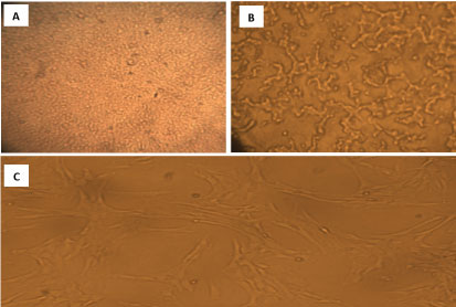 Figure 1: A) Mesenchymal stem cells culture at day 1 which show the morphological spindle shape of the MSCs; B) Mesenchymal stem cells culture after the first passage; C) Mesenchymal stem cells culture after the second passage.
View Figure 1
Figure 1: A) Mesenchymal stem cells culture at day 1 which show the morphological spindle shape of the MSCs; B) Mesenchymal stem cells culture after the first passage; C) Mesenchymal stem cells culture after the second passage.
View Figure 1
Flow cytometric analysis of cell surface markers in mesenchymal stem cells (MSCs) expressed CD105, but did not express CD34, CD45. The surface markers expression pattern corresponds to umbilical cord derived mesenchymal stem cells (UCB-MSCs) as shown in Figure 2.
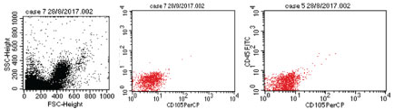 Figure 2: Characteristics of UCB-MSCs. Cells were stained with the CD45, CD34 and CD105 antibodies and analyzed by flow cytometry showed as a dot plot. The expression levels of CD45-ve, CD34 -ve and CD105 +ve of UCB-MSCs are presented.
View Figure 2
Figure 2: Characteristics of UCB-MSCs. Cells were stained with the CD45, CD34 and CD105 antibodies and analyzed by flow cytometry showed as a dot plot. The expression levels of CD45-ve, CD34 -ve and CD105 +ve of UCB-MSCs are presented.
View Figure 2
Sperm examination in our study showed head and tail abnormalities. The mice group treated with honey-bee products, G-CSF, bovine colostrum, (UCB-MSCs) and wheat germ oil had few of head and tail abnormalities as compared to control group as shown in Figure 3. After induction by CP decreased mean value of the sperm count and motility significantly into (0.14 ± 0.06), (0.40 ± 0.20) respectively as compared to the negative control group (27.62 ± 1.12, 77.23 ± 2.01) (𝑃 < 0.02).
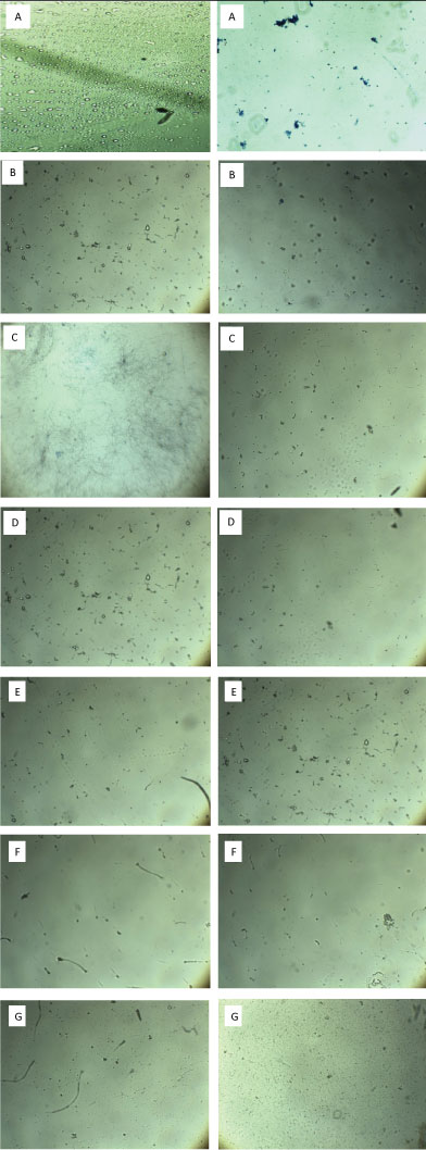 Figure 3: A) Induction by cyclophosphamide showing death of sperms as it stained with trypan blue dye; B) Sperm after treatment with honey showing large number of sperms and spermatogenic cells; C) Sperm after treatment with UCB-MSCs showing best count and motility; D) sperm after treatment with Wheat germ oil; E) Sperm after treatment with G-CSF; F) Sperm after treatment with Bovine colostrum showing lower count of sperm than other groups; G) Normal sperm in negative control group.
View Figure 3
Figure 3: A) Induction by cyclophosphamide showing death of sperms as it stained with trypan blue dye; B) Sperm after treatment with honey showing large number of sperms and spermatogenic cells; C) Sperm after treatment with UCB-MSCs showing best count and motility; D) sperm after treatment with Wheat germ oil; E) Sperm after treatment with G-CSF; F) Sperm after treatment with Bovine colostrum showing lower count of sperm than other groups; G) Normal sperm in negative control group.
View Figure 3
While treatment with MSCs, Wheat germ, Honey, Bovine colostrum and G-CSF showed increase in the mean value of sperm count and motility (27.46 ± 0.42, 13.82 ± 0.44, 18.7 ± 0.22, 6.97 ± 0.22, 9.87 ± 0.45), (74.96 ± 1.53, 60.95 ± 2.03, 68.07 ± 1.81, 50.83 ± 2.99, 58.53 ± 1.59) respectively by percent change (195%, 97.7%, 132.5%, 48.7%, 69.5%), (186.4%, 151.3%, 169%, 126%, 145%) respectively as explained in Table 1.
Table 1: The effect of MSCs, WGO, Honey, Bovine colostrum and G-CSF treatments on sperm count (*106/ml), Sperm motility(%), LH level (mIU/ml), FSH level (mIU/ml) and Testosterone level (ng/ml) in mice compared with positive and negative control. View Table 1
Also treatment with MSCs, Wheat germ, Honey, Bovine colostrum and G-CSF showed increase in the mean value of body weight, the testis and epididymis weight to (28.75 ± 0.26, 27.56 ± 0.29, 28.59 ± 0.35, 27.1 ± 0.07, 28.14 ± 0.22), (3.95 ± 0.05, 3.56 ± 0.09, 3.65 ± 0.07, 3.1 ± 0.009, 3.28 ± 0.03), (1.33 ± 0.05, 1.18 ± 0.03, 1.42 ± 0.03, 1.35 ± 0.12, 1.28 ± 0.03) respectively by percent change (0.07%, 0.03%, 0.07%, 0.01%, 0.05%), (0.2%, 0.11%, 0.14%, 0.02%, 0.03%), (0.04%, 0.007%, 0.11%, 0.06%, 0.007%) respectively when compared with the CP positive control group that showed decrease in mean value of body weight, the testis and epididymis weight to (26.68 ± 0.28, 3.19 ± 0.15, 1.27 ± 0.17) as explained in Table 1.
Also hormonal analysis for serum samples from the treated and non-treated groups of LH, FSH and Testosterone Hormone showed the lowest mean value of LH, FSH and testosterone levels respectively in positive control group as (0.27 ± 0.07), (0.31 ± 0.06), (1.57 ± 0.25) while the mean value increased in treated groups as follow: Wheat germ (3.95 ± 0.25), (4.76 ± 0.58), (4.77 ± 0.33) with percent change (13.6%, 14.3%, 2%), honey (4.63 ± 0.23), (5.39 ± 0.64), (5.8 ± 0.58) with percent change (16.1%, 16.3%, 2.6%) bovine colostrum (2.67 ± 0.24), (4.02 ± 0.31), (5.33 ± 0.47) with percent change (8.8%, 11.9%, 2.3%) and G-CSF (4.92 ± 0.35), (4.59 ± 0.48), (5.62 ± 0.59) with percent change (17.2%, 13.8%, 2.5%) but showed the highest levels in UCB-MSCs treated group (5.66 ± 0.33), (6.86 ± 0.68), (9.19 ± 0.49) with percent change (19.9%, 21%, 4.8%) indicated that the drugs honey, bovine colostrum and G-CSF homed HSCs from their niche to atrophied tissue and regenerated the testis tissue to reproduce these hormones that play important role in spermatogenesis and also these results indicated that treatment with exogenous MSCs isolated from umbilical cord blood improve the sperm parameters and hormonal level greatly more than treatment with pharmatheuticals widely used in medicine for treatment of male infertility such as wheat germ as explained in Table 1.
These microscopic photos showing that sperm shape abnormalities in CP treated group is increased as sperm observed at these times were presumably exposed to the chemical while they were spermatids, early primary spermatocytes, and spermatogonia respectively. As cyclophosphamide known to be mutagenic, induces abnormalities but the level and duration are much less pronounced than those observed with the other alkylating agents, these abnormalities disappeared when mice treated with UCB-MSCs and honey also decreased to lowest level in bovine colostrum,wheat germ and G-CSF treated groups as shown in Figure 3.
Results of the present study revealed the normal structure of the testis tissue in control group. It was also revealed that Cyclophosphamide had a marked damaging effect on testis tissue. This effect was greatly ameliorated by using honey-bee products, bovine colostrum, G-CSF, wheat germ and MSCs. In this study, the regeneration of the testes can be observed through the group of honey leading to the occurrence of testicular tissue repair. Improvements were identified based on the regeneration seminiferous tubules regenerate intact. Photomicrographs of H & E-stained sections of testes of negative control mice showing normal histological structure of the mature active seminiferous tubules with complete spermatogenic sense, epididymis of mice in the control group showing normal histological structure of the tubules and impacted by mature sperm. Photomicrographs of H & E stained sections of CP group after 7 days showing degeneration in seminiferous and atrophy and seminiferous tubules free from sperm. Photomicrographs of testes post 21 days of UCB-MSCs, honey, wheat germ, G-CSF showing giant spermatogonial formation of some seminiferous tubules and epididymal tubular lumen as shown in Figure 4.
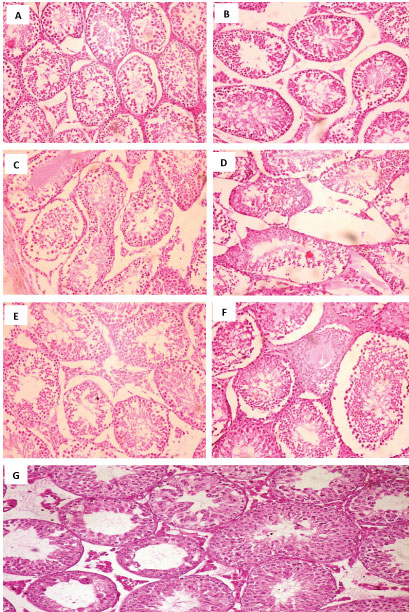 Figure 4: A) Testes from 10% honey treated group showing normal seminefrous tubules with normal cells; spermatogonia, spermatocytes, and spermatozoa (H&E × 200); B) Testes from 10% Bovine colostrum treated group showing edema in between seminefrous tubules (arrow head), (H&E × 200); C) Testes from wheat germ treated group showing sloughing of spermatogonia cells and spermatocytes, (arrow), (H&E × 200); D) Testes from control positive group showing degeneration and necrosis of the cells; spermatogonia, spermatocytes, and spermatozoa (arrow head), (H&E × 200); E) Testes from G-CSF treated group showing edema inbetween seminefrous tubules (arrow head), (H&E × 200); F) Testes from Mscs treated group showing hyperplastic activity of the lining cells; spermatogonia, spermatocytes, and spermatozoa (arrow head), (H&E × 200); G) Testes from control negative group showing normal seminefrous tubules with normal spermatogonia, spermatocytes, and spermatozoa (H&E × 200).
View Figure 4
Figure 4: A) Testes from 10% honey treated group showing normal seminefrous tubules with normal cells; spermatogonia, spermatocytes, and spermatozoa (H&E × 200); B) Testes from 10% Bovine colostrum treated group showing edema in between seminefrous tubules (arrow head), (H&E × 200); C) Testes from wheat germ treated group showing sloughing of spermatogonia cells and spermatocytes, (arrow), (H&E × 200); D) Testes from control positive group showing degeneration and necrosis of the cells; spermatogonia, spermatocytes, and spermatozoa (arrow head), (H&E × 200); E) Testes from G-CSF treated group showing edema inbetween seminefrous tubules (arrow head), (H&E × 200); F) Testes from Mscs treated group showing hyperplastic activity of the lining cells; spermatogonia, spermatocytes, and spermatozoa (arrow head), (H&E × 200); G) Testes from control negative group showing normal seminefrous tubules with normal spermatogonia, spermatocytes, and spermatozoa (H&E × 200).
View Figure 4
Mobilization of HSCs was analyzed with flow cytometry based on increased CD34 and CD45 concentrations. Flow cytometric analysis of cell surface markers in haematopoietc stem cells (HSCs) expressed CD34, but did not express CD105, CD45 in blood and tissue samples in the treated and non-treated groups as shown in Figure 5, Figure 6, Figure 7, Figure 8, Figure 9, Figure 10, Figure 11, Figure 12, Figure 13 and Figure 14.
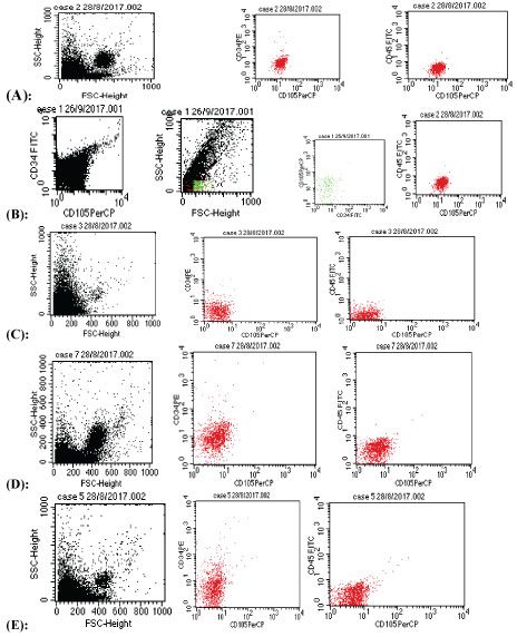 Figure 5: Mobilization of HSCs based on increased CD34 concentrations. Flow cytometric analysis of cell surface markers in haematopoietc stem cells (HSCs) expressed CD34, but did not express CD105,CD45 in blood samples in the treated and non-treated groups A) Negative control; B) Positive control; C) Bovine colostrums; D) G-CSF group; E) Honey group.
View Figure 5
Figure 5: Mobilization of HSCs based on increased CD34 concentrations. Flow cytometric analysis of cell surface markers in haematopoietc stem cells (HSCs) expressed CD34, but did not express CD105,CD45 in blood samples in the treated and non-treated groups A) Negative control; B) Positive control; C) Bovine colostrums; D) G-CSF group; E) Honey group.
View Figure 5
 Figure 6: Mobilization of HSCs based on increased CD34 concentrations. Flow cytometric analysis of cell surface markers in haematopoietc stem cells (HSCs) expressed CD34, but did not express CD105, CD45 in testis tissue samples in negative control group.
View Figure 6
Figure 6: Mobilization of HSCs based on increased CD34 concentrations. Flow cytometric analysis of cell surface markers in haematopoietc stem cells (HSCs) expressed CD34, but did not express CD105, CD45 in testis tissue samples in negative control group.
View Figure 6
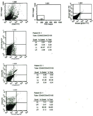 Figure 7: Mobilization of HSCs based on increased CD34 concentrations. Flow cytometric analysis of cell surface markers in haematopoietc stem cells (HSCs) expressed CD34, but did not express CD105, CD45 in testis tissue samples in positive control group.
View Figure 7
Figure 7: Mobilization of HSCs based on increased CD34 concentrations. Flow cytometric analysis of cell surface markers in haematopoietc stem cells (HSCs) expressed CD34, but did not express CD105, CD45 in testis tissue samples in positive control group.
View Figure 7
 Figure 8: Mobilization of HSCs based on increased CD34 concentrations. Flow cytometric analysis of cell surface markers in haematopoietc stem cells (HSCs) expressed CD34, but did not express CD105, CD45 in testis tissue samples in bovine colostrum group.
View Figure 8
Figure 8: Mobilization of HSCs based on increased CD34 concentrations. Flow cytometric analysis of cell surface markers in haematopoietc stem cells (HSCs) expressed CD34, but did not express CD105, CD45 in testis tissue samples in bovine colostrum group.
View Figure 8
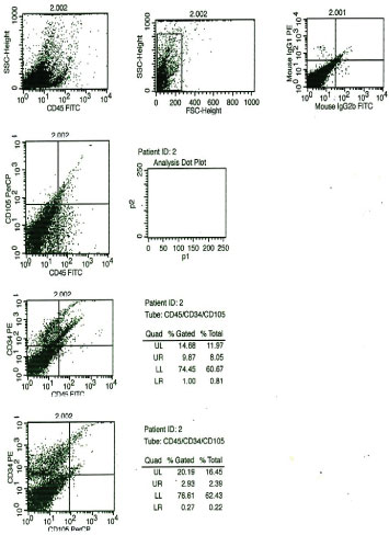 Figure 9: Mobilization of HSCs based on increased CD34 concentrations. Flow cytometric analysis of cell surface markers in haematopoietc stem cells (HSCs) expressed CD34, but did not express CD105, CD45 in testis tissue samples in G-CSF group.
View Figure 9
Figure 9: Mobilization of HSCs based on increased CD34 concentrations. Flow cytometric analysis of cell surface markers in haematopoietc stem cells (HSCs) expressed CD34, but did not express CD105, CD45 in testis tissue samples in G-CSF group.
View Figure 9
 Figure 10: Mobilization of HSCs based on increased CD34 concentrations. Flow cytometric analysis of cell surface markers in haematopoietc stem cells (HSCs) expressed CD34, but did not express CD105, CD45 in testis tissue samples in honey group.
View Figure 10
Figure 10: Mobilization of HSCs based on increased CD34 concentrations. Flow cytometric analysis of cell surface markers in haematopoietc stem cells (HSCs) expressed CD34, but did not express CD105, CD45 in testis tissue samples in honey group.
View Figure 10
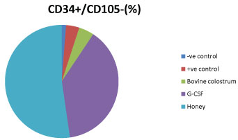 Figure 11: Percentage of positively expressed CD34 cells in blood samples is the highest in honey treated group than bovine colostrum, G-CSF treated groups indicated the larger mobilization of HSCs in honey treated group.
View Figure 11
Figure 11: Percentage of positively expressed CD34 cells in blood samples is the highest in honey treated group than bovine colostrum, G-CSF treated groups indicated the larger mobilization of HSCs in honey treated group.
View Figure 11
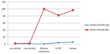 Figure 12: Percentage of positively expressed CD34 cells in blood samples is the highest in honey treated group than bovine colostrum, G-CSF treated groups indicated the larger mobilization of HSCs in honey treated group.
View Figure 12
Figure 12: Percentage of positively expressed CD34 cells in blood samples is the highest in honey treated group than bovine colostrum, G-CSF treated groups indicated the larger mobilization of HSCs in honey treated group.
View Figure 12
 Figure 13: Percentage of positively expressed CD34 cells in tissue samples is the highest in honey treated group than bovine colostrum, G-CSF treated groups indicated the larger mobilization of HSCs in honey treated group.
View Figure 13
Figure 13: Percentage of positively expressed CD34 cells in tissue samples is the highest in honey treated group than bovine colostrum, G-CSF treated groups indicated the larger mobilization of HSCs in honey treated group.
View Figure 13
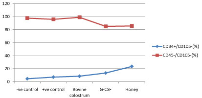 Figure 14: Percentage of positively expressed CD34 cells in tissue samples is the highest in honey treated group than bovine colostrum, G-CSF treated groups indicated the larger mobilization of HSCs in honey treated group.
View Figure 14
Figure 14: Percentage of positively expressed CD34 cells in tissue samples is the highest in honey treated group than bovine colostrum, G-CSF treated groups indicated the larger mobilization of HSCs in honey treated group.
View Figure 14
From molecular aspect different genes were expressed in testis tissue such as Prohibitin, Protamine, GFRa, SCP-3 and Sca-1 using GAPDH as control gene. Sca-1 is the most common marker used to enrich adult murine hematopoietic stem cells (HSCs) and can be used to isolate a nearly pure HSC population when used in conjunction with additional markers as shown in Figure 15.
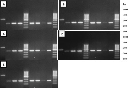 Figure 15: Agarose gel electrophoresis of RNA extracted from mice testis in different groups: After end of treatment with MSCs (lane 6), honey (lane 7), bovine colostrum (lane 4), G-CSF (lane 9) and wheat germ (lane 3) compared with positive control group (lane 8) and negative control group (lane 2) using ladder marker at (lane 5, 10) and control gene GAPDH (lane 1) at 570 bp for gene expression of prohibitin at 280bp A); SCP-3 at 350 bp B); Sca-1 at 445 bp C); Gfra-1 at 400 bp D); protamine at 220 bp E).
View Figure 15
Figure 15: Agarose gel electrophoresis of RNA extracted from mice testis in different groups: After end of treatment with MSCs (lane 6), honey (lane 7), bovine colostrum (lane 4), G-CSF (lane 9) and wheat germ (lane 3) compared with positive control group (lane 8) and negative control group (lane 2) using ladder marker at (lane 5, 10) and control gene GAPDH (lane 1) at 570 bp for gene expression of prohibitin at 280bp A); SCP-3 at 350 bp B); Sca-1 at 445 bp C); Gfra-1 at 400 bp D); protamine at 220 bp E).
View Figure 15
The results of sperm count analysis showed higher significance in honey bee treated group (p � 0.08) than G-CSF, bovine colostrum (p � 0.05), (p � 0.004) respectively indicating that honey bee is more effective in improving the sperm count due to its higher ability in homing of HSCs to testis tissue based on SSCs formation as a result of the differentiation of the spermatogonia while UCB-MSCs treated group showed higher significance (p � 0.06) than wheat germ treated group (p � 0.02) assuring the regenerative tendency of MSCs into new testis tissue to reproduce sperm and improve their motility (p � 0.03) and (p � 0.07) in honey group as shown. Also in the same time these results is certificated as the body weight, testis and epididymis weight showed higher significance in honey group (p � 0.09), (p � 0.07), (p � 0.05) respectively than other groups (p � 0.04) and (p � 0.06) due to higher nutritional value of honey bee. On the second hand MSCs treated group showed higher significance (p � 0.07), (p � 0.07), (p � 0.05) respectively than wheat germ (p � 0.05) as explained in Table 2.
Table 2: Statistical analysis showed P value for effect of different drugs such as UCB-MSCs, WGO, Honey, Bovine colostrum and G-CSF on sperm parameters and hormonal level including LH level (mIU/ml), FSH level (mIU/ml) and Testosterone level (ng/ml) compared with positive and negative control group. View Table 2
Male infertility causes significant duress to couples. Defects in spermatogenesis are the most common reasons for male infertility. At present, hormonal treatment or medical treatment is used to treat infertile men with spermatogenic defects, but clinical results are limited especially for patients with idiopathic failure of spermatogenesis [23]. In addition the effect of external factors on the reproductive system in male which shoots the testicular spermatogenesis and spermatozoa within the epididymis to produce toxicity on reproduction [24]. In the present study UCB-MSCs are used as one of the most available sources of adult stem cells. Besides hematopoietic stem cells, CB contains both endothelial progenitor cells and mesenchymal stem cells (MSC). So, to isolate MSC from cord blood, CB is obtained in a sterile citrate-phosphate-dextrose (CPD) bag.
The scientific interest in mobilization of immature cells is fuelled by its clinical relevance. Its importance in self repair mechanisms was assured after partial irradiation radiation-depleted marrow is recollected from different strange non-irradiated marrow sites, by the mobilized stem cells [25]. Quantitatively, the collection of mobilized cells by apheresis, allow transfer or cryopreservation of autologous progenitor cells for hematopoietic transplantation [26,27].
Many of mobilization protocols that were reported are followed in our study in which we used different drugs ranging from natural products to chemical drugs to mobilize the HSCs from their niche to the atrophied tissue to make regeneration of damaged seminiferous tubules in infertile mice.
The ways of injection used in this study were selected with intensive care such as intraperitoneal (IP) injection which is more common for small animals who cannot receive medications and fluids by other methods. With animals, an IP injection may be the only way to deliver medication and reduce the accumulation of MSCs within filtering organs [14]. Also oral injection is preferred method for delivering of nutrients to the small mice to get most of their nutritional effect beside their pharmaceutical effect [28].
In our study, epididymal sperm count and sperm motility decreased in mice to its minimum level after injection of mice with cyclophosphamide intraperitoneally daily for a week which then raised again to high level after treatment with bovine colostrum, wheat germ and G-CSF, while raised to its highest levels that become the nearest to the normal mice when treated with UCB-MSCs and to lesser extent with honey. Honey contains large number of biologically active components like melittin and phospholipase-A2 (PLA2) that trigger process of (VEGF-1) which is homing signal. Furthermore, VEGF-1 binds to VEGF Receptor-1 (VEGFR-1). VEGF is a component of Extracellular Matrix (ECM) from stem cells supports a conductive microenvironment for stem cells. So by triggering presence of VEGF-1-VEGFR-1 would pass a series of signals that activate Stem Cell Factor (SCF) interstitial. SCF is a mechanic in the niche signaling protein that produce more communication. Colostrum is rich in bioactive compounds that activate immune cells such as cytokines, interleukins, interferon's growth factors and peptides like proline rich polypeptides (PRP). Many of the growth factors including human growth hormone (HGH), insulin-like growth factors (lGF-1a and IGF-2), epidermal growth factor (EGF), fibroblast growth factor, platelet derived growth factor and granulocyte colony stimulating factor which stimulate stem cell homing and proliferation and activation, has an effective release of hematopoietic stem cells into the blood. G-CSF could mobilize hematopoietic cells in large numbers from the marrow into the circulation with increased progenitor cells, neurotransmitters have a direct effect on HSC as human CD34+ cells express adrenergic and dopamine receptors, that enhances responses to the chemokine CXCL12. HSCs get out by migration through the bone marrow-blood barrier, into the circulation. HSCs may remobilized into a target tissue. HSC mobilization is characterized by loss of cell-cell contacts and ignore of chemokine signaling, especially the SDF-1/CXCR4 axis [29-32].
Hormonal analysis for serum samples from the treated and non-treated groups of Leutinizing Hormone, Follicle Stimulating Hormone and Testosterone Hormone showed that the lowest levels of LH, FSH and testosterone in positive control group while the level increased in treated groups and showed the highest levels in UCB-MSCs treated group indicated that honey, bovine colostrum and G-CSF homed HSCs from their niche to atrophied tissue and regenerated the testis tissue to reproduce these hormones that play important role in spermatogenesis and also these results indicated that treatment with exogenous MSCs isolated from umbilical cord blood improve the sperm parameters and hormonal level greatly more than treatment with pharmatheuticals widely used in medicine for treatment of male infertility such as wheat germ.
These results were assured by flow cytometric analysis for CD45, CD34, CD105 to detect mobilization of HSCs into blood and testis tissue which is positively express CD34 while negatively express CD45, CD105. All these results accomplished by histological test for testis tissues to ensure that the injected MSCs treated the atrophied tissues of testis and seminiferous tubules also the natural drugs mobilized HSCs from their niche to exhibit its regenerative effect on damaged testis and improve male fertility.
The concluded result was the recovery and improvement of male infertility by increasing the sperm count and motility, also regeneration of damaged seminiferous tubules in treated groups with drugs homed HSCs or treated group with UCB-MSCs than the positive control group [33] approved that the abnormalities in sperm-shape significantly elevated in mice injected with CP, assuring that CP have induced toxicity in testes. Also, [34] indicated that in CP-treated rats, a defect in sperm quality, increase in DNA damage and decrease of chromatin quality. Moreover, Januchowski R, et al. [29,30] demonstrated the toxic effect of CP on reproductive tissues in male rats, and their results showed that CP treatment led to significant decreases in sperm count and motility with an increase of sperm-shape abnormalities. That was due to production of free radicals. Jedrzejczak P, et al. [29-32,35] showed that the observed seminiferous tubules in animals treated with Busulfan alone and animals that received G-CSF treatment contained normal spermatogenesis similar to control animals that did not receive any busulfan or G-CSF. These seminiferous tubules with complete spermatogenesis have normal histology.
Kim SH, et al. [36] proved that bone marrow-derived cells survive in host testes for more than 12 weeks after transplantation. The donor cells containing hematopoietic stem cells, progenitor cells, MSCs, are capable of differentiating into germ cells and Leydig cells in the testis. Kumar GL, et al. [37] showed that from 20 to 60 days the seminiferous tubules showed active developing stage and newly formed spermatogenic cells arranged inside the tubules. After 60 days all stages of spermatogenesis were visible and spreaded in the lumen of the tubules as in the control.
A recent study undergo extrinsic spermatogonial transplantation has demonstrated that the spermatogenesis cycle is under the control. It can be seen also from our results that MSCs can decrease alteration in DNA and protect the testis tissues from apoptotic damage. Also, MSCs relatively improve the histopathological changes induced in the testis of mice. This protection includes modulation of the oxidative stress reaction, tissue damage and repair. In this respect Lambard S, et al. [38] have reported that pretreatment with MSCs attenuates lipopolysaccharide-induced acute lung injury in rats through inhibition of neutrophilic recruitment, inflammation, oxidative stress and apoptosis. Depending on theory of tissue repair holds that organ injury is detected by stem cells that move to the site of damage and differentiate into organ specific cells, promoting structural and functional repair [39]. Because these dead cells are not able to divide, other cells must replace them to repair the tissue and maintain organ homeostasis. Reported that MSCs were present both in and outside seminiferous tubules, supporting the idea that MSCs might have functioned in reestablishment of spermatogenesis in two ways: MSCs' differentiation into sperm or maintenance of the spermatogonial stem cells.
These results showed that the MSCs are a rich and functional source for infertility treatment. In this study, the regeneration of the testes can be observed through the method of histopathology anatomy (HPA) with hematoxylin and eosin (HE) staining. Microscopic examination showed that the use of honey, UCB-MSCs, bovine colostrum, G-CSF and wheat germ leading to the testicular tissue repair. Improvements are identified based on the regeneration of seminiferous tubules. These improvements can be compared with a control group of normal testis who did not experience testicular degeneration, which remains in normal condition [38]. Hematopoietic stem cells are defined by their capacity for long-term multilineage growth potential and a capacity for self-renewal [31].
These results showed that there are different nutritional materials such as honey and colostrum that help in homing of HSCs from their niche to the defected organ such as testis and regenerate its damaged tissues without the side effects that associated with different chemical drugs. Also, we founded that using external stem cells such as UCB-MSCs is faster and more effective than the internal stem cells.
I want to acknowledge Stem cell unit and Clinical Pathology Department, at Faculty of Medicine, Zagazig university, Egypt also the Animal House Lab at Faculty of science, Zagazig University, Egypt. Compliance with Ethical Standards.
The authors declare that they have no conflict of interest.
All applicable international, national, and institutional guidelines for the care and use of animals were followed. All procedures performed in studies involving animals were in accordance with the ethical standards of the institution or practice at which the studies were conducted.
Not applicable.