Coil embolization is an effective and safe treatment of pelvic congestion syndrome. Cases of coil pulmonary migration have been reported after pelvic embolization. However, most cases are usually asymptomatic and do not require specific treatment. We present the first case of pulmonary coil migration after pelvic embolization and concomitant acute pulmonary embolism in a symptomatic patient. The patient was treated with standard anticoagulation and conservative measures without incidences after hospital discharge.
NTPE: Nonthrombotic Pulmonary Embolism; ED: Emergency Department; PA: Posteroanterior; PE: Pulmonary Embolism; CT: Computed Tomography; HU: Hounsfield Units; MIP: Maximum Intensity Projection
Nonthrombotic pulmonary embolism (NTPE) is a partial or complete occlusion of the pulmonary vasculature by organic or inorganic materials [1]. The most frequent causes associated include cancer, infective agents, fat, amniotic fluid, gases and foreign materials. The incidence of these cases is relatively low but NTPE is considered a life-threatening pathology [2].
NTPE can cause local and systemic inflammation causing endothelial and pulmonary parenchymal injury [3]. Therefore, NTPE may present as completely asymptomatic, with unspecific symptoms or even cause sudden death [4]. The diagnosis is challenging due to the non-specificity of symptoms, laboratory and radiological findings. The identification of a potential underlying disease and the clinical history are important to establish the differential diagnosis [2].
Clinical findings are often nonspecific and radiologic images can be overlooked in routine examinations. Therefore, chest CT should be performed in these cases to confirm the diagnosis.
There are no reported cases of coil migration after a pelvic embolization and concomitant acute pulmonary embolism. Most cases present as incidental radiological findings in asymptomatic patients.
We present the first case of coil migration after pelvic embolization and symptomatic acute pulmonary embolism treated with conservative measures and standard anticoagulation.
A 38-year-old woman with a history of ovarian varicose veins presented to the emergency department (ED) with progressive dyspnea. Two weeks before this presentation, the patient underwent ovarian vein obliteration by endovascular coil embolization.
On physical examination, the temperature was 37.0 ℃, the blood pressure 125/80 mm Hg, the pulse 90 beats per minute, the respiratory rate 12 breaths per minute, and oxygen saturation 95% while the patient was breathing room air. Both lungs were clear, and the remainder of the physical examination was normal.
The complete blood count and differential count, and results of cardiac troponin I and brain natriuretic peptide were normal.
Wells score showed an intermediate probability of pulmonary embolism. D-dimer was requested with a positive result (650 ng/mL, reference range < 500).
Posteroanterior (PA) chest x-ray (Figure 1) showed two metallic opacities in the left lower zone. They were suspected to be the coils coming from vein embolization that was previously performed.
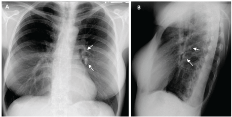 Figure 1: A and B, PA chest x-ray demonstrated two radiopaque images with tubular morphology, projected on left segmental vascular branches suggesting a metallic foreign body.
View Figure 1
Figure 1: A and B, PA chest x-ray demonstrated two radiopaque images with tubular morphology, projected on left segmental vascular branches suggesting a metallic foreign body.
View Figure 1
Contrast-enhanced pulmonary embolism (PE)-protocol helical chest computed tomography (CT) (Figure 2) revealed metallic opacities (2000 Hounsfield Units [HU]) at the origin of the lateral and posterior left basilar segmental arteries, and the posterior left segmental artery. In addition, the CT scan showed a small filling defect.
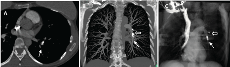 Figure 2: A. CT mediastinal window demonstrated a pulmonary embolism in the segmental basal arteries secondary to the metallic foreign body migration; B and C. Coronal maximum intensity projection (MIP) and 3D reconstructions of contrast-enhanced pulmonary embolism-protocol helical chest computed tomography showed metallic devices located at the origin of the lateral and posterior basal segmental arteries (arrow) and another located in the posterior segmental artery (empty arrow).
View Figure 2
Figure 2: A. CT mediastinal window demonstrated a pulmonary embolism in the segmental basal arteries secondary to the metallic foreign body migration; B and C. Coronal maximum intensity projection (MIP) and 3D reconstructions of contrast-enhanced pulmonary embolism-protocol helical chest computed tomography showed metallic devices located at the origin of the lateral and posterior basal segmental arteries (arrow) and another located in the posterior segmental artery (empty arrow).
View Figure 2
These findings suggested migration of the coils and concomitant acute symptomatic PE. Transthoracic echocardiography and lower limb ultrasound testing revealed no abnormalities.
The patient was discharged after a week of hospitalization with standard anticoagulant treatment. She did not report respiratory complaints in subsequent medical consultation.
The pattern presented in chest CT will depend on the type of embolization. Disperse radiopaque/metallic small densities, centrilobular ground glass opacities, and micronodules/miliary nodules, as well as fibrotic/consolidative opacities, may indicate NTPE [1].
It's important to carefully evaluate the CT lung window configuration, especially when a foreign body embolism is not initially suspected. Most abnormalities can be detected in lung windows, including calcific and metallic foreign materials [1].
Multidetector CT has the ability to create optimal multiplanar and MIP images in axial, coronal, or sagittal planes. These post-processing tools are immensely valuable in accurately locating small intravascular particles [1].
Coil migration can occur after pelvic vein embolization and cause a NTPE. The chest x-ray may show a linear density at an unusual anatomic location, but CT is the gold standard to confirm the position of the foreign body [5].
Interventional extraction should be considered immediately when the foreign body adheres firmly to the vascular wall. This approach should be performed using the same jugular access or additional femoral venous access [4].
Complications after pelvic embolization are rare [6]. A case report published by D' Amato R, et al. in 2016, presented a 49-year-old woman with a history of varicose pelvic vein embolization 15 years ago. She was derived after an incidental radiological finding of a nodular image of metal density in the left hemithorax. They proceeded to perform a CT scan of the thorax and abdomen that showed a foreign body consistent with a coil in the left lower lobe segmental branch. The patient was asymptomatic, so she was treated conservatively with clinical and image subsequent examinations [7].
Cases of coil migration have been described in men as well. In 1993 Moriel E, et al. published a case report that presented a 35-year-old man with an asymptomatic pulmonary migration of a coil after a pelvic embolization due to the treatment of erectile dysfunction [8].
As mentioned before, most of the published cases of coil migration after pelvic embolization are asymptomatic or incidental radiological findings. Our case is the first to report the migration of coils after a pelvic embolization and a concomitant acute pulmonary embolism in a symptomatic patient. This is important because the patient was treated with standard anticoagulation and conservative measures, with subsequent clinical and radiological evaluations without incidents.
The pulmonary migration of coils after a pelvic embolization is a rare complication. However, it should be considered as a differential diagnosis in patients with thoracic discomfort and previously described medical history.
Financial/nonfinancial disclosures: None declared.