Parasite, Mango fly, Surgical removal, Drying cloths
We report a 35-year-old patient that presented himself to volunteering doctors in the slums of Nairobi, Kenya. The patient has been suffering of a sensation of crawling on his back and the dorsal side of his arms. Furthermore, he mentioned multiple painful swelling increasing in size. Upon clinical status, multiple noduli were discovered in differing clinical stages. The vast majority was appearing as furuncular subcutaneous noduli with a cratered center, where others appeared as healing wounds with crusts or granulomes excreting pus (Figure 1).
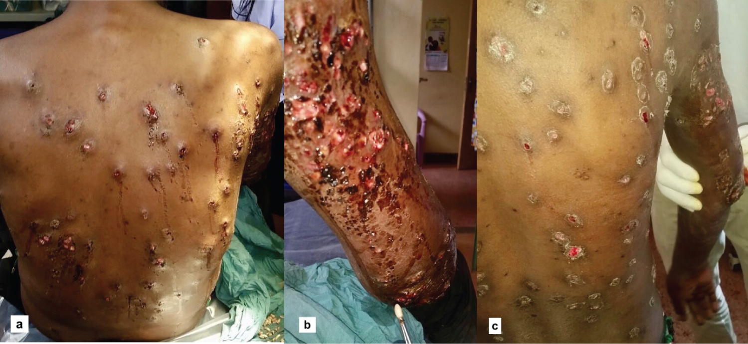 Figure 1: a) Clinical image of the patients back at first presentation; b) Clinical image of the backside of the right upper arm, after removal of maggots; c) Clinical image after one week after removal of the larva.
View Figure 1
Figure 1: a) Clinical image of the patients back at first presentation; b) Clinical image of the backside of the right upper arm, after removal of maggots; c) Clinical image after one week after removal of the larva.
View Figure 1
Anamnestically the patient revealed that his hand washed laundry was regularly left to dry in local vegetation, such as bushes and grass. This information was leading us to our working hypothesis of dermatomyiasis.
After thorough disinfection of the patients back and upper extremities, white and yellow appearing larva of about 10-15 mm in size could be removed by the use of a forceps and manual compression.
After removal of 99 maggots, antiseptic wound dressings were applied to protect the wounds from further infections. Additionally antibiotic therapy with Unacid was given for 2 days. Under closer inspection of the larvae they could be identified as the once of the mango fly (Cordylobia anthropophaga). The parasital invasion causing Myiasis by the mango fly is commonly seen in sub-Saharan Africa [1]. Typically the female fly places their eggs on sandy soil for further population. However, a common way of infection is happening through cloths that are dried outside on the ground, and are contaminated with urine, excreta or sweat. The female fly then places the fly-eggs on the spread out cloths.
Immediately after skin contact is made the larva invades the skin [2]. Most commonly an invasion of the back, gluteal region or the backside of the extremities is seen [3,4]. Alternative to the surgical removal of the larva, the skin opening can be closed by petroleum jelly or oil to hinder the maggot from breathing, leading to an escape of the skin in the next 48h [1]. Interestingly a similar effect can be achieved by closure of the opening by bacon, leading the larva to flee the skin [1,5]. To prevent secondary infection after surgical removal of the maggots surgically can be applied in case of skin damage or damage of the larva [1].
There are no conflicts of interests.