

International Journal of Clinical Cardiology
Chiari Network: An Embryological Remnant - A Case Report and Review
Shivadeep S1*, Anandaraja S2,3 and Devi Jansirani D4
1Specialist, Department of Radiodiagnosis, Badr-Al-Samaa Polyclinic, Oman
2Consultant, Department of Cardiology and Cardiac Electrophysiology, Indira Gandhi Government General Hospital, India
3Postgraduate Insitute, Puducherry, India
4Lecturer, Department of Human Structure and Neurobiology, Oman Medical College, Oman
*Corresponding author: Shivadeep S, Specialist, Department of Radiodiagnosis, Badr-Al-Samaa Polyclinic, Sohar, Oman, E-mail: drshivadeep@gmail.com
Int J Clin Cardiol, IJCC-2-038, (Volume 2, Issue 3), Case Report; ISSN: 2378-2951
Received: April 23, 2015 | Accepted: June 28, 2015 | Published: June 30, 2015
Citation: Shivadeep S, Anandaraja S, Devi Jansirani D (2015) Chiari Network: An Embryological Remnant - A Case Report and Review. Int J Clin Cardiol 2:038. 10.23937/2378-2951/1410038
Copyright: © 2015 Shivadeep S, et al. This is an open-access article distributed under the terms of the Creative Commons Attribution License, which permits unrestricted use, distribution, and reproduction in any medium, provided the original author and source are credited.
Abstract
Chiari network is a fenestrated membranous structure most commonly observed near the valve of inferior vena cava. It is considered as an embryological remnant of right valve of sinus venosus. Inspite of its benignity, its complications have been reported in the literature. This article contributes a detailed review compiling the association of Chiari network with various pathological conditions and its complications.
We report a case of Chiari network diagnosed in a 36-year old female by trans-esophageal echocardiography. The anomaly was associated with patent foramen ovale and there was a chance of impending paradoxical embolism. This case is reported here for its rarity and even rare association with paradoxical embolism.
Keywords
Chiari network, Paradoxical embolism, Eustachian valve
Introduction
Chiari network is considered as an embryological remnant formed by incomplete resorption of right valve of sinus venosus. It may be characterized as reticular network of fine strands attached to right atrium [1] or as membranous fenestration involving valve of inferior vena cavae or coronary sinus, extending to crista terminalis and interatrial septum [2]. Its incidence varies from 1.5-3% [3,4]. Although Chiari network is often considered as a benign anatomical variant, it may be associated with various clinical manifestations [3].
This case is reported here for its rarity and even rarer occurrence of associated impending pulmonary embolism.
Case Report
A 36-year-old female presented with complaints of difficulty in breathing and chest pain for past 10 days. She was a known diabetic and hypertensive since 3 months and on regular medications. There was no past history of fever, cough, expectoration, difficulty in breathing and symptoms suggestive of exertional angina. There was no history of recent surgery or child birth. She was not taking any oral contraceptive pills. There was no history of malignancy and coagulation disorder. There was no episode of deep vein thrombosis in the past. Examination of cardiovascular and respiratory systems showed no significant abnormality. ECG was within normal limits and chest radiograph was normal.
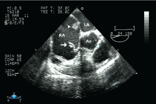
Transthoracic echocardiogram showed dilated right atrium and right ventricle. There was mild tricuspid regurgitation. The calculated peak systolic gradient between right atrium and right ventricle was 64mmHg. Transesophageal echocardiogram (Figure 1) revealed web-like structure with numerous thread-like components attached to the wall of right atrium near the site of entry of inferior vena cava. The structure showed characteristic whip like movements with each cardiac cycle. A small thrombus was found to be attached to the inter-atrial septum in the region of patent foramen ovale. The thrombus was found to enter into the foramen ovale with a part of it in the left atrial side. Severe eccentric tricuspid regurgitation was seen. Severe pulmonary hypertension was noted. There was no thrombus found either in the left atrium or in the main pulmonary artery and its bifurcation.
.
Figure 1: Trans-esophageal echocardiogram revealed web-like structure (white arrow) with numerous thread-like components attached to the wall of right atrium (RA) near the site of entry of inferior vena cava. A small thrombus (white arrowhead) was found to be attached to the inter-atrial septum in the region of patent foramen ovale. The thrombus was found to enter into the foramen ovale with a part of it in the left atrial side (LA). The right atrium was dilated.
View Figure 1
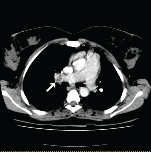
Contrast enhanced CT scan of the thorax showed dilatation of main pulmonary artery (31mm) with normal caliber of aorta (25mm). The ratio between pulmonary artery to aorta was more than one. The diameter of right and left pulmonary artery was 23mm and 22mm respectively, thereby confirming dilatation of both the vessels. There was a thrombus of size 1.4 x 0.9 x 1cm in the right pulmonary artery at the level of hilum of right lung causing partial obstruction of the lumen (Figure 2). Another thrombus was found in one of the branches of pulmonary artery, supplying lower lobe of left lung. There was no focal lesion in both lungs. Doppler of lower limb veins showed evidence of deep venous thrombosis in right superficial femoral vein.
.
Figure 2: Axial section of Contrast enhanced CT scan of thorax revealed dilated main pulmonary artery as well as right and left pulmonary artery branches. Arrow shows thrombus in the right pulmonary artery at the level of right hilum causing partial obstruction of the lumen.
View Figure 2
Patient was treated with anticoagulants and recovered. Transthoracic echocardiogram was repeated. There was no evidence of thrombus found in the inter-atrial septum.
Discussion
Embryological basis
During development of heart, when right horn of sinus venosus is absorbed into the primitive atrium, the sino-atrial orifice is guarded by right and left venous valve. Left venous valve fuses with septum secundum and blends with interatrial septum. The right valve regresses between 9th to 15th week of gestation, its cephalic portion remaining as the crista terminalis and its caudal portion dividing to form the Eustachian and The besian valves [5]. Failure of regression of right valve of sinus venosus gives rise to the fenestrated membranes or fine thread like strands attached to right atrium as Chiari's network, involving valve of inferior vena cavae or coronary sinus [1,2].
Incidence
The incidence of Chiari network in echocardiography has been reported as 1.5-3% [3,4]. The association of Chiari network with a thrombus entrapped in the channel of a patent foramen ovale with impending paradoxical embolism, as in the present case is an uncommon finding.
Morphology
This congenital remnant was first discovered by Hans Chiari in 1897 in an autopsy [3]. It is a web-like structure with variable number of thread like components. Its filaments are attached to wall of the right atrium in close proximity to the entrance of the inferior vena cava [6]. The distal attachment may extend into coronary sinus, crista terminalis and interatrial septum [1,2].
Echocardiographic findings
On echocardiography, this network appears to be a highly mobile, highly reflectant echo target in the right atrium near the opening of inferior vena cava and coronary sinus [7]. However, it can present as an unusual cystic mass in right atrium [8]. Yet another case of cystic mass in right atrium observed through transthoracic echocardiography had been confirmed as a pseudocystic representation of Chiari netwok in transoesophageal echocardiography [9], thereby emphasizing the importance of transoesophageal echocardiography.
Differential diagnosis
Chiari network possess diagnostic difficulties as it can be confused as right atrial thrombi, right heart vegetation, flail tricuspid leaflet [3], valve disruption, ruptured chordae tendinae of tricuspid apparatus [4] or even with a pedunculated right heart tumor [10] which need immediate surgery.
In the present case, the normal appearing three valves of tricuspid valve excluded the chance of valve disruption. The highly mobile, bright, cyclic echocardiographic target was not moving into the right ventricle inflow tract, thus it was least likely to be tricuspid valve vegetations. In four chamber view, the echogenic structure was typically located in posterolateral aspect and moving anteroinferior and medial direction inside the right atrium thus confirming the presence of Chiari network at the level of inferior vena caval opening.
Therefore knowledge about echocardiographic appearance of this normal variant is mandatory to avoid unwanted surgery due to misinterpretation especially in those cases with history of febrile illness, intravenous drug abuse and congestive cardiac failure.
Associated pathological conditions
Though Chiari network is considered as anatomical variant, it is reported in the literature that it was associated with infective endocarditis [11], fibroelastic papilloma [12], tricuspid atresia [5], fetal hydrops [13,14], platypnea-orthodeoxia with atrial septal hypertrophy [15], atrial septal aneurysm [1], atrial fibrillation [6], Bechet's disease [16]. Entrapment of a Cardiac catheter [2] and entanglement of an Atrial septal defect Occluder device [17] by strands of Chiari network had been reported. On auscultation, Chiari network can create low thronging murmur [18,19] and split heart sound [13].
Chiari network can act as a site of thrombus formation. This can be primary thrombus formed in the network itself or capture of venous thrombus formed elsewhere in the body [20]. Right atrial thrombus formed in this manner, can lead to pulmonary emboli and even cerebral and peripheral arterial emboli when associated with patent foramen ovale [1].
Chiari network and paradoxical embolism
Since Chiari network is considered as embryological remnant, it maintains the embryologic right atrial flow pattern and directs the blood from right to left, favoring the persistence of patent foramen ovale; thereby creating cyanosis [13,21,22], atrial septal aneurysm and paradoxical embolism [1].
In the present case, Chiari network was associated with patent foramen ovale and impending paradoxical embolism. There is even a recent case report, wherein a patient who had embolic stroke was diagnosed with a giant Chiari network creating paradoxical embolism for which surgical resection of the network was done. The author also cited that 83% of the Chiari network was associated with patent foramen ovale [23].
First described by Cohnheim in 1877, paradoxical embolism involves the passage of venous embolic material through a right-to-left intracardiac shunt into the arterial circulation, leading to a systemic thromboembolic manifestation such as stroke, kidney infarction, or acute limb infarction. It can be treated with anticoagulants and thrombolytic agents. Surgical intra-cardiac embolectomy and closure of patent foramen ovale may be curative [24].
In the present case, association of Chiari network with a thrombus in inter-atrial septum which was projecting through the patent foramen ovale, was a situation of impending paradoxical embolism. Had it not been diagnosed by transoesophageal echo and thrombolysed earlier, patient may undergo an episode of systemic embolic manifestation. Prompt diagnosis helped in immediate treatment of the patient with anti-coagulants and had saved the patient from disastrous paradoxical systemic thrombo-embolic events like catastrophic stroke or limb infarct.
Is Chiari network a nidusfor thrombi OR a congenital protective filter?
Chiari network was mentioned as a congenital inferior vena cava filter [1] and prevented pulmonary embolism in a case of polycythemia patient. However, it had been cited that once the filter like effect of Chiari network was overcome, it could result in fatal massive pulmonary embolism [25]. Another author opines that Chiari network removes emboli from the circulation purely by chance, and further emboli may likely reach the lung [6].
In this case, it is an issue of debate whether the thrombus formed in the right atrium is due to disturbed hemodynamics created by Chiari network itself or due to deep vein thrombosis getting dislodged and filtered by Chiari network.
Whatever the possibilities may be, in all cases of Chiari network, it is recommended to scrutinize for any impending thrombus in the region of patent foramen ovale. Chiari network should no longer be treated as innocuous.
Conclusion
To prevent catastrophic thromboembolic events, anticoagulation therapy is highly recommended for all patients having Chiari network associated with patent foramen ovale, irrespective of whether they are symptomatic or not. In all stroke patients, especially young, one should look for Chiari network and associated patent foramen ovale. Proper diagnosis and prompt management can save the patients from serious life threatening thromboembolic events.
References
-
Goedde TA, Conetta D, Rumisek JD (1990) Chiari network entrapment of thromboemboli: congenital inferior vena cava filter. Ann Thorac Surg 49: 317-318.
-
Goldschlager A, Goldschlager N, Brewster H, Kaplan J (1972) Catheter entrapment in a Chiari network involving an atrial septal defect. Chest 62: 345-346.
-
Bekar L, Onalan O, Altunkas F, Atmaca H, Atasoy I, et al. (2008) A prominent Chiari network prolapsing into right ventricle. Anadolu Kardiyol Derg 8: E27.
-
Werner JA, Cheitlin MD, Gross BW, Speck SM, Ivey TD (1981) Echocardiographic appearance of the Chiari network: differentiation from right-heart pathology. Circulation 63: 1104-1109.
-
McMahon CJ, Nihill MR, Kovalchin JP, Lewin MB (2000) Echocardiographic features of Chiari's network in association with tricuspid atresia. Tex Heart Inst J 27: 312-313.
-
Zuzana M, Petr W, Dana B, Martin P, Hana L, et al. (2010) An embolus in the right atrium caught in the Chiari network and resistant to thrombolysis. BMJ Case Rep 2010.
-
Islam AK, Sayami LA, Zaman S (2013) Chiari network: A case report and brief overview. J Saudi Heart Assoc 25: 225-229.
-
Bae CH, Kwon OC, Lee S, Lee CH, Cho JW (2012) Cystic Mass on Right Atrium of Unusual Form of Chiari's Network: A Case Report. Korean J Thorac Cardiovasc Surg 45: 254-256.
-
Walpot J, Sahin-Arpaci G, Sadreddini M (2015) A chiari network mimicking a cystic structure. Neth Heart J 23: 70-71.
-
Tanaka H, Ueda K, Murakami M, Hasegawa S, Sunamori M (2002) A prominent Chiari network. Ann Thorac Surg 73: 1985.
-
Payne DM, Baskett RJ, Hirsch GM (2003) Infectious endocarditis of a Chiari network. Ann Thorac Surg 76: 1303-1305.
-
Wasdahl DA, Wasdahl WA, Edwards WD (1992) Fibroelastic papilloma arising in a Chiari network. Clin Cardiol 15: 45-47.
-
Bendadi F, van Tijn DA, Pistorius L, Freund MW (2012) Chiari's network as a cause of fetal and neonatal pathology. Pediatr Cardiol 33: 188-191.
-
Aypar E, Sert A, Odabas D (2013) Unusually prominent Chiari's network prolapsing into the right ventricle in an asymptomatic newborn. Pediatr Cardiol 34: 1017-1019.
-
Shakur R, Ryding A, Timperley J, Becher H, Leeson P (2008) Late emergence of platypnea orthodeoxia: Chiari network and atrial septal hypertrophy demonstrated with transoesophageal echocardiography. Eur J Echocardiogr 9: 694-696.
-
Alonso G, Santos E, Fuertes A, Jim�nez A, Guti�rrez JA (2007) Beh�et's disease and Chiari's network. Clin Rheumatol 26: 2189-2190.
-
Cooke JC, Gelman JS, Harper RW (1999) Chiari network entanglement and herniation into the left atrium by an atrial septal defect occluder device. J Am Soc Echocardiogr 12: 601-603.
-
Alvarez J, Hermann G (1931) Unusual signs from an expansive Chiari network along with signs of a syphilitic aortic regurgitation. Am J Syph 15: 532.
-
Wilson R, Charleston SC (1938) A case of Chiari's network associated with a murmur resembling the bruit de Roger. J Am Med Assoc 11: 917-918.
-
Schneider B, Hofmann T, Justen MH, Meinertz T (1995) Chiari's network: normal anatomic variant or risk factor for arterial embolic events? J Am Coll Cardiol 26: 203-210.
-
Lanzarini L, Lucca E, Fontana A, Foresti S (2001) Cortriatriatumdextrum resulting from the persistence of embryonic remnants of the right valve of the sinus venosus: prevalence and echocardiographic aspects in a large consecutive non-selected patient population. Ital Heart J 2: 1209-1216.
-
Gussenhoven WJ, Essed CE, Bos E (1982) Persistent right sinus venosus valve. Br Heart J 47: 183-185.
-
Laguna G, Arce N, Blanco M. (2015) Giant Chiari network, foramen ovale and paradoxical embolism. Rev Esp Cardiol 68: 250.
-
Tang CE (2004) Paradoxical embolism: a rare life- and limb-threatening emergency. CJEM 6: 40-44.
-
Obaji SG, Cooper R, Somauroo J (2012) Chiari network: a protective filter against pulmonary embolism in a case of polycythaemia. BMJ Case Rep 2012.
