

International Journal of Clinical Cardiology
Circulating Endothelial Cells, Flow Mediated Dilatation % as Markers of Endothelial Dysfunction in Paroxysmal Lone Atrial Fibrillation
Rania Gaber*, Wessam Salah Ibrahim and Manal Hamesa
Department of Cardiology, Clinical pathology and Radiology Tanta University, Egypt
*Corresponding author: Rania Gaber, Department of Cardiology, Clinical pathology and Radiology Tanta University, Egypt, E-mail: raniagaber2009@hotmail.com
Int J Clin Cardiol, IJCC-3-082, (Volume 3, Issue 2), Research Article; ISSN: 2378-2951
Received: March 17, 2016 | Accepted: July 27, 2016 | Published: July 30, 2016
Citation: Gaber R, Ibrahim WS, Hamesa M (2016) Circulating Endothelial Cells, Flow Mediated Dilatation % as Markers of Endothelial Dysfunction in Paroxysmal Lone Atrial Fibrillation. Int J Clin Cardiol 3:082. 10.23937/2378-2951/1410082
Copyright: © 2016 Gaber R, et al. This is an open-access article distributed under the terms of the Creative Commons Attribution License, which permits unrestricted use, distribution, and reproduction in any medium, provided the original author and source are credited.
Abstract
Background: Atrial fibrillation (AF) is the most prevalent sustained cardiac arrhythmia in adult population. Aim of the present study is to evaluate the association of paroxysmal lone AF with endothelial dysfunction in young patients.
Methods: Two groups of participants were prospectively enrolled. The first group comprised of 70 patients with recurrent paroxysmal lone AF. The second group comprised of 20 healthy controls in sinus rhythm matched by age and gender.
All the participants underwent physical examination, laboratory analysis (including determination of C-reactive protein (CRP)), standard echocardiography, exercise-stress testing, brachial artery Flow Mediated Dilatation (FMD) % and Circulating Endothelial cells (CECs) by flow cytometry were assessed.
Results: There were no differences between the 2 groups regarding age, gender and most clinical, laboratory and echocardiographic characteristics (all p > 0.05 except CRP level). FMD % of lone AF patients was significantly lower 6.4 ± 1.6 versus 9.2 ± 2.4 (p < 0.0001) than FMD of healthy controls. CECs count was significantly elevated in lone AF patients compared to controls 24.7 ± 7.2 versus 13.2 ± 3.8 (p < 0.0001). In the multivariate analysis, the independent FMD %, CECs determinants in our study population were the duration of attacks and CRP level.
Conclusion: Paroxysmal lone AF is associated with systemic endothelial dysfunction. Duration of the attack and high level CRP are independent contributors to lower FMD % and higher CECs which may confer the risk for more profound endothelial damage.
Keywords
AF, Endothelial dysfunction
Abbreviations
CECs: Circulating Endothelial Cells; FMD %: Flow Mediated Dilatation%; AF: Atrial Fibrillation; LA: Left Atrium; CRP: C - Reactive Protein
Introduction
Atrial Fibrillation (AF) is the commonest arrhythmia encountered in clinical practice and is increasingly considered as an emerging health epidemic. Despite rapidly evolving treatment strategies, AF presents a complex management challenge to the physician and is now attracting substantial clinical and academic interest because of a strong association with substantial mortality and morbidity [1]. Over the past decade, systemic arterial endothelial dysfunction has been demonstrated both experimentally and clinically in various subsets of AF patients [2-4].
Circulating endothelial cells (CECs) have emerged as markers of vascular damage. While present in very small numbers in healthy individuals, CECs increase dramatically in diseases with vascular damage, such as cardiovascular disease, specific infections, and vasculitis [5-8].
Assessment of flow-mediated dilatation (FMD %) of the brachial artery is a reliable non-invasive tool to evaluate endothelial function. The technique provokes the release of nitric oxide, resulting in vasodilation that can be quantified as an index of vasomotor function [9].
Lone AF is defined as the occurrence of AF in subjects younger than < 60 years without associated comorbidities or recognized risk factors [10,11]. Lone AF is considered a benign condition with favorable long-term prognosis [12,13]. However, even in patients with persistent lone AF, an evidence of damage/dysfunction of atrial endocardium, platelet activation and increased inflammatory and oxidative stress has been found [14,15] and also no available studies concerning endothelial function in paroxysmal lone AF patients. The aim of this study was to evaluate the association of paroxysmal lone AF with endothelial dysfunction by comparing CECs, brachial artery FMD % of younger patients with paroxysmal lone AF with FMD % of healthy control subjects in sinus rhythm.
Patients and Methods
The study was conducted between September 2011 and April 2013. Ethical committee of Tanta university approval was taken and all the study group 70 patients and 20 volunteers approval for study was taken after full explanation. Patients with recurrent paroxysmal lone AF (sinus rhythm at examination) and healthy volunteers without history of arrhythmia, matched by age, gender and no AF risk factors. The patients were eligible if developed recurrent attacks of short duration AF, relieved without treatment. AF was confirmed by 12-lead ECG. AF duration was determined as accurately as possible according to patient-reported symptom on set and available medical documentation. AF was considered lone in patients younger than 60 years of age if there were no known associated cardiovascular disorders, or precipitating factors for AF.
Exclusion Criteria
History of hypertension, diabetes mellitus, smoking or other cardiovascular disorders priorto AF, thyroid dysfunction, LA more than 40 mm, chronic pulmonary diseases, acute or chronic inflammatory disorders, malignancy, recent body trauma or surgery. No regular medications except rate control during attacks (verapamil or beta blockers).
All the Study Group underwent the Following
1. Complete history taking
2. Clinical evaluation
3. Routine laboratory investigations which include: Complete blood count, urine analysis, kidney function tests, thyroid function assessment, determination of C-reactive protein (CRP) levels (by a commercially available immunoassay for high-sensitivity detection - detection limit 0.1 mg/L), liver function test, lipid profile which include: Total cholesterol, Triglyceride, Low-density lipoprotein and high -density lipoprotein.
4. 12-lead electrocardiogram (ECG), exercise stress testing and standard transthoracic echocardiographic examination.
5. Assessment of endothelial function by flow mediated dilatation (FMD %):
1. Endothelial function was assessed with high-resolution B-mode Doppler (ATL HDI 5000 with a 7.4 MHz linear-array transducer). The brachial artery was examined using the standard protocol [16,17]. The test was performed in the morning in quiet, low light room.
2. Subjects had fasted and the brachial artery was scanned 5-15 cm above cubital fossa. Resting diameter was measured, then blood pressure cuff was inflated to 300 mmHg around forearm and further scanning was done 1 min during occlusion then 1 min after occlusion (cuff release). FMD % was calculated as: [(post deflation diameter - resting diameter)/resting diameter] × 100.
6. Inter, intra-observer variability: Vascular studies were successful in all the participants. Inter- and intra-observer variations for baseline brachial artery measurements in our laboratory are 0.04 ± 0.01 mm and 0.05 ± 0.02 mm, respectively.
7. Immunophenotyping of CECs by flow-cytometry
Venous blood samples (10 ml) were separated into 2 tubes: one tube (5 ml) was collected into EDTA tube and transferred into anticoagulant then analyzed by flow-cytometry [18]. Freshly isolated peripheral blood mononuclear cells were washed and separated from blood of patients and healthy control using lysis solution for erythrocytes lysis then re-suspended in phosphate buffered saline (pH 7.4) containing 20 uL of the appropriate antibody and cells were double stained with mouse anti-human fluorescein isothiocyanate conjugated CD45 antibody and mouse anti-human phycoerythrin conjugated CD146 antibody (BD Biosciences) to identify CD45 - and CD146 + respectively [19]. The Iso-type control was used to determine nonspecific c binding of the lymphocyte subset-specific c antibodies and to set the cut-off between fluorescence-negative and fluorescence-positive staining. Stained cells were washed 3 times with 1% bovine serum albumin, pH 7.2, and then 7AAD was added to stain dead cells. The cells were analyzed within 15 min after addition of 7AAD using a fluorescence-activated cell scanner and Cell Quest software [FACS Caliber, Becton-Dickinson]. Cells were plotted according to forward scatter and side scatter profiles and a region was drawn around the small, live cell population containing the lymphocyte. The cell population data obtained from the quadrant statistics (2-color staining) was standardized for the number of mature CEC using the sum of CD45 -, CD146 + and 7-AAD negative (Live) cells within this region (i. e., CD45 -, CD146 + and 7-AAD + cells were not accounted). Normal CEC count by flow-cytometry was < 20 cells/ml [20].
Statistics
Statistical presentation and analysis of the present study was conducted, using the mean and standard deviation, unpaired t- test used to compare. Linear regression analysis was used in correlation between CEC, FMD % and different parameters done.
Results
Demographic, clinical and laboratory data are summarized in table 1. There were no differences between the 2 groups regarding age, gender and most clinical, laboratory and echocardiographic characteristics, CRP level was significantly higher in paroxysmal lone AF group comparing to controls 1.12 ± 0.44 versus 0.84 ± 0.35 (Table 1 and Table 2).
![]()
Table 1: Demographic, clinical and laboratory parameters.
View Table 1
![]()
Table 2: M -mode echocardiographic data of study group and controls.
View Table 2
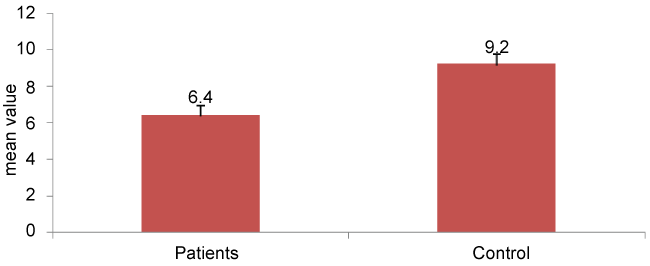
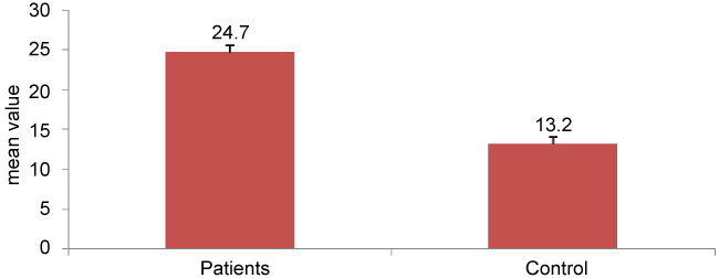
FMD % of AF patients was significantly lower than FMD of healthy controls 6.4 ± 1.6 versus 9.2 ± 2.4 (p < 0.0001). CECs count was significantly elevated in paroxysmal lone AF patients compared to controls 24.7 ± 7.2 versus 13.2 ± 3.8, p < 0.0001 (Table 3, Figure 1 and Figure 2).
.
Figure 1: Shows the differences between the mean values of FMD % in lone AF patients group and controls. FMD % = Flow Mediated Dilatation, AF = Atrial Fibrillation
View Figure 1
.
Figure 2: Shows the differences between the mean values of CECs in lone AF patients group and controls. CECs = Circulating Endothelial Cells, AF = Atrial Fibrillation
View Figure 2
In the multivariate analysis, the independent FMD %, CECs determinants in our study population were the duration of the attack and CRP level (Table 4).
![]()
Table 3: FMD %, CECs of study group and controls.
View Table 3
![]()
Table 4: Correlation in between FMD %, CECs and different parameters in AF group using multi-linear regression analysis.
View Table 4
Discussion
Lone atrial fibrillation (AF) is a term commonly used to denote AF occurring in a small subset (∼3%) of patients without identifiable cardiovascular and extra-cardiac comorbidities or triggering factors [21,22]. It has been recognized that circulating indices of endothelial damage are related to increased risk of stroke in AF and endothelial dysfunction in peripheral vessels has been associated with adverse vascular events [23,24]. However, the systemic endothelial dysfunctions in paroxysmal lone AF young patients are still not fully investigated. Therefore, the aim of this study was to evaluate the association of paroxysmal lone AF with endothelial dysfunction by comparing brachial artery FMD %, CECs of younger patients with paroxysmal lone AF with FMD % of healthy controls.
In the present study two groups of participants were prospectively enrolled. The first group comprised of 70 patients with recurrent paroxysmal lone AF (sinus at time of examination). The second group comprised of 20 healthy controls without history of arrhythmia matched by age and gender. All the participants underwent physical examination, laboratory analysis (including determination of C-reactive protein (CRP)), standard echocardiography, exercise-stress testing, brachial artery FMD % and CECs by flow-cytometry were assessed. There were no differences between the 2 groups regarding age and gender. FMD % of AF patients was significantly lower (p < 0.001) than FMD of healthy controls. CECs count was significantly elevated in lone AF patients compared to controls. In the multivariate analysis, the independent FMD %, CECs determinants in our study population were the duration of the attacks and CRP level.
Although, current evidence indicates that chronic low-grade inflammation could represent a link between AF and subclinical vascular disease and Increased plasma levels of inflammatory markers (e.g., C-reactive protein) have been reported in subjects with lone AF compared to healthy individuals in sinus rhythm same as our study results, the mechanism of endothelial dysfunction in young patients with lone AF is not clear. To our knowledge, the present study is the first study emphasis upon endothelial dysfunction in young patient experienced paroxysmal lone AF without any recognizable risk factor for endothelial dysfunction or presence of LA dilatation.
Previous studies showing that the FMD % technique could be reliably utilized for endothelial function assessment in AF. These trials invariably demonstrated impaired FMD % in the AF patients in comparison with the healthy subjects. The implication of these findings was that AF presence could be regarded as a risk factor for systemic endothelial dysfunction. However, most of these trials have been conducted in patients with underlying comorbidities, most often hypertension, coronary artery disease and diabetes, which are recognized risk factors for endothelial damage. There have been a few studies that enrolled a relatively small subset of predominantly older patients with idiopathic AF that also confirmed impaired FMD [3,25-30]. Recently Polivina M et al. [31] demonstrated impaired FMD % in relatively young patients (mean age 45 years) with persistent lone AF (more than 7 days) and low cardiovascular risk profile. Two well recognized risk factors for endothelial damage, i.e., smoking and serum cholesterol levels were independent predictors of lower FMD % in the study, also LA dimension up to 45 mm patients were included in the study.
Study Limitations and Recommendations
- Asymptomatic AF not included as they don't seek medical advice and absence of continuous monitoring of the patients during study duration (more than one year).
- Duration of attacks before arrival to hospital was dependent on patient assessment due to absence of continuous monitoring of the patients during study duration (more than one year).
- Lack of long term follow up of this group of patients.
Further larger clinical and pathological studies are needed for full understanding the mechanism of endothelial dysfunction in young patients with lone AF.
Conclusion
Paroxysmal lone AF is associated with systemic endothelial dysfunction even in relatively young patients with no cardiovascular disorders or risk factors. Duration of the attack and high level CRP are independent contributors to lower FMD % and higher CECs which may confer the risk for more profound endothelial damage.
References
-
Stewart S, Murphy NF, Walker A, McGuire A, McMurray JJ (2004) Cost of an emerging epidemic: an economic analysis of atrial fibrillation in the UK. Heart 90: 286-292.
-
Cai H, Li Z, Goette A, Mera F, Honeycutt C, et al. (2002) Downregulation of endocardial nitric oxide synthase expressionand nitric oxide production in atrial fibrillation. Circulation 106: 2854-2858.
-
Takahashi N, Ishibashi Y, Shimada T, Sakane T, Ohata S, et al. (2001) Atrial fibrillation impairs endothelial function of forearm vessels in humans. J Card Fail 7: 45-54.
-
Freestone B, Chong AY, Nuttall S, Blann AD, Lip GY (2007) Soluble E-selectin, von Willebrand factor, soluble thrombomodulin, and total body nitrate/nitrite product as indices of endothelial damage/dysfunction in paroxysmal, persistent, and permanent atrial fibrillation. Chest 132: 1253-1258.
-
Khan SS, Solomon MA, McCoy JP Jr (2005) Detection of circulating endothelial cells and endothelial progenitor cells by flow cytometry. Cytometry B Clin Cytom 64: 1-8.
-
Nakatani K, Takeshita S, Tsujimoto H, Kawaura Y, Tokutomi T, et al. (2003) Circulating endothelial cells in Kawasaki disease. Clin Exp Immunol 131: 536-540.
-
Woywodt A, Streiber F, de Groot K, Regelsberger H, Haller H, et al. (2003) Circulating endothelial cells as markers for ANCA-associated small-vessel vasculitis. Lancet 361: 206-210.
-
McClung JA, Naseer N, Saleem M, Rossi GP, Abrarham NG, et al. (2005) Circulating endothelial cells are elevated in patients with type 2 diabetes mellitus independently of HbA (1)c. Diabetologia 48: 345-350.
-
Corretti MC, Anderson TJ, Benjamin EJ Celermajer D, Charbonneau F, et al. (2002) Guidelines for the ultrasound assessment of endothelial-dependent flow-mediated vasodilatation of the brachial artery: a report of the International Brachialartery Reactivity Task Force. J Am Coll Cardiol 16: 257-265.
-
Camm JA, Kirchhof P, Lip GYH, Schotten U, Savelieva I, et al. (2010) Guidelines for the management of atrial fibrillation. The Task Force for the Management of Atrial Fibrillation of the European Society of cardiology (ESC). Eur Heart J 31: 2369-2429.
-
Potpara TS, Lip GY (2011) Lone atrial fibrillation: what is known and what is to come. Int J Clin Pract 65: 446-457.
-
Jahingir A, Lee V, Friedman PA, Trusty JM, Hodge DO, et al. (2007) Long-term prognosis and outcomes with aging in patients with lone atrial fibrillation: a 30-yeare follow-up study. Circulation 115: 3050-3056.
-
Potpara T, Grujic M, Marinkovic J, Vujisic-Tesic B, Ostojic M, et al. (2010) Mortality of patients with lone and idiopathic atrial fibrillation is similar to mortality in general population of Serbia. Vojnosanit Pregl 67: 132-135.
-
Mondillo S, Sabatini L, Agricola E, Ammaturo T, Guerrini F, et al. (2000) Correlation between left atrial size, prothrombotic state and markers of endothelial dysfunction in patients with lone chronic nonrheumatic atrial fibrillation. Int J Cardiol 75: 227-232.
-
Goette A, Weber M, Lendeckel U, Welte T, Lutze G, et al. (2004) Effect of physical exercise on platelet activity and the von-Willebrand-factor in patients with persistent lone atrial fibrillation. J Interv Card Electrophysiol 10: 139-146.
-
Rajagopalan S, Brook R, Mehta RH, Supiano M, Pitt B (2002) Effect of losartan in aging-related endothelial impairment. Am J Cardiol 89: 562-566.
-
Zhang X, Zhao SP, Li XP, Gao M, Zhou QC (2000) Endothelium-dependent and -independent functions are impaired in patients with coronary heart disease. Atherosclerosis 149: 19-24.
-
Macey M, McCarthy D, Azam U, Milne T, Golledge P, et al. (2003) Ethylene diaminetetraacetic acidplus citrate-theophylline-adenosine-dipyridamole (EDTA-CTAD): a novel anticoagulant for the flow cytometric assessment of platelet and neutrophil activation ex vivo in whole blood. Cytometry B Clin Cytom 51: 30-40.
-
Goon P, Boos C, Stonelake S, Blann AD, Gregory YH (2006) Detection and quantification of mature circulating endothelial cells using flow cytometry and immunomagnetic beads: A methodological comparison. Thromb Haemost 96: 45-52.
-
Woywodt A, Blann AD, Kirsch T, Erdbruegger U, Banzet N, et al. (2006) Isolation and enumeration of circulating endothelial cells by immunomagnetic isolation: proposal of a definition and a consensus protocol. J Thromb Haemost 4: 671-677.
-
Camm AJ, Kirchhof P, Lip GY, Schotten U, Savelieva I, et al. (2010) Guidelines forthe management of atrial fibrillation: the Task Force for the Management of Atrial Fibrillation of the European Society of Cardiology (ESC). Europace 12: 1360-1420.
-
Potpara TS, Lip GY (2014) Lone atrial fibrillation - an overview. Int J Clin Pract 68: 418-433.
-
Lip GY, Lane D, Van Walraven C, Hart RG (2006) Additive role of plasma von Willebrand factor levels to clinical factors for risk stratification of patients with atrial fibrillation. Stroke 37: 2294-2300.
-
Lind L, Berglund L, Larsson A, Sundström J (2011) Endothelial function in resistance and conduit arteries and 5-year risk of cardiovascular disease. Circulation 123: 1545-1551.
-
Freestone B, Chong A, Nuttall S, Lip GY (2008) Impaired flow mediated dilation as evidence of endothelial dysfunction in chronic atrial fibrillation: relationship to plasma von Willebrand factor and soluble E-selectin levels. Thromb Res 122: 85-90.
-
Guazzi M, Belletti S, Tumminello G, Fiorentini C, Guazzi MD (2004) Exercise hyperventilation, dyspnea sensation, and ergoreflex activation in lone atrial fibrillation. Am J Physiol Heart Circ Physiol 287: H2899-2905.
-
Guazzi M, Belletti S, Bianco E, Lenatti L, Guazzi MD (2006) Endothelial dysfunction and exercise performance in lone atrial fibrillation or associated with hypertension or diabetes: different results with cardioversion. Am J Physiol Heart Circ Physiol 291: H921-928.
-
Guazzi M, Belletti S, Lenatti L, Bianco E, Guazzi MD (2007) Effects of cardioversion of atrial fibrillation on endothelial function in hypertension or diabetes. Eur J Clin Invest 37: 26-34.
-
Skalidis EI, Zacharis EA, Tsetis DK, Pagonidis K, Chlouverakis G, et al. (2007) Endothelial cell function during atrial fibrillation and after restoration of sinus rhythm. Am J Cardiol 99: 1258-1262.
-
Shin SY, Na JO, Lim HE, Choi CU, Choi JI, et al. (2011) Improved endothelial function in patients with atrial fibrillation through maintenance of sinus rhythm by successful catheter ablation. J Cardiovasc Electrophysiol 22: 376-382.
-
Polovina M, Potpara T, Giga V, Stepanovic J, Ostojic M (2013) Impaired endothelial function in lone atrial fibrillation. Vojnosanit Pregl 70: 908-914.
