

International Journal of Oral and Dental Health
Effect of Placement Techniques on Sealant Efficacy
Apa Juntavee1*, Niwut Juntavee1, Jiranan Paleekoup1, Phillip Millstein2 and Behrouz Abedian3*
1Faculty of Dentistry, Khon Kaen University, Khon Kaen, Thailand
2School of Dental Medicine, Harvard University, Boston, MA, USA
3Department of Mechanical Engineering, Tufts University, Medford, MA, USA
*Corresponding author: Apa Juntavee, Faculty of Dentistry, Khon Kaen University, Khon Kaen 40002, Thailand, E-mail: apa.edu@hotmail.com
Behrouz Abedian, Department of Mechanical Engineering, Tufts University, Medford, MA 02155, USA, E-mail: Behrouz.abedian@tufts.edu
Int J Oral Dent Health, IJODH-1-006, (Volume 1, Issue 1), Research Article; ISSN: 2469-5734
Received: March 10, 2015 | Accepted: April 09, 2015 | Published: April 11, 2015
Citation: Juntavee A, Juntavee N, Paleekoup J, Millstein P, Abedian B (2015) Effect of Placement Techniques on Sealant Efficacy. Int J Oral Dent Health 1:006. 10.23937/2469-5734/1510006
Copyright: © 2015 Juntavee A, et al. This is an open-access article distributed under the terms of the Creative Commons Attribution License, which permits unrestricted use, distribution, and reproduction in any medium, provided the original author and source are credited.
Abstract
This in-vitro study evaluated and compared two techniques for placement of dental sealants. It is shown that sealant bondage to occlusal areas, a key factor in success and effectiveness of dental sealants, can be greatly influenced by the placement technique used in such operations. Custom-made flow-burs (Abedian and Millstein, J. Oper. Dent. 2006) of different dimensions were designed, constructed and compared to a conventional applicator for sealant efficacy. A moisture-tolerant resin-based sealant was applied to occlusal surfaces of caries-free upper and lower premolars extracted teeth, and manipulated by using a conventional applicator or flow-bur. Microleakage and depth of penetration studies on sealant integrity were conducted after subjecting the treated samples to thermocycling. Image processing was conducted on the sectioned teeth for all cases and SPSS statistical analysis of the data indicated that use of flow-burs in placing dental sealants can reduce microleakage by 50% as well as 66% reduction of unfilled voids. With advantages in consistency, convenience and better marginal adaptation, the use of such flow-burs may reduce occurrence of caries after the sealant application and ultimately improve sealant clinical success.
Keywords
Microleakage, Applicators, Sealants, Marginal adaptation, Dental caries
Introduction
Fissure sealants are one of the effective preventive tools in reducing incidence of caries for children and adolescents [1-6] and their applications in the U.S. alone cover significant portion of the population across all age groups and all income levels [7]. An ineffective marginal seal between the resin and the enamel is one of the most important factors causing sealant failure [8]. To increase sealant success rate, most recent attempts have focused on the chemical treatments such as acid etching in the preparation step before applying the sealant [9-11].
Efficacy of sealants also strongly depends on their mechanical penetration and filling into occlusal areas [12]. Surface depressions have restricted access: hard to clean making them more susceptible to caries, and hard to reach when applying the sealant. In an ideal application, the low-viscosity resin sealant fills all irregular occlusal areas. But penetrating deep into an occlusal area also give rise to a stronger physical bond between the sealant and the substrate which may reduce secondary caries [13], and providing longer lasting and more successful applications. Accordingly, a better marginal adaptation and deeper occlusal penetration will be key in improving the rate of success of this operation.
In present clinical practice, many sealants are often injected with a syringe onto the occlusal surfaces of teeth. For better adaptation, they are then shaped and spread around with a commercial applicator; however, upon withdrawing, the applicator leaves excess sealant which is light cured. Evidence of placement difficulty with a commercial applicator for other dental sealants has been shown previously [14]. This study introduces a method of delivery that uses an oscillating applicator that momentarily thins the sealant so that it will flow more readily without sticking to the applicator. The excess sealant in the treated tooth depends on the technique by which the sealant is applied.
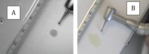
It's been shown that asymmetric rotating burs can provide superior marginal adaptation [15]. The purpose of this study is to show that better marginal adaptations with the application of such burs ultimately can reduce microleakage, resulting in more successful sealant application. The effect of a vibrating tool to control microleakage and sealant penetration using a microvibe vibrating probe has been studied previously and no significant difference was detected [16]. No physical reason was provided on how such microprobes are ineffective and no indication if such tools have better spreading ability. However, the burs that we have designed and used in this study may spread, deform and manipulate the sealant much more effectively than a conventional applicator (Figure 1) and could be more effective in dental sealant placement.
.
Figure 1: Spreading ability of a dental adhesive (Esthet-X flow). Using a flow-burs A - before application; B - After application
View Figure 1
When an asymmetric rotating bur 1-3 mm in size (flow-bur) with a moderate rotational speed (100 < ω < 1000 rpm, with ω the rotational speed) is used to deform an adhesive material, the bur will not stick to the material as opposed to a conventional plugger [14]. At such moderate speeds, the rotating bur will induce surface oscillations at high enough frequencies such that the interfacial interactions between the surface of the adhesive and the rotating bur will be minimal due to Deborah-number effects. Accordingly, the sealant may not stick to or wet the bur, as the bur deforms the sealant. The shortest times are often a Deborah number of unity, which brings about the low end of the working speeds of our flow-burs. If a non-oscillating conventional applicator is used to deform the adhesive, the material will stick to this plugger and any motion by the plugger will not result in better marginal adaptation. Induced oscillations of the dental adhesives have another beneficial effect as such materials exhibit pseudo-plastic behavior. With the application of rotating bur to deform the sealant, the sealant possesses less viscosity and might more completely wet occlusal surfaces with better adaptation of the interior surfaces.
Microleakage studies are a standard method for sealant efficacy [16-19], either in vivo or in-vitro. In in-vitro studies, it is possible to assess marginal leakage and overall sealant capacity to predict how such sealant operations are successful and how long they might last. Many factors influences microleakage between enamel surface and the sealant including anatomical constrictions, aprismatic enamel structure, enamel surface preparation, moisture contamination, errors in applying the sealant and temperature effects. In this study, we conducted microleakage and penetration investigation for two placement techniques with all other factors that influence adhesion and penetration remaining unchanged.
We hypothesize that using a flow bur when applying the sealant, by a dental operator who is familiar and comfortable with this technique, has several important advantages, compared to using a conventional dental applicator:
• Less operational errors
• Improved marginal adaptation
• Improved penetration into occlusal areas
The present study examines the effectiveness of two techniques in applying a dental sealant on how it adapt and seal to the occlusal surfaces. To evaluate some of these advantages, we utilize a comparative microleakage and depth of penetration study for two application procedures one with a conventional applicator and the other with a set of flow-burs that is designed and constructed to conduct these tests.
Materials and Methods
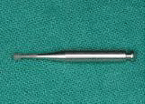
Custom-made Flow-burs for this comparative study were constructed in-house by modifying the tip of commercial round cutting burs. The burs were initially sand blasted to ensure better retention of the composite at the tip. Light-cured flowable composite dental material (Esthet-Xflow) was applied on the sand blasted tip and then shaped manually by hand such that it forms asymmetrical bi-paddle-type geometry without any sharp corners, as used in previous studies [14,15]. Once the desired shape was achieved, the tip material was light cured to harden with better grip on the remainder of the bur. The burs were then tested on their spreading ability of a dental adhesive on a PVC flat surface to confirm their functionality. Different size custom-made burs with maximum diagonal dimension of 1-3 mm were constructed. A flow-bur used in this study shown in Figure 2. If one follows this recipe, such burs could be made in a well-equipped dentist office.
Sample preparation
Eighty caries free upper and lower premolars, extracted for orthodontics reasons, were selected. In the post- extraction stage, the teeth were stored in 0.1% thymol solution in a dark container at room temperature. For the experiments, each specimen was embedded in a stone model and randomly divided into two groups of forty teeth each. Each group was further divided into two subgroups of twenty teeth per group. Subgroups 1 and 3 were assigned to be sealed with the oscillating flow- bur application technique; subgroups 2 and 4 were assigned to be sealed using the conventional applicator. In all the experiments, the rotational speed of the handpiece was set to approximately 300 rpm.
Occlusal surfaces of the samples were thoroughly cleaned for 20 seconds with a pumice slurry in a low speed micro-motor handpiece. The surfaces were then rinsed with air-water for 10 seconds and dried. The dried teeth were subsequently etched for 15 seconds with a phosphoric etchant (Etch-Rite™, Pulpdent), and rinsed for 10 seconds. The teeth to be sealed were air dried but not desiccated. Forty teeth (Gr1, Gr3) were sealed by the constructed flow-bur with Embrace™ Wetbond™ (Pulpdent, Watertown, MA) with an oscillating bur application technique while two groups of control (Gr2, Gr4) were sealed by using the conventional supplied cannula tip. Teeth were cured with UV light (3M™ ESPE™ Elipar™ 2500 Halogen Curing Light) for 20 second.
Processing of the samples
The teeth with sealant treatments were stored in distilled water at 37°C for 24 hours. Subsequently, the samples were subjected to a thermocycling (SPC20, Yamatake Honeywell, HW B 332R) regimen of 500 cycles between 5 and 55°C with a dwell time of 30 s in each bath
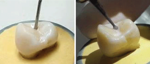
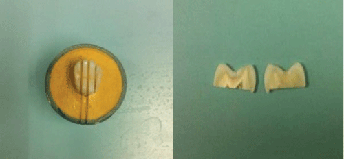
All samples in Groups 1 and 2 were covered with sticky wax and the surface of each sample was covered with two layers of nail varnish leaving a 1-mm window around the margin of the sealant and immersed in a dye solution (methylene blue 0.5%; Merck4279, Darmstadt, Germany) for 4 hours at 37°C. Following immersion in the dye solution, the teeth were subsequently rinsed under running tap water for 30s. All samples from Groups 1-4 were fixed and sectioned longitudinally in a bucco-lingual direction with a slow speed rotating diamond blade (Model 1000 ™ISOMET, Illinois, USA), providing two 1 mm-sections per tooth, shown in Figure 3a,3b.
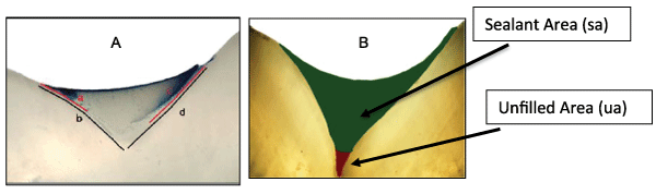
The two sides of each sectioned sample were inspected by polarized light and subjected to image analysis software (Image J: NIH) (50× magnification) to measure microleakage and penetration data. Measurements, analysis, and recording were performed by the same evaluator. After calibration to a millimeter ruler, the microleakage was evaluated quantitatively by measuring the length of dye-penetration. For each tooth-section sample that was considered, the fraction of microleakage was calculated according to where the lengths a and c are measured interfacial length of the penetrated dye on the two sides of the cavity and b and d are the respective total length of the sealant interface with enamel, as shown in Figure 4A.
.
Figure 4: Measuring microleakage and penetration. (A): Lengths a and c are length of microleakage, and b and d represent the length of sealant on two opposite side. (B): The fissure sealant area (sa) and unfilled area (ua).
View Figure 4
The penetration ability was assessed as the proportional area of the fissure which is unfilled by sealant relative to the whole fissure area. The fraction of sealant area is also determined by the ratio with sa and ua the areas of the sealant and unfilled void, respectively, as shown in Figure 4B.
During a month period, a random portion of all sectioned samples were re-examined (10% of the sections). The intra-examiner reliability values of microleakage and unfilled area proportions using Cohen's kappa coefficient were 0.85 and 0.87 respectively. With SPSS Statistical Analysis, the data were primarily explored to assess the distribution. The Levene's test and a Mann-Whitney U test were used for the comparison. The level of significance was set at P< 0.05.
Results
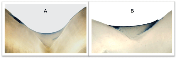
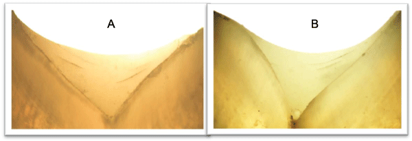
A total of 320 images were investigated for microleakage and unfilled area fractions. The results for microleakage and unfilled area fractional values are shown in table 1. Images of microleakage tests and unfilled areas for the two placement techniques are shown in Figures 5 and 6 respectively.
.
Figure 5: Images of microleakage tests for the two placement techniques using: A - Flow-bur; B- conventional applicator.
View Figure 5
.
Figure 6: Images of sealant penetration for the two placement techniques using: A - Flow-bur; B- conventional applicator.
View Figure 6
![]()
Table 1: Statistial Data on Microleakage and occlusal penetration
View Table 1
Comparison between groups showed that group 1 had significantly (P< 0.05, P=0.004) less microleakage (mean: 0.0883 ± -0.0503(SD)) than group 2 (mean: 0.1594 ± 0.0889 (SD)). Regarding the unfilled area proportions (penetration ability), group 3 (samples were sealed by oscillating bur application technique) showed the least unfilled area proportion (mean: 0.9899 ± 0.0082 (SD)) in comparison to group 4 (mean: 0.9686 ± 0.0498 (SD)) (P=.017).
Discussions
Sealants have gained wide acceptance by pediatric clinicians as an effective preventive method to inhibit the occurrence of caries. However, recent comprehensive studies have concluded that roughly 10% of such operations will fail [6] and indicates that failure of this operation on an annual basis can be as high as 5 million in the US alone [7]. The present study indicates that the use of custom-made flow-burs that were constructed in-house for applying a sealant have much improved marginal adaptation with less unfilled fissure volume and much less microleakage compared with a conventional applicator. The use of flow-bur in this study and our statistical analysis indicate that there is a 50% reduction in microleakage for filling an occlusal area and 66% reduction of unfilled areas inside the fissures. While the authors are confident that there is a definite advantage in applying the sealant and for a more successful sealant filling operation, additional long-term studies are needed to demonstrate if such advantages may result in less failure when such flow-burs are used.
Statistical data for microleakage and penetration has another significant advantage in using a flow-bur for sealant placement, besides their average values that we discussed above. And this has to do with consistency of the operations on a routine basis. The standard deviation of data for use of flow-bur for microleakage is 45% less than the corresponding value for a non-vibrating applicator. For penetration and adaption data, the result also show that the flow-bur data Group 3 are far less scattered than the data for group 4 that used the applicator. Accordingly, we conclude that not only use of flow-bur will result in less microleakage, superior adhesion between the sealant and enamel, and deeper penetration, but also such usage will yield more consistent result with less operational errors.
Use of flow-burs for sealant placement may have another remarkable effect that has been envisioned for these operations. While we have not obtained quantitative data, it is been a general observation that the flow-bur may use less sealant material to effectively cover occlusal areas, in comparison to the standard applicator. This result would indicate that each fill can be performed even more economically and more importantly if sealant material is limited or scarce, this technique can cover multiple operations with the same amount of material that is used for a single operation. However, more studies would be needed to quantify the material use for such operations.
In a developing tooth, occlusal areas are not static but dynamic, as they continuously deformed outwardly. Thus, use of flow-bur with less adhesive in the fissure area will generate less sealant-enamel-interfacial resistance for occlusal enamel deformation and ultimately a healthier tooth.
Use of flow-burs will allow a dental practitioner to more effectively manipulate the sealant in occlusal areas for stronger sealant-dentin bonding, with superior consistency using less material for dental sealants. Such attributes will surely generate more confidence for the operator; yet, a wide clinical adaptation of this placement technique may require additional studies, before it can be fully implemented. In a future study, one can see if other dental sealants can also exhibit the similar improved penetration and marginal adaptation that we have observed with one sealant. The other concern is the longevity of these treatments. The thermocycling of the specimen is to ensure that in-vitro analysis can also be applied to in vivo experience. However, to ensure the longevity of this technique a long-term clinical study should be in performed using a variety of sealants used in present day practice.
It should also be emphasized that for construction of these in-house made flow-burs was not optimized in any form, beyond ensuring that they adhere to design principles for such burs [14] and their overall spreading functionality. Inducing an oscillation to the interface of the sealant, not only is affected by the frequency of these oscillations, but also by the amplitude of these vibrations. The amplitude of oscillations induced by the flow-burs is dependent on the asymmetricity of the tip and the aspect ratio of maximum and minimal dimensions at the bur tip. Once such optimizations for the design of the flow-burs are achieved, it is anticipated that use of flow burs can yield even better results that we reported in this paper.
The effectiveness of flow-burs that were designed and tested in this study to deform and manipulate an adhesive depends not only on the frequency and amplitude of these oscillations, but also on rheological and interfacial properties of such materials. In our studies we have shown such burs are effective for one commercial sealant. It is expected that other sealants on the market should have similar rheological properties as the one we have tested [20]. Accordingly, one expects that flow burs should be successful with other sealants not considered in our studies. Nonetheless, we recommend in-vitro microleakage and penetration studies before adaptation of flow burs for other sealants.
This study has been performed exclusively for the effective placement of sealants in dental fissures. A previous study has already demonstrated that a needle vibrating tool was effective in in cleansing and etching occlusal pits and fissures [21]. Accordingly, one may extend this study and show that flow-burs, constructed in a dentist office, can also be used for etching and cleaning, as well as sealant placement.
Conclusions
We have reported comparative microleakage and depth of penetration studies on dental sealants using two placement techniques. It is shown that dental sealant placement technique can have a decisive effect on occlusal adhesion and penetration. A set of flow-burs with identical tip geometry as used previously for lining of flowable composites were constructed and used to manipulate the sealant in the occlusal area of extracted healthy teeth. The results were compared with similar data using a conventional dental applicator. The comparisons indicate that the use of flow-burs resulted in 50% reduction in microleakage on average for filling a tight area and 66% reduction of unfilled voids inside the treated fissures. With superior marginal adaptation and deeper penetration, the analysis also indicated that the use of an oscillating bur as used in this study to deform the sealant was more consistent with significantly less operational errors. It is concluded that use of flow-burs may have a significant advantage in successful placements of dental sealants.
Acknowledgements
The authors thank Dr. Franklin Garcia Godoy for helpful discussions and comments. The experimental portion of this work was supported financially by Khon Kaen University in Thailand. All opinions, interpretations, conclusions, and recommendations belong to the authors. The authors also acknowledge that they don't have any financial conflict of interest on this subject that they have studied.
References
-
Beauchamp J, Caufield PW, Crall JJ, Donly K, Feigal R, et al. (2008) Evidence-based clinical recommendations for the use of pit-and-fissure sealants: a report of the American Dental Association Council on Scientific Affairs J Am Dent Assoc 139: 257-268.
-
(1984) Consensus development conference statement on dental sealants in the prevention of tooth decay. J Am Dent Assoc 108: 233-236.
-
Llodra JC, Bravo M, Delgado-Rodriguez M, Baca P, Galvez R (1993) Factors influencing the effectiveness of sealants--a meta-analysis. Community Dent Oral Epidemiol 21: 261-268.
-
Task Force on Community Preventive Services (2002) Recommendations on selected interventions to prevent dental caries, oral and pharyngeal cancers, and sport-related craniofacial injuries. Am J Prev Med 23: 16-20.
-
Griffin SO, Griffin PM, Gooch BF, Barker LK (2002) Comparing the costs of three sealant delivery strategies. J Dent Res 81: 641-645.
-
Beltrán-Aguilar ED, Barker LK, Canto MT, Dye BA, Gooch BF, et al. (2005) Surveillance for Dental Caries, Dental Sealants, Tooth Retention, Edentulism, and Enamel Fluorosis --- United States, 1988--1994 and 1999-2002. CDC.gov, 2005.
-
National Center for Health Statistics - CDC (2007) Trends in Oral Health Status: US 1988-94 and 1999-2004 Vital and Health Statistics. Series 11, No. 248, US DHHS, Hyattsville, Maryland, April 2007.
-
Barata JS, Casagrande L, Pitoni CM, De Araujo FB, Garcia-Godoy F, et al. (2012) Influence of gaps in adhesive restorations in the development of secondary caries lesions: an in situ evaluation. Am J Dent 25: 244-248.
-
Perry AO, Rueggeberg FA (2003) The effect of acid primer or conventional acid etching on microleakage in a photoactivated sealant. Pediatr Dent 25: 127-131.
-
Celiberti P, Lussi A (2005) Use of a self-etching adhesive on previously etched intact enamel and its effect on sealant microleakage and tag formation. J Dent 33: 163-171.
-
Blackwood JA, Dilley DC, Roberts MW, Swift EJ Jr (2002) Evaluation of pumice, fissure enameloplasty and air abrasion on sealant microleakage. Pediatr Dent 24: 199-203.
-
Geiger SB, Gulayev S, Weiss EI (2000) Improving fissure sealant quality: mechanical preparation and filling level. J Dent 28: 407-412.
-
Hannig M, Gräfe A, Atalay S, Bott B (2004) Microleakage and SEM evaluation of fissure sealants placed by use of self-etching priming agents. J Dent 32: 75-81.
-
Li L, Abedian B (2001) Adhesion frequency in Interfacial dynamics of copolymers. J Adhe Sci 75: 325-340.
-
Abedian B, Millstein P (2006) An effective method for spreading flowable composites in resin-based restorations. Oper Dent 31: 151-154.
-
Hosoya Y, García-Godoy F, Summitt JB (2004) Microleakage and sealant penetration using a vibrating probe. Am J Dent 17: 427-432.
-
17. Bonifacio CC, Navarro RS, Sardenberg F, Imparato JC, de Carvalho RC, et al. (2009) Microleakage of an adhesive system used as a fissure sealant. J Contemp Dent Pract 10: 26-33.
-
Khogli AE, Cauwels R, Vercruysse C, Verbeeck R, Martens L (2013) Microleakage and penetration of a hydrophilic sealant and a conventional resin-based sealant as a function of preparation techniques: a laboratory study. Int J Pediatr Dentistry 23: 13-22.
-
Celiberti P, Lussi A (2007) Penetration ability and microleakage of a fissure sealant applied on artificial and natural enamel fissure caries. J Dent 35: 59-67.
-
Prabhakar AR, Sankriti AM, Sugandhan S (2011) Comparative evaluation of the length of resin tags, viscosity and microleakage of pit and fissure sealants - an in vitro scanning electron microscope study. Contemporary Clinical Dentistry 2: 324-330.
-
Tadokoro Y, Iwaku M, Fusayama T (1982) A laboratory report on vibration etching for fissure sealants. J Dent Res 61: 780-785.
