

International Journal of Oral and Dental Health
Stability of Temporary Orthodontic Implants after Dynamic Load Cycles
Flavia Almeida Barbosa1, Alexandre Barboza1, Daniel Jogaib Fernandes2* and Carlos Nelson Elias2
1Universidade São Leopoldo Mandic, Rio de Janeiro, Brazil
2Laboratory of Biomaterials, Military Institute of Engineering, Rio de Janeiro, Brazil
*Corresponding author:
Daniel Jogaib Fernandes, Laboratory of Biomaterials, Military Institute of Engineering, Praca General Tibúrcio, 80/sala 2030, Praia Vermelha, Rio de Janeiro, RJ 22290-270, Brazil, Tel: +55-21-2546-7080 (ext.6848), E-mail: djfernandes@ime.eb.br
Int J Oral Dent Health, IJODH-2-035, (Volume 2, Issue 4), Research Article; ISSN: 2469-5734
Received: September 28, 2016 | Accepted: November 19, 2016 | Published: November 22, 2016
Citation: Barbosa FA, Barboza A, Fernandes DJ, Elias CN (2016) Stability of Temporary Orthodontic Implants after Dynamic Load Cycles. Int J Oral Dent Health 2:035. 10.23937/2469-5734/1510035
Copyright: © 2016 Barbosa FA, et al. This is an open-access article distributed under the terms of the Creative Commons Attribution License, which permits unrestricted use, distribution, and reproduction in any medium, provided the original author and source are credited.
Abstract
Objective: This work analyzed the influence of dynamic load in temporary orthodontic screws stability.
Materials and methods: Self-drilling titanium (Ti6Al4V) screws (6 × 1.5 mm) for orthodontic anchorage were inserted and removed from pig ribs. Screws were loaded by a static load of 2 N for 5 weeks. Dynamic force was applied during 56,000 cycles for simulations of a patient’s opening-closing mouth movements. The dynamic applied load ranged from 2-5 N under a frequency of 1 Hz. Vertical (pull out) and horizontal (pulling) components of the dynamic load was 15-25% and 96-98% higher than static load, respectively. The insertion and removal torques were measured before and after static and dynamic cycles. Results: No significant (p = 0.2165) difference was observed between insertion and removal torque in the static load (11.51 ± 2.11 N cm vs. 11.37 ± 4.02 N cm). Statistical analysis (p = 0.0026) showed that after dynamic cycles the removal torque (4.2 ± 2.8 N cm) was lower than insertion torque (10.18 ± 1.17 N cm).
Conclusions: Dynamic loads demonstrated detrimental effects onto stability of screws, showing that the currently intermittent force usually used by intermaxillary biomechanics might be applied wisely to not lost temporary screws during clinical procedures.
Keywords
Orthodontic implants, Miniscrew, Orthodontic anchorage, Stability, Orthodontics, Biomaterial
Introduction
Titanium alloy (Ti-6Al-4V) screws are multi-purpose devices used in different fields of medical and dentistry [1-3]. The excellent corrosion resistance and adequate mechanical properties lead these devices to applications since permanent placement in orthopedic spine surgeries until temporary use as in fixation of bone fractures or temporary bone anchorage for dental movements.
Depending on application, different materials can be addressed for screws, including commercially pure titanium when osseointegration is desired or different other types of titanium alloys when just a temporary implantation period is planned. Because in most cases, these devices are outlined for limited time, primary stability is prioritized instead of osseointegration potential. To achieve an adequate mechanical retention, diverse factors are considered regarding screw design including length, diameter, thread shape (geometry, depth and pitch), self-drilling or self-tapping and tapered or cylindrical shape [4,5]. Still in screws influence, placement technique, head design, elastic modulus (stress shield) and the efficiency of load transference to cortical shell must also be considered. From host perspective, the main influence is ruled by bone features, headed by its quality and quantity. Lekholm and Zarb organized jaw bone tissue in 4 types, in coherence with its thickness, density and quality [6]. Bone D1, mainly homogeneous compact cortical bone; D2, a thick layer of compact cortical bone surrounding a dense trabecular bone core; D3, a thin layer of cortical bone bordering a lower dense trabecular bone core and, D4, a thin layer of cortical bone environing a reduced density trabecular core.
Different authors have stated the importance of cortical bone thickness into primary stability of temporary screws, since the screw-bone interlock played a major role in primary stability [7-9]. Although a positive correlation was identified between cortical thickness and primary stability, an excessive thick and dense cortical thickness can contribute to screws failure [7]. Different studies in maxillofacial region stated that a predominant failure rate occurred in the thickest and densest cortical areas available for intraoral placement of the screws, as observed in the posterior buccal region of mandible [10-13]. Distinct authors confirmed the use of placement torque as an adequate method to evaluate the immediate stability after screw insertion [4,5,14-18].
The great approval by surgeons for the intraoral use of temporary screws for orthodontic purposes is addressed to it reduced size which reflects in minimal anatomic limitations, minor surgery and less patient discomfort [11,19]. If tooth movements are desired, these appliances provided an immediate load possibility, which allow a great array of biomechanical applications, include even retractions of an entire arch, composed for more than ten teeth, at once. However, for limiting applications that deserve multiple teeth movements, remarkable loads would be applied on temporary screws, which might compromise its stability [20]. The most critical biomechanical situation, that reduces temporary screws stability, should be the application of cyclic loads when the movement of several teeth is planned by the activation of elastomers connected from one jaw to a temporary screw placed on the other jaw. Although this clinical situation represents an unexceptional demand for biomechanics, from our knowledge, the response of temporary screws stability has never been evaluated upon the simulation of the cyclic loads imposed by the opening and closing movements of the patient's mouth. The evaluation of the temporary screws stability when submitted to dynamic load cycles is the objective of this work.
Materials and Methods
Twenty-five temporary self-drilling titanium grade 5 (Ti-6Al-4V) screws (Conexão Sistema de Próteses, SP, Brazil) were divided into three groups, whereas static (2 N) and dynamic (between 2 N and 5 N) loads were applied. The groups were labeled as static (n = 10), dynamic (n = 5) and control (n = 10). Ten screws were attached as control for the possible influence of rib bones inhomogeneity in screws stability. All screws had the same taper design and dimensions: 6 mm long and 1.5 mm at maximum diameter. Sample size calculations were performed at a power of 0.8 and alpha at 5%.
Eleven fresh pig rib bone segments with 30 mm long were harvested at the same day of the pig euthanasia from a local butcher. The slaughter age was around 36 months. The ribs were completely cleaned, all adhered tissues were removed and bones were sliced by a precision band saw machine in transverse direction. Samples were cut and cortical thickness selections were done, in order to reduce variations in density, mineral content and cortical thickness (bone type). All samples have a 2 mm in thickness cortical shell, identified from the borders. The influence of bone anatomy on insertion and removal torques was evaluated by statistical comparison with control group.
Twenty-five self-drilling screws were attached with perpendicular orientation into ribs, after a 1.0 mm-diameter pre-drilled hole at a speed of 5 rpm. The procedure was in accordance with ASTM F117 (ASTM F117, Test Method for Driving Torque of Medical Bone Screws) and F543 (F543-13e1, Standard Specification and Test Methods for Metallic Medical Bone Screws). The same protocol was used in (9). Placement and removal torque peaks were measured with a digital torquemeter (Lutron TQ-8800, Taipei, Taiwan).
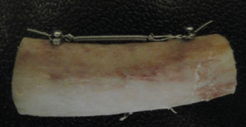
In group 1 (static), five samples of two screws were attached into pig ribs at distance of 20 mm. A 6.0 mm long stainless steel close spring was stretched until a 2 N was applied to screws. The force was measured by a digital data collection module Pasco Xplorer GLX PS-2002 (Pasco, CA, USA). The static load was maintained for 5 weeks (Figure 1) in order to simulate the time period expected between consecutive clinical appointments. The static load applied to screws was able to reproduce the moments imposed during a different range of orthodontic movements addressed directly to screw head.
.
Figure 1: Static (left) and dynamic (right) loads applied for 5 weeks in temporary screws attached in pig rib bones. Vertical (pull out) and horizontal (pulling) components of the dynamic load (L), and the angle (θ) between L and a horizontal baseline were showed. Θ and |L| were 10° and 2 N during closed mouth and 15° and 5 N during opened mouth simulations. Static load remained stable for 5 weeks at 2 N.
View Figure 1
In group 2 (dynamic), five screws were placed into pig ribs at a distance of 15 mm from each other. A mechanical machine was developed to simulate opening-closing mouth movements by up-and-down axial movements. The loads before cycles were measured by a digital data collection module Pasco Xplorer GLX PS-2002 (Pasco, CA, USA) at minimum (2 N) and maximum (5 N) extensions. Dynamic loads were applied at an angle of 10° and 15°, during simulations of mouth closed and opened, respectively. A total of 56,000 cycles were applied to each screw, at a frequency of 1 Hz, to reproduce the amount of patient's mouth movements referred to a time period of 5 weeks (Figure 1). In both groups, placement and removal torque were measured, as a reference of the screws stability before and after static and dynamic loads. The dynamic load could reproduce intermittent forces if the screws were used to anchorage intermaxillary mechanics.
In group 3 (control), ten screws were placed into pig ribs at a distance of 15 mm from each other. The screws of this have not been submitted to a load.
Statistical tests involved a one-way ANOVA with Tukey's multiple comparisons at p = 0.05. Paired comparisons were performed before and after static and dynamic loads to evaluate the influence of loading in screws stability. Placement torque values were compared from both groups with control to identify possible variations in bone sources and its influence in stability.
Results
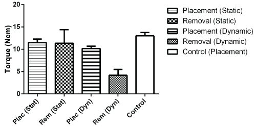
Table 1 and figure 2 showed comparisons among different placement and removal torque values after static and dynamic loads. The placement torque (11.51 ± 2.11 N cm) of screws after a static load of 20 N for 5 weeks was a slight higher than removal torque (11.37 ± 4.02 N cm), without statistical significant difference in that reduction (p = 0.2165). The same behavior was observed in control group, whereas the placement and removal torque were 13.02 ± 1.67 N cm and 12.82 ± 1.75 N cm. No statistical difference was also seen between these groups (p = 0.8581).
.
Figure 2: Mean peak torque values (N cm) after static and dynamic loads cycles. Control group showed placement torque to identify any influence of pig rib bones in stability of the different screws evaluated.
View Figure 2
![]()
Table 1: Torque peak mean values (N.cm) and standard deviation from screws placement and removal after static and dynamic loads.
View Table 1
After dynamic loads, a significant reduction was seen in torque means at p = 0.0026. Placement torque (10.18 ± 1.17 N cm) was higher than removal torque (4.2 ± 2.8 N cm). This removal torque measured might represent the loss of the temporary screws, since this screw stability is far away from the clinical demand for screws as bone anchorage.
Dynamic load applied was composed in 2 components, according to the vertical and horizontal force components. The load force vector was decomposed, based on the angle (θ) formed between the dynamic load vector (Ḹ) and a horizontal reference baseline. Θ angle was settled at 10° and 15° during mouth simulations for closed and opened situations, respectively. The two components of dynamic load (|L|) were exposed in terms of |L|.sinθ for vertical (pull out force) component and |L|.cosθ for horizontal (pulling force) component. Calculations indicated a percentage in range of 17-25% of static load |L| for vertical forces and 96-98% of static load |L| for horizontal forces, during closed and opened mouth simulations in dynamic cycles.
The placement torques of static (11.51 ± 2.11 Ncm), dynamic (11.37 ± 4.02 N cm) and control group (13.02 ± 1.67 N cm) did not show statistical difference. Similarities were observed among the placement torque values for static and control group (p = 0.2165), dynamic and control group (p = 0.2337), and also between static and dynamic loads (p = 0.1869). The pig rib bone quality and morphology did not influence in the results of this work.
Discussion
The implant primary stability is an accepted indicator of the initial interlock or mechanical attachment attained between screw surface and bone architecture. The primary stability is analyzed in terms of the placement torque, mainly in situations whereas non-osseointegration device response is planned for temporary purposes [4,5,15-18, 21-24]. In this work, torque values were measured before and after two different loads conditions. The proposed was simulating distinct orthodontic loading where temporary screws are demanded for dental anchorage.
Orthodontic treatment load with a stainless steel spring is a common application for biomechanics when a tooth movement is desired, instead of use another tooth as backing. The mechanics can be supported by different kinds of devices, from elastomers linked to temporary screws, through extra oral appliances when greater forces are needed [20,25,26]. We selected stainless steel springs just to minimize the force degradation commonly observed during polymers degradation. The load proposed (2 N) is in accordance with literature for a movement of a group of 3-4 teeth; a commonly situation when a temporary screw is suggested to achieve faster results for improvements in dental treatment progress [26,27].
Although bone turnover could not be simulated from this work, the results collected by the insertion and removal torque after static loads showed the maintenance of the stability achieved after screws placement [28]. The insertion torque was also considered as a relevant information of the quality of bone, as showed in studies of [15,29]. The maintenance of the screws stability after 5 weeks under an immediate static load of 2 N, demonstrated an adequate device stability to support grouped teeth movements during dental treatments. The screw placement torque of the present work was in line with literature, and were considered safe to prevent the transmission of excessive strains and the appearance of cracks, which contribute to the reduction of the device stability [4,30].
The choice of pig ribs as bone sources for screws placement was justified by the intention of the authors to test the influence of different load situations under the most critical bone quality possible. Pig ribs are recognized as a low density bone substrate compared to human bone D4 type, which represent a limit situation, normally faced by dentists when adult patients demanded for temporary screws use. These devices are planned to allow specific tooth movements in situations where insufficient dental support is available to be used as anchorage [30]. Even though a low density bone source were selected, a pilot hole of 1 mm in diameter were performed to prevent any kind of cracks formation in cortical shell that could influence the removal torque values after the different load conditions proposed. Other studies have also performed pre-drilling holes, with the objective to reduce bone tissue microstrain, preventing microfractures identified in SEM images [20,31-34]. Furthermore, authors decided to perform pilot holes, in favor to prevent the propagation of cracks, which would be intensified by the different load protocols proposed in this work.
The possibility of bone quality, density variation and its influence on the placement torque were appraised by statistical test, whereas multiple comparisons of insertion torque peak values were performed among all groups. Comparisons were established considering the different distances among screw positions in each testing group, whereby the two outer screws from control group were compared just with static load group, while the three central screws were confronted with devices in dynamic load group. Similarities in placement torque have discarded any concern upon heterogeneity influence of bone source architecture into results acquired. The absence of significant differences was disclosed between static and control group (p = 0.2165), dynamic and control group (p = 0.2337), and also between static and dynamic group (p = 0.1869). Although statistical tests ensured similarities in different bone sources, bone samples collected from different ribs might lead to significant influence on torque values due to the high anisotropy regarding variation in bone quality and density. That was reason why dynamic group was comprised only by 5 screws. The use of ribs was also observed in [29], and supported our decision of use this source for all testing groups.
One of the highlights of this paper is the influence of the dynamic load in temporary screws stability. From our knowledge, the dynamic protocol proposed has never been reported in literature. The protocol considered the simulation of patient's opening-closing mouth, imposing relevant loads onto screws, that ranged from 2 N (mouth closed) through 5 N (mouth opened). The relevance of this simulation is coherent with intermaxillary biomechanics, when a huge block of teeth, or even an entire jaw, is desired to be moved. Examples of these biomechanics were presented in [26,27]. Currently available devices involve the use of several springs, with similar design to the stainless steel used in this study. The spring description is accessible on [35,36].
Comparing the torque insertion and removal showed that after dynamic loading a great reduction occurs in the removal torque. This result shows the main role of the pulling forces in the screws stability. This decrease was not observed in static load group. Different works have simulated the same conditions of the static state applied by opened springs between two temporary screws, in laboratory and in vivo from animals and human approaches [25, 37-39]. The same absence of statistically significance of the static load influence in stability was disclosed by [37,39]. Çehreli and Arman-Ozcirpici ranked insertion torque values in different ranges according to clinical needs, being the values lower than 5 N cm considered poor for clinical applications [40]. The values of our removal torque after dynamic loads were below 5 N cm (4.2 ± 2.8 N cm), which demonstrated the detrimental effect of the intermittent loads applied by the dynamic cycles in the primary stability of the temporary screws. Although this approach has not been reported yet, authors believed that the main difference between the static and dynamic load protocols pertained on the intermittent load applied and on the pull out force imposed by the vertical component of the dynamic load. However, the influence of vertical component was limited (just 15-17% of the dynamic load), Largura, et al. stated that the strain distribution of deformation was not uniform throughout the interface host bone-screw surface [41] and thus, we admit that a heterogenic stress transmitted by an intermittent source might have potentially influence on the stability loss of the temporary screws. This statement is in agreement with notes from Ferrara and Ryken upon different factors which interfere into the holding strength of a screw [42]. From these factors, we considered that the pull out parameters might be compared with our dynamic load protocol, since the continuous cycles applied have also a vertical component of the dynamic force. From this perspective, Ferrara and Ryken stated the importance of the speed at which the screw is withdrawn and the angle of screw insertion; and these factors could be redefined in terms of our dynamic protocol as the frequency of the cycles and the angulation of the force submission [42]. Other factors described were acceptable controlled in our work, regarding screw design and bone architecture, since we used the same screw designs in all experiments and pig ribs homogeneity was proved by similarities in statistical comparisons. Only the intermittence of the dynamic load had not been considered by works with reference to pull out strengths, and we believe that is one of the major reasons of the significant decrease in screws stability after dynamic load cycles.
The influence of the dynamic load on the screws stability can be decomposed in two vectors, based on the angle between the load direction and a bottom baseline (Figure 1). Vertical vector is addressed to pull out component and horizontal vector to pulling forces responsible for screw tip. The angle resulted from the load application direction ranged from 10° (closed mouth simulation) to 15° (open mouth situation). From this perspective, pull out influence is limited, since the value of sine influence is 17-25% smaller than the entire applied load (|L|). Horizontal component represented by cosine had an influence of 96-98% from the |L| force applies. Therefore, if a 5 N load was transferred to screws, just 0.85-1.25 N were resulted in vertical orientation, while 4.5-4.8 N were transmitted pulling the screw in horizontal direction. Different works have considered pull out strength when evaluating the stability of temporary screws [42-45]. This parameter is a complementary indicator of primary stability, apart from placement torque. However, in our work due to the attempt to simulate dynamic load cycles, the complete influence of vertical forces was not complete evaluated for comparisons with other works which analyzed the pull out strength influence in stability. Furthermore, the direction of load applied can efficiently transform pull out forces in shear forces, due to the inclination of screws and the amount of contacting bone. The same approach was reported by Xu, et al. during evaluations of angulated temporary screws stability in dogs [46]. In the same model, Xu, et al. reported that slight angulated temporary screws demonstrated enhanced stability than vertical or extremely tilted screws. The results of the present work supported the development of our dynamic model loaded by pulling forces applied under angles between 10-15° [46].
Conclusions
The stability of orthodontic temporary screws after static and dynamic loads allowed the following statements:
• Limited influence of a static load of 2 N in temporary screws were identified;
• Dynamic loads applied under 56,000 cycles showed relevant influence in screws stability loss;
• A poor stability was identified after the dynamic protocol, showing that an intermittent force usually employed by intermaxillary biomechanics might be applied wisely to not lost temporary screws during clinical procedures.
References
-
Mihara H, Cheng BC, David SM, Ohnari K, Zdeblick TA (2001) Biomechanical comparison of posterior cervical fixation. Spine (Phila Pa 1976) 26: 1662-1667.
-
Calvert GC, Lawrence BD, Abtahi AM, Bachus KN, Brodke DS (2015) Cortical screws used to rescue failed lumbar pedicle screw construct: a biomechanical analysis. J Neurosurg Spine 22: 166-172.
-
Elias CN, de Oliveira Ruellas AC, Fernandes DJ (2012) Orthodontic implants: concepts for the orthodontic practitioner. Int J Dent 2012: 549761.
-
Chang JZ, Chen YJ, Tung YY, Chiang YY, Lai EH, et al. (2012) Effects of thread depth, taper shape, and taper length on the mechanical properties of mini-implants. Am J Orthod Dentofacial Orthop 141: 279-288.
-
Tabuchi M, Ikeda T, Nakagawa K, Hirota M, Park W, et al. (2015) Ultraviolet photofunctionalization increases removal torque values and horizontal stability of orthodontic miniscrews. Am J Orthod Dentofacial Orthop 148: 274-282.
-
Lekholm U, Zarb GA (1985) Patient selection and preparation. In: Zarb GA, Albrektsson T Branemark PI, Tissue integrated prostheses: osseointegration in clinical dentistry. Chicago, Quintessence,: 199-209.
-
Alrbata RH, Yu W, Kyung HM (2014) Biomechanical effectiveness of cortical bone thickness on orthodontic microimplant stability: an evaluation based on the load share between cortical and cancellous bone. Am J Orthod Dentofacial Orthop 146: 175-182.
-
da Cunha AC, Marquezan M, Lima I, Lopes RT, Nojima LI, et al. (2015) Influence of bone architecture on the primary stability of different mini-implant designs. Am J Orthod Dentofacial Orthop 147: 45-51.
-
Fernandes DJ, Elias CN, Ruellas ACO (2015) Influence of Screw Length and Bone Thickness on the Stability of Temporary Implants. Materials 8: 6558-6569.
-
Farnsworth D, Rossouw PE, Ceen RF, Buschang PH (2011) Cortical bone thickness at common miniscrew implant placement sites. Am J Orthod Dentofacial Orthop 139: 495-503.
-
Migliorati M, Signori A, Silvestrini-Biavati A (2012) Temporary anchorage device stability: an evaluation of thread shape factor. Eur J Orthod 34: 582-586.
-
Brettin BT, Grosland NM, Qian F, Southard KA, Stuntz TD, et al. (2008) Bicortical vs monocortical orthodontic skeletal anchorage. Am J Orthod Dentofacial Orthop 134: 625-635.
-
Ozdemir F, Tozlu M, Germec-Cakan D (2013) Cortical bone thickness of the alveolar process measured with cone-beam computed tomography in patients with different facial types. Am J Orthod Dentofacial Orthop 143: 190-196.
-
Yoo SH, Park YC, Hwang CJ, Kim JY, Choi EH, et al. (2014) A comparison of tapered and cylindrical miniscrew stability. Eur J Orthod 36: 557-562.
-
Suzuki EY, Suzuki B (2011) Placement and removal torque values of orthodontic miniscrew implants. Am J Orthod Dentofacial Orthop 139: 669-678.
-
Cha J, Hwang C, Kwon SH, Jung H, Kim K, et al. (2015) Strain of bone-implant interface and insertion torque regarding different miniscrew thread designs using an artificial bone model. Eur J Orthod 37: 268-274.
-
Lim HJ, Choi YJ, Evans CA, Hwang HS (2011) Predictors of initial stability of orthodontic miniscrew implants. Eur J Orthod 33: 528-532.
-
McManus MM, Qian F, Grosland NM, Marshall SD, Southard TE (2011) Effect of miniscrew placement torque on resistance to miniscrew movement under load. Am J Orthod Dentofacial Orthop 140: e93-98.
-
Reynders R, Ronchi L, Bipat S (2009) Mini-implants in orthodontics: a systematic review of the literature. Am J Orthod Dentofacial Orthop 135: 564.
-
Carney LO, Campbell PM, Spears R, Ceen RF, Melo AC, et al. (2014) Effects of pilot holes on longitudinal miniscrew stability and bony adaptation. Am J Orthod Dentofacial Orthop 146: 554-564.
-
Suzuki M, Deguchi T, Watanabe H, Seiryu M, Iikubo M, et al. (2013) Evaluation of optimal length and insertion torque for miniscrews. Am J Orthod Dentofacial Orthop 144: 251-259.
-
Inoue M, Kuroda S, Yasue A, Horiuchi S, Kyung HM, et al. (2014) Torque ratio as a predictable factor on primary stability of orthodontic miniscrew implants. Implant Dent 23: 576-581.
-
Santos RF, Ruellas ACO, Fernandes DJ, Elias CN (2014) Insertion torque versus mechanical resistance of mini- implants inserted in different cortical thickness. Dental Press J Orthod 19: 90-94.
-
Vilani GN, Ruellas AC, Mattos CT, Fernandes DJ, Elias CN (2015) Influence of cortical thickness on the stability of mini-implants with microthreads. Braz Oral Res 29.
-
El-Dawlatly MM, Abou-El-Ezz AM, El-Sharaby FA, Mostafa YA (2014) Zygomatic mini-implant for Class II correction in growing patients. J Orofac Orthop 75: 213-225.
-
Kim GT, Kim SH, Choi YS, Park YJ, Chung KR, et al. (2009) Cone-beam computed tomography evaluation of orthodontic miniplate anchoring screws in the posterior maxilla. Am J Orthod Dentofacial Orthop 136: 628.e1-628.e10.
-
Esenlik E, Ağlarcı C, Albayrak GE, Fındık Y (2015) Maxillary protraction using skeletal anchorage and intermaxillary elastics in Skeletal Class III patients. Korean J Orthod 45: 95-101.
-
Woodall N, Tadepalli SC, Qian F, Grosland NM, Marshall SD, et al. (2011) Effect of miniscrew angulation on anchorage resistance. Am J Orthod Dentofacial Orthop 139: e147-152.
-
Motoyoshi M, Yoshida T, Ono A, Shimizu N (2007) Effect of cortical bone thickness and implant placement torque on stability of orthodontic mini-implants. Int J Oral Maxillofac Implants 22: 779-784.
-
Oliscovicz NF, Shimano AC, Marcantonio Junior, Lepri CP, dos Reis AC (2013) Analysis of Primary Stability of Dental Implants Inserted in Different Substrates Using the Pullout Test and Insertion Torque. Int J Dent 2013: e1-e5.
-
Uemura M, Motoyoshi M, Yano S, Sakaguchi M, Igarashi Y, et al. (2012) Orthodontic mini-implant stability and the ratio of pilot hole implant diameter. Eur J Orthod 34: 52-56.
-
Cho KC, Baek SH (2012) Effects of predrilling depth and implant shape on the mechanical properties of orthodontic mini-implants during the insertion procedure. Angle Orthod 82: 618-624.
-
Son S, Motoyoshi M, Uchida Y, Shimizu N (2014) Comparative study of the primary stability of self-drilling and self-tapping orthodontic miniscrews. Am J Orthod Dentofacial Orthop 145: 480-485.
-
Shin YS, Ahn HW, Park YG, Kim SH, Chung KR, et al. (2012) Effects of predrilling on the osseointegration potential of mini-implants. Angle Orthod 82: 1008-1013.
-
Armstrong MM (1970) Coiled wire spring appliances for use in orthodontics. Patent US 3618214 A.
-
Lewis-Klapper RG (1998) Orthodontic device for correcting overbite and underbite. Patent US 5846074 A.
-
Brown RN, Sexton BE, Chu TG, Katona TR, Stewart KT, et al. (2014) Comparison of stainless steel and titanium alloy orthodontic miniscrew implants: A mechanical and histologic analysis. Am J Orthod Dentofacial Orthop 145: 496-504.
-
Catharino PC, Dominguez GC, Pinto Ddos S Jr, Morea C (2014) Histologic, Histomorphometric, and Radiographic Monitoring of Bone Healing Around In-Office-Sterilized Orthodontic Mini-implants With or Without Immediate Load: Study in Rabbit Tibiae. Int J Oral Maxillofac Implants 29: 321-330.
-
Chen Y, Shin HI, Kyung HM (2008) Biomechanical and histological comparison of self-drilling and self-tapping orthodontic microimplants in dogs. Am J Orthod Dentofacial Orthop 133: 44-50.
-
Cehreli S, Arman-Özcırpıcı A (2012) Primary stability and histomorphometric bone-implant contact of self-drilling and self-tapping orthodontic microimplants. Am J Orthod Dentofacial Orthop 141: 187-195.
-
Largura LZ, Argenta MA, Sakima MT, Camargo ES, Guariza-Filho O, et al. (2014) Bone stress and strain after use of a miniplate for molar protraction and uprighting: a 3-dimensional finite element analysis. Am J Orthod Dentofacial Orthop 146: 198-206.
-
Ferrara LA, Ryken TC (2000) Screw Pullout Testing. In: Draughn RA, An YH, Mechanical testing of bone and the bone-implant interface. Boca Raton, CRC Press.
-
Wu JH, Wang HC, Chen CM, Lu PC, Lai ST, et al. (2011) Pullout strengths of orthodontic palatal mini-implants tested in vitro. J Dent Sci 6: 200-204.
-
Crum MS, Young FA, An YH (2000) Screw pullout test for evaluating mechanical properties of bone. In: Draughn RA, An YH, Mechanical testing of bone and the bone-implant interface. Boca Raton, CRC Press.
-
Huja SS, Litsky AS, Beck FM, Johnson KA, Larsen PE (2005) Pull-out strength of monocortical screws placed in the maxillae and mandibles of dogs. Am J Orthod Dentofacial Orthop 127: 307-313.
-
Xu Z, Wu Y, Zhao L, Zhou Y, Wei X, et al. (2013) Effect of placement angle on the stability of loaded titanium microscrews in beagle jaws. Angle Orthod 83: 659-666.
