
International Journal of Sports and Exercise Medicine
Changes in Endothelial Markers during a Summer Ultra-Endurance Road Cycling Event in the Heat
Kupchak BR1*, Kazman JB1, Umeda EA1, Vingren JL2, Lee EC3, Armstrong LE3 and Deuster PA1
1Department of Military & Emergency Medicine, Uniformed Services University of Health Sciences, USA
2Department of Kinesiology, Health Promotion and Recreation, University of North Texas, USA
3Human Performance Laboratory, Department of Kinesiology, University of Connecticut, USA
*Corresponding author: Kupchak BR, Department of Military & Emergency Medicine, Uniformed Services University of Health Sciences, 4301 Jones Bridge Road, Bethesda, MD 20814, USA, E-mail: brian.kupchak@usuhs.edu
Int J Sports Exerc Med, IJSEM-2-045, (Volume 2, Issue 3), Original Article; ISSN: 2469-5718
Received: April 26, 2016 | Accepted: August 19, 2016 | Published: August 22, 2016
Citation: Kupchak BR, Kazman JB, Umeda EA, Vingren JL, Lee EC, et al. (2016) Changes in Endothelial Markers during a Summer Ultra-Endurance Road Cycling Event in the Heat. Int J Sports Exerc Med 2:045. 10.23937/2469-5718/1510045
Copyright: © 2016 Kupchak BR, et al. This is an open-access article distributed under the terms of the Creative Commons Attribution License, which permits unrestricted use, distribution, and reproduction in any medium, provided the original author and source are credited.
Abstract
Purpose: To assess the impact of completing a 164 km road cycling event performed in a hot environment (Wichita Falls, Texas in August), on endothelial biomarkers and resultant risk of blood clots in men and women.
Methods: 37 event participants (28 men and 9 women; 51.8 ± 9.5 y) completed the ride. Plasma samples were collected in the morning before (PRE) and immediately after (IP) completing the ride. Concentrations of endothelial cell markers - endothelin-1 (ET-1), p-selectin, and intercellular adhesion molecule 1 (ICAM1), were measured and associations between changes from PRE- to IP-ride were examined as a function of race event completion time and participant characteristics (demographics and anthropometrics).
Results: All of the endothelial cell markers (ET-1, p-selectin, and I-CAM1) increased significantly from PRE to IP. After controlling for PRE values, completion time was positively correlated with ET-1 (r = 0.42, p < 0.01) and negatively related to p-selectin (r = -0.42, p < 0.05). Waist circumference was positively related to ICAM1 (r = 0.34, p < 0.01). In addition, males had greater concentrations of ICAM1 (d = 1.32, p < 0.01) and p-selectin (d = 0.84, p < 0.05) than females.
Conclusion: Completing a 164 km road cycling event in hot conditions resulted in increased concentrations of endothelial cell markers in men and women. Although this result may suggest endothelial cell injury, it is unclear whether this response is indicative of an increased risk of blood clot formation.
Keywords
Cycling, Heat stress, Endothelium
Acronyms
BMI: Body Mass Index; HHH: Hotter'N Hell Hundred; PRE: Pre-race; IP: Immediate Post-race; ET-1: Endothelin-1; ICAM1: Intercellular Adhesion Molecule 1
Introduction
Over the last decade, ultra-endurance running and cycling events have become more popular [1]. The Hotter 'N Hell (HHH) is a 164 km road cycling event held each August in Wichita Falls, Texas. The temperature during the ride regularly exceeds 37°C. Each year more than 12,000 recreational and competitive athletes participate in this event. HHH provides a unique opportunity to study the impact of a hot environment combined with prolonged exercise on markers of vascular shear stress and endothelial activation/injury. This study aimed to investigate whether participation in such events may carry an increased risk for vascular thrombotic events [2,3].
The endothelium is a thin layer of squamous cells that line the internal surface of blood and lymphatic vessels. It acts as semi-selective barrier between the vessel lumen and surrounding tissue, provides a non-thrombogenic surface that inactivates several factors in the coagulation cascade, plays a major role in inflammation, as well as provides control of blood pressure through vasodilation and vasoconstriction. The vascular endothelium plays a major role in hemostatic balance by synthesizing and secreting a multitude of modulators. Two of these molecules, ICAM1 and p-selectin are cellular adhesion molecules, which are secreted by endothelial cells.ICAM1 plays a role in the leukocyte-endothelial cell interaction in the regulation of vascular permeability [4], and is up-regulated during atherosclerosis [5]. While p-selectin, is not only expressed by endothelium cells, but also expressed by platelets, plays an essential role in the initial recruitment of leukocytes to the site of injury during inflammation [6], and elevated levels are known to be linked to thrombotic and cardiovascular risk [7,8]. ET-1 is another protein released mostly from the vascular endothelial cells, and is a potent constrictor of smooth muscle [9]. ET-1 primary function is to preserve vascular tone and may help achieve the redistribution of blood flow to active skeletal muscle during exercise via vasoconstriction [10,11].
Prolonged strenuous exercise or rigorous physical exertion such as that encountered during a marathon or in intensive military training can negatively affect many physiological systems, particularly when conducted in a hot environment [12]. Completion of a marathon run under thermo-neutral conditions has been shown to increase endothelial activation with subsequent increases in p-selectin [13] and intercellular adhesion molecule 1 (ICAM1) [13,14]. Heat stress alone can also lead to changes in blood homeostasis. Prior research has also demonstrated that even without exercise, exposure to hot temperatures for prolonged periods of time can cause endothelium activation/injury [15]. Bouchoma, et al. [15] found evidence of endothelial dysfunction and increases in concentrations of ICAM1 in subjects exposed to hyper-thermic conditions. Since, heat stress and prolonged exercise can independently activate the endothelium, the combination of heat stress and prolonged exercise has the potential to further enhance the risk of clot formation, which may in turn lead to vascular thrombotic events.
Although many studies have examined the effect of prolonged exercise on endothelial activation, only a few have explored the impact of hyperthermia on endothelial activation/injury. To our knowledge, no one has examined the combination of heat stress and prolonged exercise on parameters of endothelial activation. The present investigation specifically evaluated the combination of prolonged exercise and heat exposure on endothelial response in male and female recreational cyclists participating in the HHH. Additionally, it was examined whether anthropometrics were related to changes in endothelial markers in response to the event. We hypothesized that endothelial cell factors would be up-regulated following the HHH.
Methods
Subjects
Subjects were recruited through the HHH website, via an email sent to all HHH entrants and at the Multi-Purpose Event Center (Wichita Falls, TX) during the HHH Exposition held August 27-28, 2015. Inclusion criteria included, 21-65 years of age, participating in the 164 km cycling event, and previous completion of an organized bike ride of at least 164 km. Exclusion criteria consisted of the following: (1) inability to speak or understand English; (2) current tobacco use; (3) use of cholesterol-lowering, vasoactive, and/or anticoagulant medications (e.g., Coumadin); (4) current musculoskeletal injury; (5) diagnosis of cardiovascular, liver, kidney, blood or gastrointestinal disease or severe metabolic or endocrine disorders (e.g., Type II Diabetes); (6) presence of a known medical condition or currently taking medication that would alter fluid balance; (7) history of exertional heatstroke or exercise-heat intolerance; (8) self-reported use of hormonal substances including testosterone, anabolic steroids, or growth hormone. The Institutional Review Board of the Uniformed Services University approved this study and all procedures prior to subject recruitment. All subjects provided written informed consent after having the study risks and benefits carefully explained to them. For the study, 83 individuals were screened, 49 were enrolled (34 were excluded for not participating in the 164 km cycling event or unwillingness to have phlebotomy performed on themselves), and 37 completed the cycling event (12 did not complete cycling event or did not start the event).
Anthropometrics, monitoring gastrointestinal temperature and environmental conditions: Anthropometric measurements were completed 1-2 days prior to the event. Body mass was measured on a floor scale, accurate to 0.1 kg, while subjects wore only t-shirt and shorts. Height was measured with the subject standing against a tape measure attached to the wall. Body mass index (BMI) was calculated as body mass (kg) height (m-2). Circumference (waist and hip) and percent body fat measurements were performed by a trained investigator using a Gulick tape measure (Perform Better, Cranston, RI, USA) and calibrated skinfold calibers (Body care Harpenden Caliper, England), respectively. Waist and hip circumference were measured according to ACSM guidelines by a trained investigator, where the same male or female technician performed the measurements on the participant. Waist circumference was measured at a level midway between the lowest rib and iliac crest, while hip circumference was measured at the maximal circumference of the hip, just below the gluteal fold with legs slightly apart (10 cm) [16]. Skin- fold thickness measurements were performed in duplicate on the participant's right side and recorded to the nearest mm. Body-fat percentage (%BF) was estimated using a three-site skinfold equation [17].
Gastrointestinal temperature (TGI) was measured via an ingestible thermistor (CorTemp®, HQ Inc., Palmetto, FL, USA), which was swallowed by each volunteer at 2100 h on the night before the event. Ingestion was verified by a telephone call from an investigator. The TGI was measured with a hand-held digital thermometer, positioned near the posterior lumbar curve. Since some individuals' ingestible thermistor could not be detected, TGI was only obtained from 24 participants. A Kestrel Meter 4400 Heat Meter (Nielson-Kellerman Co., Boothwyn, PA, USA) was used to monitor environmental conditions at the start/finish line of the event. Both wet-bulb globe temperature and dry-bulb temperature were measured hourly throughout the race, from 0700 to 1700 h.
Ride characteristics:The 164 km HHH course is a relatively flat, single-loop course, with a peak elevation of 290.5 m. The event, started on August 29, 2015 and began at 0700 h, and starts and ends in Wichita Falls, Texas.
Urine and blood collection:Urine and blood samples were collected before the start of the ride (PRE; 04:30 - 06:30 h; August 29, 2015) and immediately post ride (IP; within 10 min of finishing the ride). Hydration state was evaluated via a urine sample, and was measured via urine refractometry (Model Reichert TS400); a urine specific gravity of < 1.025 g.mL-1 was considered dehydrated. In addition, urine color was assessed from the sample [18]. Urine was only collected from 33 participants, due to the participants unavailable to void at IP.
Participants were not fasted for the blood collection. Whole blood was collected from an antecubital vein without stasis into evacuated vacutainers (containing EDTA) while the subject was in a seated position. The vacutainers were then placed in a 4°C refrigerator until they were centrifuged. (Centrifugation occurred within 40 min of blood collection.) The EDTA vial was centrifuged at 1,500 g for 15 min and the resultant plasma was separated into several individual cryovials and flash frozen in liquid nitrogen. The frozen cryovials were immediately stored in a -80°C ultra-low freezer prior to being shipped on dry ice. Upon arrival at the Uniformed Services University/Consortium for Health and Military Performance (CHAMP), the specimens were stored at -80°C ultra-low freezer prior to biochemical analysis.
Biochemical analysis:Endothelin-1 (ET-1) and Intercellular Adhesion Molecule 1 (ICAM1) (R&D Systems; Minneapolis, MN, USA) were determined in duplicate by Enzyme Linked Immunosorbent Assay (ELISA) in EDTA plasma. The intra-assay coefficients of variation (CVs) were 3.6 and 2.9%, and inter-assay CVs were 4.7 and 5.8%, respectively. Soluble p-selectin was determined from EDTA-plasma (Invitrogen Corporation; Camarillo, CA, USA) by ELISA, and had an intra-assay CV of 4.3% and inter-assay CV of 6.0%. For all ELISAs, absorbance was measured using a BIO-RAD iMark microplate reader (Hercules, CA, USA) at the appropriate wavelength for that particular assay.
Statistical analysis:Subject characteristics and endothelial markers were assessed for assumptions of parametric statistics. Outliers and influential cases were examined using standard case diagnostics. One female participant was removed based on particularly high and influential levels of IP-ICAM1 (z = 3.2) and IP-p-selectin (z = 2.3), yielding a final sample size of 37. In order to determine whether endothelial markers changed significantly from PRE to IP, dependent-samples t-tests were conducted. Cohen's d was calculated and used to estimate effect size in order to indicate mean-change in standard deviation units (of change values). Next, associations between subject characteristics (demographics, anthropometrics, and event completion time) and IP endothelium marker concentrations at IP were examined. These were analyzed using partial correlations between subject characteristics and IP endothelial marker levels while controlling for PRE endothelium marker concentrations. Partial correlations were used instead of simple PRE-IP difference-scores, because the former requires fewer assumptions about correlations between PRE and IP markers [19]. Lastly, gender differences in subject characteristics and endothelial markers were explored. The level of significance was set at p < 0.05. Data are presented as mean ± SD unless otherwise indicated.
Results
Twenty-eight men and nine women completed the HHH in < 9 h with a mean finishing time of 6.6 h. Subject characteristics are provided in table 1. Participants' hydration and internal temperature parameters are listed in table 2; among these, only TGI was significantly changed (p = 0.013) from PRE to IP. Environmental conditions on August 29, 2015 were recorded at the start/finish line. The dry bulb temperature ranged from 26.1 to 39.3°C, while the wet bulb globe temperature ranged from 22.3 to 34.6°C. Both dry bulb and wet bulb globe temperature peaked at 14:20 h. Percent relative humidity was 56.5% at 0800 h and fell to 26.8% by 1300 h. Wind speed ranged from 0 to 6.1 kmph. During the event, partial cloud cover was observed.
![]()
Table 1: Subject characteristics.
View Table 1
![]()
Table 2: Subject urine and internal temperature characteristics.
View Table 2
Results from dependent t-tests (Table 3) indicated statistically significant (p < 0.001) changes in all three endothelial factors from PRE to IP. The largest change was observed in ET-1, which changed by 1.5 effect size units; p-selectin and ICAM1 both changed by 1.0 effect size units.
![]()
Table 3: Endothelial markers pre- and post-ride.
View Table 3
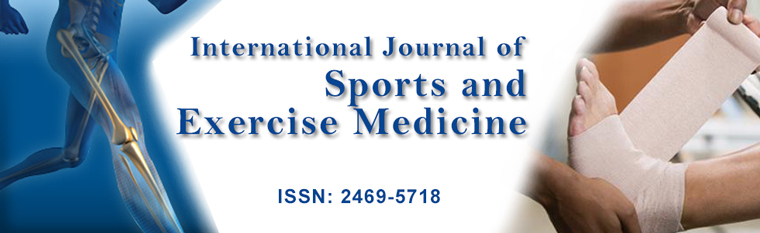
Partial correlations between subject characteristics and IP endothelial marker concentrations, controlling for PRE endothelial concentrations, are presented in table 4. Significant correlations were noted for event completion time and %BF with endothelial markers: Completion time was positively related to ET-1 (r = 0.42, p < 0.05) and negatively to p-selectin (r = -0.42, p < 0.05); while BF% exhibited a positive trend to ET-1 (r = 0.33, p = 0.06) and was negatively correlated to p-selectin (r = -0.40, p < 0.01) and to ICAM1 (r = -0.34, p < 0.01). Furthermore, waist circumference was positively correlated with ICAM1 (r = 0.34, p < 0.010). No relations were noted between endothelial markers and IP hydration and internal temperature measures (TGI, USG, urine color) (Data not shown). Taken together, these results indicate that lower fitness (as indicated by longer completion time) and higher %BF correlated with greater increases in ET-1 and decreases in p-selectin in response to the ride figure 1.
.
Figure 1: Relationship between percentage change in ET-1 and percent body fat (left)/race time (right), by sex.
View Figure 1
![]()
Table 4: Correlations between post-ride (controlling for PRE) endothelial markers and participants' characteristics.
View Table 4
Of note, event completion time positively correlated with %BF (r = 0.64, p < 0.001), but not with BMI (r = 0.11, p = 0.53) or waist circumference (r = -0.06, p = 0.74). PRE concentrations of endothelial markers did not correlate with any subject characteristics.
The strength of correlations between PRE- and IP-concentrations of endothelial markers differed among marker: PRE and IP-ET-1 correlated moderately (r = 0.35, p < 0.05); PRE and IP-p-selectin correlated strongly (r = 0.56, p < 0.001); and PRE and IP-ICAM1 correlated very strongly (r = 0.96, p < 0.001). These relations did not appear to differ by subject characteristics.
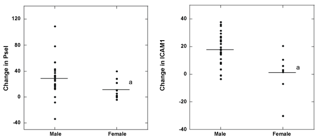
Overall, it is difficult to separate the roles of gender and other participant characteristics in endothelial marker levels, due to the inter-relations between these variables and the small number of females in the sample. Gender differences were noted for most of the participant characteristics. Compared to males, females were younger (d = 1.0, p < 0.05; males = 53.9y, females = 45.6y) and had a higher BF% (d = 2.5, p < 0.001; males = 19.1%, females = 29.6%) and longer race times (d = 0.8, p < 0.05; males = 6.42 h, females = 7.29 h). Furthermore, controlling for PRE levels, females had lower IP levels of p-selectin (d = 0.9, p < 0.05) and ICAM1 (d = 1.2, p < 0.01) than men (Figure 2).
.
Figure 2: Comparison of individual changes of PRE to IP concentrations for p-selectin (left) and ICAM1 (right) by sex; a significantly different from males.
View Figure 2
Discussion
Few studies have examined the effects of prolonged endurance exercise on endothelial cell markers. Moreover, no study appears to have examined these factors during a long-distance cycling event in a hot environment. Thus, the present study investigated affected endothelial cell markers response to a 164 km recreational road cycling event held during the hot Texas summer. Participants who completed this event demonstrated increased concentrations of ET-1, p-selectin, and ICAM1. Furthermore, individuals exhibited a positive relationship between ICAM1 concentrations and waist circumference, suggesting a greater predisposition for blood clot formation in those with a larger waist circumference when cycling in the heat for prolonged period of time.
Intensive military training or strenuous exercise in the heat can lead to heatstroke, where an individual's core temperature can vary from approximately 41°C to 47°C during heat stroke [20-22]. Although our subjects averaged 37.9°C, no one topped 39.5°C or showed any signs of heat stroke. One of the complications seen during thermal injury is its activation of the vascular endothelium [15,23], and even though no individual in our study exhibited heat stroke, the continuous, prolonged exercise in the heat during this event could theoretically have led to vascular endothelium damage followed by initiation of coagulation.
The vascular endothelium contributes to hemostatic balance by synthesizing and secreting of a multitude of hemostatic modulators. One of these parameters is p-selectin, which is primarily located in endothelium cells and storage granules in platelets and translocated to the cell surface upon activation of the endothelium [24]. The concentrations of p-selectin is increased in individuals with various immunological, inflammatory, and cardiovascular disease ([25,26] and increases after high intensity exercise [13,27]. It has been suggested that age is associated with greater concentrations of p-selectin immediately following prolonged endurance exercise events [13]. The 28% increase in p-selectin concentrations from PRE to IP observed in the present study supports previous findings of exercise-induced activation of the endothelium [13,27], and adds to these findings by demonstrating that this activation is also present after prolonged cycling in the heat. However, unlike Parker, et al. [13], we found no relationship between changes in p-selectin and changes in age of the riders in our study. Nor did we find a correlation between changes in core temperature and p-selectin.
ET-1 has not only been shown to be derived from endothelial cells, but from cardiomyocytes [28] and is thought to be involved in exercise mediated cardiac hypertrophy [29]. Its main function is to act as a potent and long lasting vasoconstrictor that plays a key role in the regulation of vascular tonus [30,31], therefore changes often reflect variations in blood pressure regulation and blood flow. Previous studies have shown exercise performed during thermo-neutral conditions have been inconclusive with findings of both increasing [32-34], and no change in ET-1 concentrations after exercise [35,36]. It appears that exercise duration and intensity is an important factor for inducing an increase in ET-1: the exercise protocols that did not show a change in ET-1 lasted less than 30 minutes and were performed at a moderate intensity; whereas, the studies that found an increase in ET-1 were of longer duration (marathon run) [37] or intensity (5 km of cross country skiing) [32].
ET-1 has also been found to increase during and after (2-hr into recovery) exercise in a hot environment [34,38], although it was suggested that this increase in ET-1 was due to dehydration [34]. In the present study, we found a greater than two-fold increase in ET-1 concentrations immediately after exercise. It is unlikely that the rise in ET-1 is attributed to dehydration because PRE and IP indices of hydration status (USG and urine color) were very similar, and were not related to change in ET-1. Therefore, we do not attribute the rise in ET-1 concentrations observed in the present study to dehydration, but the combination of prolonged exercising and environmental elements experienced during the HHH. Mechanistically, increases in ET-1 have been attributed to vasoactive hormones, shear stress, free radicals, and hypoxia [38,39], and ET-1 release can be stimulated by thrombin, and epinephrine in vitro [9], and by arginine vasopressin in vivo [40]. In a study involving a prior HHH event, we showed an increased formation of thrombin as measured by thrombin-antithrombin complexes [41] following exercise; thus, we propose that the increase in ET-1 observed in the present study following prolonged cycling in the heat is due, at least in part, to increases in thrombin formation.
Increased concentrations of ET-1 have been associated with aging in healthy adults [42] and in cultured endothelial cells extracted from the aorta of older and younger adults [43]. ET-1 has also been associated with age-associated diseases [44], a relationship that might be due to activation of pro-inflammatory pathways [45]. Although our participants' ages ranged from 26 to 65 years, we did not see find any relationship between ET-1 and age. However, a positive relation¬ship was observed between ET-1 concentrations and BF%. Excessive production of ET-1 is present in individuals who are obese [44], and is known to stimulate platelet aggregation thus promoting a prothrombotic state [46]. Additionally, our participants showed a positive relationship between event ride time and ET-1. This suggests that individuals who were in better cardiovascular shape (i.e., finished the event faster) experienced a smaller increase in ET-1 during the event. This observation is in agreement with previous studies, which have found that ET-1 concentration decreased following aerobic exercise training [42,47].
ICAM1 is expressed in endothelial cells, and plays an important role in the adhesion of leukocytes to the vascular endothelium in order to allow them to transmigrate into tissues. In thermoneutral conditions, circulating concentrations of ICAM1 increase following half (21.1 km) and full (42.2 km) marathons run events [14]. But not after low impact ergometer cycling [3], and has been associated with muscle damage seen during high impact exercises [14,48]. However, a previous study showed no changes in ICAM1 after a low impact cycling ergometer test [3]. However, unlike that study, our study had an added environment stress. The heat stress encountered during the HHH may be the reason behind the increase of ICAM1. Bouchoma, et al. [15] previously demonstrated that heatstroke is associated with endothelial cell activation/injury, including an increase ICAM1. Even though no one in our study exhibited heat stroke, the extended time the subjects participated in the event may explain the increase.
Body composition appears to affect ICAM1 release. Jenkins, et al. [49] found ICAM1 concentrations increased with increasing BF% in sedentary older adults, suggesting that adipose tissue can produce ICAM1. However, in the current study concentrations of ICAM1 were negatively correlated with ICAM1 BF%, suggesting that in trained cyclist individuals with lower BF% are more sensitive in releasing ICAM1 than those who have higher levels of body fat, or that individuals with lower BF% have a healthier lifestyle and lower ICAM1 concentration [50]. Conversely to these results, waist circumference exhibited a positive relationship to ICAM1. This phenomenon can be explained by waist circumference usually being utilized as a measure of central obesity, while BF% generally used as measures of overall obesity. Thus individuals who exhibit greater central obesity (located in the trunk) than overall obesity may have a more profound effect on the release of ICAM1 during exercise. In a previous study, it has been reported that waist circumference is stronger associated with cardiovascular disease risk factors than BF% [51]. Therefore, individuals who exhibit a greater waist circumference and participate in prolonged endurance exercise in extreme conditions may be at greater risk of endothelial injury that can lead to vascular thrombotic events (i.e., pulmonary embolism, stroke).
The HHH is an annual event that attracts a wide array of participants, with ages ranging from 18 to 70 y (Participants in the current study ranges from 26 to 65 y of age) and a large variety of body types. BMI, a common index to classify subjects as overweight, in our sample was 26.5 kg.m-2 (20 participants were considered overweight >25 kg.m-2; 5 were considered obese > 30 kg.m-2). However, BMI was not related to race time or any endothelial markers. Since BMI is not a measure of fatness and thus individuals with high muscle mass can incorrectly be classified as overweight or obese. We also measured waist circumference, which is both more indicative of body fatness and more predictive of cardiovascular complications due to obesity [52]. However, waist circumference did not correlate with race time. Last, we found that BF% had a positive relationship with event completion time. This suggests individuals who are considered overweight and obese had a slower time completing this race, and they may be considered to be at high risk for cardiovascular disease even though they are physically active [53].
In the present study, as in the general population, women exhibited higher BF% than men [54,55]. In addition, the women had longer event completion times than the men, again this was not surprising since women, in general, use more time to complete prolonged exercising events such as marathons than men [56-58]. Nonetheless, the women in our study exhibited lower p-selectin and ICAM1 responses to exercise compared to the men. This result indicates that the women had less endothelium activation following the prolonged exercising in the heat, perhaps because as a group they were younger than the men, or because their intensity was lower, since the men finished in a shorter time. Additionally, a major factor contributing to these results is the female hormone, estrogen. In pre-menopausal women, estrogen has both cardio- and vaso-protective effects [59,60], and there is evidence that it lowers p-selectin and [61] and ICAM1 [62]. Thus the variations in these factors between men and women may simply relate to variations in sex hormones as opposed to age or fitness. In any case, the higher ICAM1 and p-selectin concentrations demonstrated in men puts them at higher risk for cardiovascular disease than women [8,63].
In summary, the HHH places substantial physiological stress on the endothelial system. The completion of the 164 km road cycling event in hyperthermic conditions activated the endothelium in men and women, as evidenced in increase post-exercise concentrations of p-selectin, ET-1, and ICAM1. Furthermore, relationship existed between ET-1 (controlling for pre-race levels) and event completion time, while a positive relationship existed between ICAM1 and waist circumference. Additionally, we found a greater duration in completion time with BF%. Last, females demonstrated a lower p-selectin and ICAM1 concentrations (controlling for pre-race levels) compared to males. In conclusion, although this study suggests endothelial cell injury, it is unclear whether this activation leads to an increased risk of blood clot formation.
Acknowledgements
The authors gratefully acknowledge the expertise of Keith H. Williamson, MD (medical director) for his expertise and the HHH Expo committee for the use of facilities in the Wichita Falls Multi-Purpose Event Center. All funds were provided by the Center of Alliance for Nutrition and Dietary Supplementation.
Conflict of Interest
The authors declare that they have no conflict of interest. The opinions and assertions expressed herein are those of the authors and should not be construed as reflecting those of the Uniformed Services University, Department of the Army, Department of the Air Force, Department of the Navy or the United States Department of Defense.
References
-
https://secure.ultrarunning.com/archive/view-article.php?id=1428&page=25.
-
Albert CM, Mittleman MA, Chae CU, Lee IM, Hennekens CH, et al. (2000) Triggering of sudden death from cardiac causes by vigorous exertion. N Engl J Med 343: 1355-1361.
-
Siscovick DS, Weiss NS, Fletcher RH, Lasky T (1984) The incidence of primary cardiac arrest during vigorous exercise. N Engl J Med 311: 874-877.
-
Sumagin R, Lomakina E, Sarelius IH (2008) Leukocyte-endothelial cell interactions are linked to vascular permeability via ICAM-1-mediated signaling. Am J Physiol Heart Circ Physiol 295: H969-H977.
-
Nakashima Y, Raines EW, Plump AS, Breslow JL, Ross R (1998) Upregulation of VCAM-1 and ICAM-1 at atherosclerosis-prone sites on the endothelium in the ApoE-deficient mouse. Arterioscler Thromb Vasc Biol 18: 842-851.
-
Lorant DE, Topham MK, Whatley RE, McEver RP, McIntyre TM, et al. (1993) Inflammatory roles of P-selectin. J Clin Invest 92: 559-570.
-
Rectenwald JE, Myers DD Jr, Hawley AE, Longo C, Henke PK, et al. (2005) D-dimer, P-selectin, and microparticles: novel markers to predict deep venous thrombosis. A pilot study. Thromb Haemost 94: 1312-1317.
-
Blann AD, Nadar SK, Lip GY (2003) The adhesion molecule P-selectin and cardiovascular disease. Eur Heart J 24: 2166-2179.
-
Yanagisawa M, Kurihara H, Kimura S, Tomobe Y, Kobayashi M, et al. (1988) A novel potent vasoconstrictor peptide produced by vascular endothelial cells. Nature 332: 411-415.
-
Maeda S, Miyauchi T, Sakane M, Saito M, Maki S, et al. (1997) Does endothelin-1 participate in the exercise-induced changes of blood flow distribution of muscles in humans? J Appl Physiol (1985) 82: 1107-1111.
-
Maeda S, Miyauchi T, Kobayashi T, Goto K, Matsuda M (1998) Exercise causes tissue-specific enhancement of endothelin-1 mRNA expression in internal organs. J Appl Physiol (1985) 85: 425-431.
-
Pandolf KB, Sawka MN, Gonzales RR (1988) Human performance physiology and environmental medicine at terrestial extremes. Benchmark Press, Indianapolis, IN.
-
Parker BA, Augeri AL, Capizzi JA, Ballard KD, Kupchak BR, et al.(2012) Effect of marathon run and air travel on pre- and post-run soluble d-dimer, microparticle procoagulant activity, and p-selectin levels. Am J Cardiol 109: 1521-1525.
-
Nielsen HG, Lyberg T (2004) Long-distance running modulates the expression of leucocyte and endothelial adhesion molecules. Scand J Immunol 60: 356-362.
-
Bouchama A, Hammami MM, Haq A, Jackson J, al-Sedairy S (1996) Evidence for endothelial cell activation/injury in heatstroke. Crit Care Med 24: 1173-1178.
-
Callaway CW, Chumlea WC, Bouchard C, Himes JH, Lohman TG, et al. (1988) Anthropometric standardization reference manual. Human Kinetics, Champaign, IL, 39 -54.
-
Lohman TG, Roche AF, Mortorell (1988) Anthropometrics standardization reference manual. Human Kinetics Books, Champaign, IL, 55-70.
-
Armstrong LE, Maresh CM, Castellani JW, Bergeron MF, Kenefick RW, et al. (1994) Urinary indices of hydration status. Int J Sport Nutr 4: 265-279.
-
Cohen J, Cohen P, West, SG, Aiken LS (2003) Applied multiple regression/correlation analysis for the behavioral sciences, (3rd edn) Routledge, New York, NY.
-
Bouchama A, al-Sedairy S, Siddiqui S, Shail E, Rezeig M (1993) Elevated pyrogenic cytokines in heatstroke. Chest 104: 1498-1502.
-
Bouchama A, Parhar RS, el-Yazigi A, Sheth K, al-Sedairy S (1991) Endotoxemia and release of tumor necrosis factor and interleukin 1 alpha in acute heatstroke. J Appl Physiol (1985) 70: 2640-2644.
-
Hammami MM, Bouchama A, Al-Sedairy S, Shail E, AlOhaly Y, et al. (1997) Concentrations of soluble tumor necrosis factor and interleukin-6 receptors in heatstroke and heatstress. Crit Care Med 25: 1314-1319.
-
Tong H, Wan P, Zhang X, Duan P, Tang Y, et al. (2014) Vascular endothelial cell injury partly induced by mesenteric lymph in heat stroke. Inflammation 37: 27-34.
-
McEver RP, Beckstead JH, Moore KL, Marshall-Carlson L, Bainton DF (1989) GMP-140, a platelet alpha-granule membrane protein, is also synthesized by vascular endothelial cells and is localized in Weibel-Palade bodies. J Clin Invest 84: 92-99.
-
Ballantyne CM, Entman ML (2002) Soluble adhesion molecules and the search for biomarkers for atherosclerosis. Circulation 106: 766-767.
-
Jilma B, Eichler HG, Stohlawetz P, Dirnberger E, Kapiotis S, et al. (1997) Effects of exercise on circulating vascular adhesion molecules in healthy men. Immunobiology 197: 505-512.
-
Wang JS (2004) Intense exercise increases shear-induced platelet aggregation in men through enhancement of von Willbrand factor binding, glycoprotein IIb/IIIa activation, and P-selectin expression on platelets. Eur J Appl Physiol 91: 741-747.
-
Rosano GM, Gebara O, Sheiban I, Silvestri A, Wajngarten M, et al. (2007) Acute administration of 17beta-estradiol reduces endothelin-1 release during pacing-induced ischemia. Int J Cardiol 116: 34-39.
-
Maeda S, Miyauchi T, Sakai S, Kobayashi T, Goto K, et al. (1998) Endothelin-1 in the heart during exercise. J Cardiovasc Pharmacol 31: S392-394.
-
Miyauchi T, Tomobe Y, Shiba R, Ishikawa T, Yanagisawa M, et al. (1990) Involvement of endothelin in the regulation of human vascular tonus. Potent vasoconstrictor effect and existence in endothelial cells. Circulation 1990 81: 1874-1880.
-
Yang ZH, Richard V, von Segesser L, Bauer E, Stulz P, et al. (1990) Threshold concentrations of endothelin-1 potentiate contractions to norepinephrine and serotonin in human arteries. A new mechanism of vasospasm? Circulation 82: 188-195.
-
Appenzeller O, Wood SC (1992) Peptides and exercise at high and low altitudes. Int J Sports Med 13: S135-140.
-
Maeda S, Miyauchi T, Goto K, Matsuda M (1994) Alteration of plasma endothelin-1 by exercise at intensities lower and higher than ventilatory threshold. J Appl Physiol (1985) 77: 1399-1402.
-
Maeda S, Miyauchi T, Waku T, Koda Y, Kono I, et al. (1996) Plasma endothelin-1 level in athletes after exercise in a hot environment: exercise-induced dehydration contributes to increases in plasma endothelin-1. Life Sci 58: 1259-1268.
-
Tanabe K, Yamamoto A, Suzuki N, Yokoyama Y, Osada N, et al. (2000) Physiological role of endothelin-1 in nonworking muscles during exercise in healthy subjects. Jpn Circ J 64: 27-31.
-
Kullmer T, Jungmann E, Haak T, Usadel KH (1995) Modification of the responses of endothelin-1 to exhaustive physical exercise under simulated high-altitude conditions with acute hypoxia. Metabolism 44: 8-9.
-
Kim YJ, Ahn JK, Shin KA, Kim CH, Lee YH, et al. (2015) Correlation of Cardiac Markers and Biomarkers With Blood Pressure of Middle-Aged Marathon Runners. J Clin Hypertens (Greenwich) 17: 868-873.
-
Sureda A, Mestre-Alfaro A, Banquells M, Riera J, Drobnic F, et al. (2015) Exercise in a hot environment influences plasma anti-inflammatory and antioxidant status in well-trained athletes. J Therm Biol 47: 91-98.
-
Haynes WG, Webb DJ (1998) Endothelin as a regulator of cardiovascular function in health and disease. J Hypertens 16: 1081-1098.
-
Neri Serneri GG, Cecioni I, Migliorini A, Vanni S, Galanti G, et al. (1997) Both plasma and renal endothelin-1 participate in the acute cardiovascular response to exercise. Eur J Clin Invest 27: 761-766.
-
Kupchak BR, McKenzie AL, Luk HY, Saenz C, Kunces LJ, et al. (2015) Effect of cycling in the heat for 164 km on procoagulant and fibrinolytic parameters. Eur J Appl Physiol 115: 1295-1303.
-
Maeda S, Tanabe T, Miyauchi T, Otsuki T, Sugawara J, et al. (2003) Aerobic exercise training reduces plasma endothelin-1 concentration in older women. J Appl Physiol (1985) 95: 336-341.
-
Kumazaki T, Fujii T, Kobayashi M, Mitsui Y (1994) Aging- and growth-dependent modulation of endothelin-1 gene expression in human vascular endothelial cells. Exp Cell Res 211: 6-11.
-
Barton M, Carmona R, Morawietz H, d'Uscio LV, Goettsch W, et al. (2000) Obesity is associated with tissue-specific activation of renal angiotensin-converting enzyme in vivo: evidence for a regulatory role of endothelin. Hypertension 35: 329-336.
-
Howcroft TK, Campisi J, Louis GB, Smith MT, Wise B, et al. (2013) The role of inflammation in age-related disease. Aging (Albany NY) 5: 84-93.
-
Touyz RM, Larivière R, Schiffrin EL (1995) Endothelin influences pHi of human platelets through protein kinase C mediated pathways. Thromb Res 78: 55-65.
-
Maeda S, Miyauchi T, Kakiyama T, Sugawara J, Iemitsu M, et al. (2001) Effects of exercise training of 8 weeks and detraining on plasma levels of endothelium-derived factors, endothelin-1 and nitric oxide, in healthy young humans. Life Sci 69: 1005-1016
-
Akimoto T, Furudate M, Saitoh M, Sugiura K, Waku T, et al. (2002) Increased plasma concentrations of intercellular adhesion molecule-1 after strenuous exercise associated with muscle damage. Eur J Appl Physiol 86: 185-190.
-
Jenkins NT, Landers RQ, Prior SJ, Soni N, Spangenburg EE, et al. (2011) Effects of acute and chronic endurance exercise on intracellular nitric oxide and superoxide in circulating CD34+ and CD34- cells. J Appl Physiol (1985) 111: 929-937.
-
Rohde LE, Hennekens CH, Ridker PM (1999) Cross-sectional study of soluble intercellular adhesion molecule-1 and cardiovascular risk factors in apparently healthy men. Arterioscler Thromb Vasc Biol 19: 1595-1599.
-
Shen W, Punyanitya M, Chen J, Gallagher D, Albu J, et al. (2006) Waist circumference correlates with metabolic syndrome indicators better than percentage fat. Obesity (Silver Spring) 14: 727-736.
-
Yusuf S, Hawken S, Ounpuu S, Bautista L, Franzosi MG, et al. (2005) Obesity and the risk of myocardial infarction in 27,000 participants from 52 countries: a case-control study. Lancet 366: 1640-1649.
-
Barreira TV, Staiano AE, Harrington DM, Heymsfield SB, Smith SR, et al. (2012) Anthropometric correlates of total body fat, abdominal adiposity, and cardiovascular disease risk factors in a biracial sample of men and women. Mayo Clin Proc 87: 452-460.
-
Padwal R, Leslie WD, Lix LM, Majumdar SR (2016) Relationship Among Body Fat Percentage, Body Mass Index, and All-Cause Mortality: A Cohort Study. Ann Intern Med 164: 532-541.
-
Lee WS (2016) Body fatness charts based on BMI and waist circumference. Obesity (Silver Spring) 24: 245-249.
-
Lepers R, Cattagni T (2012) Do older athletes reach limits in their performance during marathon running? Age (Dordr) 34: 773-781.
-
Hunter SK, Stevens AA, Magennis K, Skelton KW, Fauth M (2011) Is there a sex difference in the age of elite marathon runners? Med Sci Sports Exerc 43: 656-664.
-
Ahmadyar B, Rüst CA, Rosemann T, Knechtle B (2015) Participation and performance trends in elderly marathoners in four of the world's largest marathons during 2004-2011. Springerplus 4: 465.
-
Cheng DY, Feng CJ, Kadowitz PJ, Gruetter CA (1994) Effects of 17 beta-estradiol on endothelium-dependent relaxation induced by acetylcholine in female rat aorta. Life Sci 55: PL187-191.
-
Dantas AP, Tostes RC, Fortes ZB, Costa SG, Nigro D, et al. (2002) In vivo evidence for antioxidant potential of estrogen in microvessels of female spontaneously hypertensive rats. Hypertension 39: 405-411.
-
Jilma B, Hildebrandt J, Kapiotis S, Wagner OF, Kitzweger E, et al. (1996) Effects of estradiol on circulating P-selectin. J Clin Endocrinol Metab 81: 2350-2355.
-
Hou X, Pei F (2015) Estradiol Inhibits Cytokine-Induced Expression of VCAM-1 and ICAM-1 in Cultured Human Endothelial Cells Via AMPK/PPARa Activation. Cell Biochem Biophys 72: 709-717.
-
van Bussel BC, Schouten F, Henry RM, Schalkwijk CG, de Boer MR, et al. (2011) Endothelial dysfunction and low-grade inflammation are associated with greater arterial stiffness over a 6-year period. Hypertension 58: 588-595.
