

International Journal of Women's Health and Wellness
Breast MRI Screening for the Clinician: Evolution to Current Evidence-Based Practice
R Jared Weinfurtner* and Jennifer Drukteinis
Breast Imaging, Moffitt Cancer Center, United States of America
*Corresponding author:
R Jared Weinfurtner, MD, Assistant Professor of Breast Imaging, Moffitt Cancer Center, 10920 N McKinley Dr, Tampa, FL 33647, United States of America, E-mail: robert.weinfurtner@moffitt.org
Int J Womens Health Wellness, IJWHW-2-030, (Volume 2, Issue 3), Review Article; ISSN: 2474-1353
Received: May 17, 2016 | Accepted: August 12, 2016 | Published: August 15, 2016
Citation: Weinfurtner RJ, Drukteinis J (2016) Breast MRI Screening for the Clinician: Evolution to Current Evidence-Based Practice. Int J Womens Health Wellness 2:030. 10.23937/2474-1353/1510030
Copyright: © 2016 Weinfurtner RJ, et al. This is an open-access article distributed under the terms of the Creative Commons Attribution License, which permits unrestricted use, distribution, and reproduction in any medium, provided the original author and source are credited.
Abstract
The utility of MRI in the detection of breast cancer has evolved over the last few decades. It is now the most sensitive imaging tool available for the diagnosis of invasive breast cancer. There are drawbacks however, namely increased cost in comparison to screening mammography and decreased specificity. Thus, it is important to tailor the use of breast MRI to specific screening populations in order to maximize the benefit to drawback ratio. In this review, the current evidence for screening MRI in specific patient populations is discussed, in order to give clinicians the information they need to confidently and appropriately utilize breast MRI as an adjunct to screening mammography.
Keywords
Breast imaging, Breast MRI, Breast cancer screening, Screening guidelines
Introduction
The utility of MRI in the evaluation of the breast was significantly limited until an evolution occurred in the 1980s. First, El Yousef et al. suggested in their study in 1985 that MRI may have a role as an adjunct to mammography screening, as MRI showed higher signal intensity for breast carcinoma than normal duct tissue [1]. However, differentiating benign from malignant disease was still limited until gadolinium-based contrast was introduced. Contrast enhancement in cancer correlates with increased vascularity and permeability exhibited in the neoplasm. The efficacy of MRI with contrast enhancement in the detection of breast cancer was first reported by Heywang et al. in 1986. They examined gadolinium enhanced breast MRIs in 20 patients with breast cancer. In these twenty patients, MRI with gadolinium showed enhancement of all breast cancers in contrast with normal breast tissue and yielded additional diagnostic information in 4 of the 20 patients when compared to mammogram or MRI without gadolinium [2]. This ushered in a new tool for the detection and further evaluation of breast cancer. Studies evaluating dynamic enhancement kinetics followed and demonstrated further diagnostic value in differentiating benign from malignant breast lesions [3-6].
Body
Breast MRI was approved by the FDA as an adjunct to mammography in the detection of breast cancer in 1991. In the intervening decades, breast MRI has been further refined and is now the most sensitive imaging tool available in the diagnosis of invasive breast cancer. There are limitations, however, and these must be weighed against the potential benefits [7]. In this perspective review, we will examine evidence-based recommendations for breast MRI as a screening tool.
To aid clinicians in deciding when to order a screening breast MRI in their patients by weighing the potential benefits with limitations inherent in MRI, the American Cancer Society (ACS) released guidelines in 2007. These guidelines, based on evidence and/or expert opinion, recommend screening breast MRI in patients with a BRCA mutation, 1st degree relatives of patients with a BRCA mutation, lifetime risk of breast cancer 20-25% or greater based on risk models, history of radiation to the chest age 10-30, Li Fraumeni syndrome and 1st degree relatives, and Cowden and Bannayan-Riley-Ruvalcaba syndromes and first degree relatives. The recommendations also mention instances when MRI screening can be considered, but where there is insufficient evidence to support or recommend against MRI screening. These include patients with lifetime risk of 15-20%, history of Lobular Carcinoma in Situ (LCIS), Atypical Lobular Hyperplasia (ALH), or Atypical Ductal Hyperplasia (ADH), mammographically dense breasts, and personal history of breast cancer. They recommend against screening in patients with less than 15% lifetime risk of breast cancer [8].
In their most recent revision of appropriateness criteria for breast MRI screening, the American College of Radiology states that they and the Society of Breast Imaging endorse the ACS recommendations. They also stress screening breast MRI should not be used as a substitute for screening mammography, but rather as an adjunct (accessed 2016). Guidelines released by the National Comprehensive Cancer Network (NCCN) in 2009 recommend considering breast MRI screening as an adjunct to mammography in patients with BRCA1 or BRCA2 mutations, patients with lifetime risk of breast cancer > 20% based on risk models highly dependent on family history, patients with a history of chest radiation between ages 10 and 30, certain genetic mutation carriers (TP53 or PTEN genes), and for patients with a history of LCIS [9]. It is important to note the NCCN stresses risk assessment based on models highly dependent on family history, which would exclude the Gail model due to this limitation [10]. In the United Kingdom, the National Institute for Health and Care Excellence (NICE) offers recommendations for breast MRI screening in patients with breast cancer family history, and it gives recommendations broken down by age group. These guidelines recommend annual MRI screening beginning at age 20 for patients with high probability of TP53 mutation and beginning at age 30 for patients with BRCA1 or BRCA2 mutations, for instance [11].
The evidence behind these recommendations for breast MRI screening continues to evolve. A number of studies have shown benefit in high risk (> 20% lifetime risk) women or genetic mutation carriers [12-18]. A meta-analysis by Warner et al. in 2008 examined 11 prospective, non-randomized studies where MRI with mammography was compared with mammography alone in annual screening of women at high genetic risk for breast cancer. The meta-analysis yielded an increase in sensitivity for the detection of breast cancer of 94% when combined with mammography compared to just 32% for mammography alone. The sensitivity ranges for the studies were 25-59% for mammography alone vs. 80-100% when combined with MRI [19]. This is similar to other studies, including the prospective multicenter trial by Sardenelli et al. comparing clinical breast examination, mammography, ultrasonography, and contrast enhanced MRI in surveillance of asymptomatic high risk women. The authors found MRI was more sensitive (91%) than clinical breast examination (18%), mammography (50%), ultrasonography (52%), or mammography plus ultrasonography (63%). In this study, 31% of cancers were detected only by MRI. This did come at the cost of decreased specificity when MRI was added to mammography, of 96% vs. 99% for mammography alone [16]. This is similar to the 95% specificity of mammography and MRI vs. 99% specificity of mammography alone in the overall meta-analysis by Warner and colleagues. In all but one of the studies analyzed, the specificity of MRI with mammography was lower than mammography alone (range 73-93% vs. 91-100%) [19]. Of note, the study by sardinella and colleagues also showed no added benefit of adding ultrasound screening in patients screened with mammogram and MRI [16]. The study by Riedl and colleagues in 2015 examining high risk screening with mammography, sonography, and MRI showed an even higher percentage of cancers detected by MRI alone at 45% vs. 5% by mammography alone and 0% detected by ultrasound alone [17].
Additional studies since the ACS released their guidelines in 2007 lend further evidence to the subset of instances where insufficient data were present to recommend for or against MRI screening. MRI screening demonstrated benefit in patients with atypia or LCIS with added cancer detection rates (ACDR) of 2-4% for cancers detected by MRI alone [20-22]. This is in line with numbers from other studies for patients at high lifetime risk. For instance, the study by Riedl et al. noted above found an added cancer detection rate of 1.3% for MRI alone translating into an additional 13 cancers detected per 1000 screening MRIs performed. Again, these added cancers detected are at the expense of additional biopsies or imaging, as reflected in the lower specificity for MRI. Schwartz and colleagues suggested in their conclusion that the PPV of 20% and the increased callback rate of 24% may not justify MRI screening in their population for the 2% ACDR [22]. In contrast, Friedlander and colleagues recommended MRI screening in their population of patients with history of LCIS as their 4% ACDR was in line with other studies of breast MRI in high risk patient populations [21]. As the evidence is still limited, additional studies will help strengthen a recommendation for or against MRI screening in patients with a history of atypia and LCIS.
In patients with a personal history of breast cancer and no other risk factors, Brennan and colleagues did a retrospective study of 1699 screening MRIs. They found a cancer detection rate of 12% where 59% were detected by MRI alone. MRI in this study yielded a PPV of 39%. Results of this study suggest a definite benefit in this population. It is important to note, a high biopsy rate was seen in this population during the study. Thirty percent (44/144) of the patients with a history of breast cancer in their screening regimen underwent biopsy to detect cancer in 17/44 biopsies [23].
Data are limited in evaluating screening MRI for patients with dense breasts. Dutch researchers have launched the Dense Tissue and Early Breast Neoplasm Screening (DENSE) trial to evaluate the cost and effectiveness of MRI screening in patients with negative mammograms and dense breasts. The trial is currently ongoing. At this time, implementation of breast MRI screening in patients with dense breasts is not supported by sufficient data [24].
While a number of studies have shown improved cancer detection with the addition of breast MRI to the screening regimen, as noted in studies above, evidence of survival improvement is limited to date. A survival analysis of breast MRI screening in high-risk women by Evans and colleagues from the MARIBS (magnetic resonance imaging breast screening) study in 2014 showed a 95% 10 year survival rate for the MRI + mammography screening group compared to 88% for the mammography alone group. This result, however, was not statistically significant. In a study by Evans and colleagues in 2016 of intensive breast screening of BRCA2 mutation carriers, patients with combined MRI + mammography had a 10 year survival of 100% compared to 86% in the mammography alone group [25]. Thus, survival benefit for breast MRI screening vs. mammography alone is an active area of investigation.
Benefits of MRI screening should be weighed against limitations inherent in MRI exams. One important limitation is increased cost compared to mammography alone. Saadatmand and colleagues evaluated data from a large prospective MRI screening study in patients with familial risk factors and estimated lifetime risk of breast cancer > 15%. They found that adding MRI to the screening regimen yearly was 2.5 times more expensive per life year gained. The benefit of this increased cost in their study was an estimated mortality reduction of 25% vs. 17% with mammography and clinical breast exam screening without MRI [26].
Another limitation of MRI noted earlier is decreased specificity in lesion evaluation with MRI screening. For instance, in a study by Raikhlin and colleagues, they report specificity of 86% for breast MRI with associated callback rates at nearly 10 times higher for MRI than mammography and biopsies nearly 5 times higher. This came, however, with the added benefit of detecting an additional 10 cancers per 1000 patients screened with MRI and mammography vs. mammography alone [27]. These data are in line with the studies noted previously relating to specificity. It is also important to note, in comparing MRI to screening ultrasound, many studies have shown the specificity of MRI to be higher [9]. Finally, limitations in sensitivity should be mentioned, where false negative MR exams have been shown to be associated more commonly with DCIS [28]. However, later studies using higher resolution images have shown higher sensitivity for detecting DCIS than mammography [9,12,29].
Addressing the cost and acquisition time limitations, active research in abbreviated protocol screening breast MRI shows promising results. In 2014, Kuhl and colleagues conducted a prospective observational reader study on an abbreviated breast MRI based on maximum intensity projection analysis of the early postcontrast T1 images (see example Figure 1D). Compared with the full protocol with an acquisition time of 17 minutes, the abbreviated protocol took 3 minutes to acquire with an average 28 seconds to read. Additionally, the abbreviated protocol showed equivalent diagnostic accuracy [30]. Since then, additional studies lend further evidence that the abbreviated screening protocols may replace the full protocols in the future without sacrificing diagnostic accuracy [31-33].
.
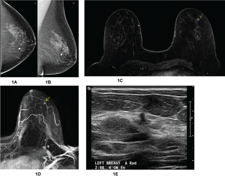
Figure 1: The CC (1A) and MLO (1B) mammogram of the left breast in a patient with personal history of breast cancer were read as normal. Subsequent breast MRI performed for history of breast cancer (1C, T1 post-contrast with fat subtraction and 1D, T1 post-contrast with fat subtraction and dynamic subtraction maximum intensity projection) showed a 6 mm mass in the left breast at the 2:00 position (yellow arrows) which was mammographically occult. Second look ultrasound (1E) showed a corresponding irregular hypoechoic mass. Subsequent ultrasound-guided biopsy yielded Invasive Ductal Carcinoma grade 2.
View Figure 1
Conclusion
In summary, breast MRI demonstrates both benefits and limitations in the detection of breast cancer when added to a screening regimen. Studies to date demonstrate in patients with estimated lifetime risk of breast cancer > 20%, benefits typically outweigh limitations. Data on survival benefits are limited, though recent studies suggest a trend toward improved survival outcomes for high-risk patients. On the other hand, in patients with lifetime risk < 15%, the limitations such as cost and decreased specificity may not justify the increase in sensitivity. New data suggest benefits may outweigh limitations in the subset of patients with prior history of breast cancer and in patients with a history of certain types of atypia on breast biopsy. In the subset of patients with lifetime risk 15-20% and in patients with dense breasts, data is still insufficient to justify for or against adding breast MRI to the annual screening regimen. Ongoing studies will continue to add information as to which subsets of patients will benefit the most from breast MRI screening. Additional studies evaluating newer tools such as diffusion weighted imaging, apparent diffusion coefficient values, and spectroscopy may also yield increases in future breast MRI specificity. The field has made significant advances in the past 4 decades, and further progress is eagerly anticipated.
References
-
el Yousef SJ, O'Connell DM, Duchesneau RH, Smith MJ, Hubay CA, et al. (1985) Benign and malignant breast disease: magnetic resonance and radiofrequency pulse sequences. AJR Am J Roentgenol 145: 1-8.
-
Heywang SH, Hahn D, Schmidt H, Krischke I, Eiermann W, et al. (1986) MR imaging of the breast using gadolinium-DTPA. J Comput Assist Tomogr 10: 199-204.
-
Kaiser WA, Zeitler E (1989) MR imaging of the breast: fast imaging sequences with and without Gd-DTPA. Preliminary observations. Radiology 170: 681-686.
-
Stack JP, Redmond OM, Codd MB, Dervan PA, Ennis JT (1990) Breast disease: tissue characterization with Gd-DTPA enhancement profiles. Radiology 174: 491-494.
-
Hickman PF, Moore NR, Shepstone BJ (1994) The indeterminate breast mass: assessment using contrast enhanced magnetic resonance imaging. Br J Radiol 67: 14-20.
-
Gilles R, Guinebretière JM, Lucidarme O, Cluzel P, Janaud G, et al. (1994) Nonpalpable breast tumors: diagnosis with contrast-enhanced subtraction dynamic MR imaging. Radiology 191: 625-631.
-
Heywang-Kobrunner SH, Hacker A, Sedlacek S (2013) Magnetic resonance imaging: the evolution of breast imaging. Breast 22: S77-82.
-
Saslow D, Boetes C, Burke W, Harms S, Leach MO, et al. (2007) American Cancer Society guidelines for breast screening with MRI as an adjunct to mammography. CA Cancer J Clin 57: 75-89.
-
Lehman CD, Smith RA (2009) The role of MRI in breast cancer screening. J Natl Compr Canc Netw 7: 1109-1115.
-
Afonso N (2009) Women at high risk for breast cancer--what the primary care provider needs to know. J Am Board Fam Med 22: 43-50.
-
(2013) Familial Breast Cancer: Classification and Care of People at Risk of Familial Breast Cancer and Management of Breast Cancer and Related Risks in People with a Family History of Breast Cancer. Cardiff (UK).
-
Lehman CD, Blume JD, Weatherall P, Thickman D, Hylton N, et al. (2005) Screening women at high risk for breast cancer with mammography and magnetic resonance imaging. Cancer 103: 1898-1905.
-
Kam JK, Naidu P, Rose AK, Mann GB (2013) Five-year analysis of magnetic resonance imaging as a screening tool in women at hereditary risk of breast cancer. J Med Imaging Radiat Oncol 57: 400-406.
-
Chiarelli AM, Prummel MV, Muradali D, Majpruz V, Horgan M, et al. (2014) Effectiveness of screening with annual magnetic resonance imaging and mammography: results of the initial screen from the ontario high risk breast screening program. J Clin Oncol 32: 2224-2230.
-
Le-Petross HT, Whitman GJ, Atchley DP, Yuan Y, Gutierrez-Barrera A, et al. (2011) Effectiveness of alternating mammography and magnetic resonance imaging for screening women with deleterious BRCA mutations at high risk of breast cancer. Cancer 117: 3900-3907.
-
Sardanelli F, Podo F, Santoro F, Manoukian S, Bergonzi S, et al. (2011) Multicenter surveillance of women at high genetic breast cancer risk using mammography, ultrasonography, and contrast-enhanced magnetic resonance imaging (the high breast cancer risk italian 1 study): final results. Invest Radiol 46: 94-105.
-
Riedl CC, Luft N, Bernhart C, Weber M, Bernathova M, et al. (2015) Triple-modality screening trial for familial breast cancer underlines the importance of magnetic resonance imaging and questions the role of mammography and ultrasound regardless of patient mutation status, age, and breast density. J Clin Oncol 33: 1128-1135.
-
Berg WA, Zhang Z, Lehrer D, Jong RA, Pisano ED, et al. (2012) Detection of breast cancer with addition of annual screening ultrasound or a single screening MRI to mammography in women with elevated breast cancer risk. Jama 307: 1394-1404.
-
Warner E, Messersmith H, Causer P, Eisen A, Shumak R, et al. (2008) Systematic review: using magnetic resonance imaging to screen women at high risk for breast cancer. Ann Intern Med 148: 671-679.
-
Port ER, Park A, Borgen PI, Morris E, Montgomery LL (2007) Results of MRI screening for breast cancer in high-risk patients with LCIS and atypical hyperplasia. Ann Surg Oncol 14: 1051-1057.
-
Friedlander LC, Roth SO, Gavenonis SC (2011) Results of MR imaging screening for breast cancer in high-risk patients with lobular carcinoma in situ. Radiology 261: 421-427.
-
Schwartz T, Cyr A, Margenthaler J (2015) Screening breast magnetic resonance imaging in women with atypia or lobular carcinoma in situ. The Journal of surgical research 193: 519-522.
-
Brennan S, Liberman L, Dershaw DD, Morris E (2010) Breast MRI screening of women with a personal history of breast cancer. AJR Am J Roentgenol 195: 510-516.
-
Emaus MJ, Bakker MF, Peeters PH, Loo CE, Mann RM, et al. (2015) MR Imaging as an Additional Screening Modality for the Detection of Breast Cancer in Women Aged 50-75 Years with Extremely Dense Breasts: The DENSE Trial Study Design. Radiology 277: 527-537.
-
Evans DG, Kesavan N, Lim Y, Gadde S, Hurley E, et al. (2014) MRI breast screening in high-risk women: cancer detection and survival analysis. Breast Cancer Res Treat 145: 663-672.
-
Saadatmand S, Tilanus-Linthorst MM, Rutgers EJ, Hoogerbrugge N, Oosterwijk JC, et al. (2013) Cost-effectiveness of screening women with familial risk for breast cancer with magnetic resonance imaging. J Natl Cancer Inst 105: 1314-1321.
-
Raikhlin A, Curpen B, Warner E, Betel C, Wright B, et al. (2015) Breast MRI as an adjunct to mammography for breast cancer screening in high-risk patients: retrospective review. AJR Am J Roentgenol 204: 889-897.
-
Kriege M, Brekelmans CT, Boetes C, Besnard PE, Zonderland HM, et al. (2004) Efficacy of MRI and mammography for breast-cancer screening in women with a familial or genetic predisposition. N Engl J Med 351: 427-437.
-
Kim JS, Lee SM, Cha ES (2014) The diagnostic sensitivity of dynamic contrast-enhanced magnetic resonance imaging and breast-specific gamma imaging in women with calcified and non-calcified DCIS. Acta Radiol 55: 668-675.
-
Kuhl CK, Schrading S, Strobel K, Schild HH, Hilgers RD, et al. (2014) Abbreviated breast magnetic resonance imaging (MRI): first postcontrast subtracted images and maximum-intensity projection-a novel approach to breast cancer screening with MRI. J Clin Oncol 32: 2304-2310.
-
Harvey SC, Di Carlo PA, Lee B, Obadina E, Sippo D, et al. (2016) An Abbreviated Protocol for High-Risk Screening Breast MRI Saves Time and Resources. J Am Coll Radiol 13: 374-380.
-
Grimm LJ, Soo MS, Yoon S, Kim C, Ghate SV, et al. (2015) Abbreviated screening protocol for breast MRI: a feasibility study. Acad Radiol 22: 1157-1162.
-
Moschetta M, Telegrafo M, Rella L, Stabile Ianora AA, Angelelli G (2016) Abbreviated Combined MR Protocol: A New Faster Strategy for Characterizing Breast Lesions. Clin Breast Cancer 16: 207-211.
