

Journal of Obesity and Weight-loss Medication
Dermatologic Manifestations of Obesity: Part I Mechanical Causes
Reid Alexander Waldman* and Anne H Kettler
School of Medicine, University of Missouri-Kansas City, Kansas City, Missouri, USA
*Corresponding author:
Reid Alexander Waldman, School of Medicine, University of Missouri-Kansas City, Kansas City, Missouri 64108, USA, Tel: 913-341-9841, E-mail: rawthf@mail.umkc.edu
J Obes Weight-Loss Medic,
JOWM-2-010, (Volume 2, Issue 1),
Review Article
Received: April 02, 2015: Accepted: January 14, 2016: Published: January 16, 2016
Citation: Waldman RA, Kettler AH (2016) Dermatologic Manifestations of Obesity: Part I Mechanical Causes. J Obes Weight-Loss Medic 1:010
Copyright: © 2016 Waldman RA, et al. This is an open-access article distributed under the terms of the Creative Commons Attribution License, which permits unrestricted use, distribution, and reproduction in any medium, provided the original author and source are credited.
Introduction
Over the past several decades, the proportion of Americans suffering from obesity has risen drastically [1]. Accompanying this increase in obesity is a concomitant increase in many of the co-morbidities associated with obesity, many of which are seen less frequently in persons with normal body weight. Notably, there has been an increase in dermatologic conditions seen in this special patient population. The skin maladies seen with increased frequency in obese patients are caused by a variety of factors, specifically: (1) the mechanical changes associated with increased weight; (2) the hyperandrogenism of obesity; and (3) the secondary hyperinsulinemia of obesity.
This three-part series of articles will review clinical dermatologic manifestations of obesity, identify clinical findings that can serve as harbingers of more serious systemic disease, help direct treatment choices, and ultimately improve patient care outcomes. Part I will explore the relationship between the mechanical changes caused by obesity and the resultant common dermatologic conditions associated with the changes.
Striae Distensae (Stretch Marks)
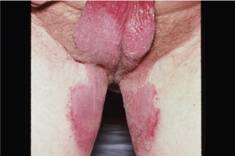
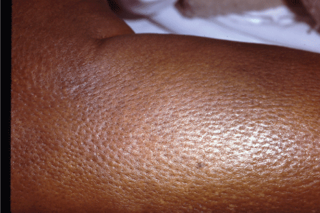
Striae distensae, also known colloquially as stretch marks, is a common dermatologic ailment that while physiologically benign, has the propensity to cause significant psychological stress for the obese patient who is often suffering from other issues of poor body image. The psychological stress associated with striae distensae is frequently exacerbated by the slow and often sub-optimal response to standard treatments. Striae distensae most commonly appear on the abdomen, thighs, and lumbosacral images in both males and females with the breasts and buttocks more commonly effected in females [2].
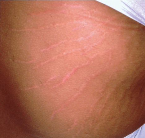
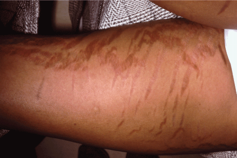
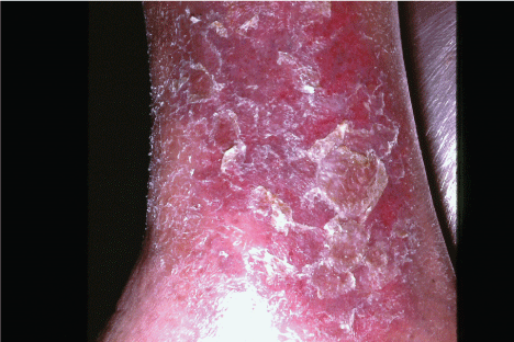
The clinical presentation of striae distensae can be divided into an acute and a chronic state with both states frequently occurring simultaneously in the same patient. The acute state which is known as striae rubra is characterized by flattened, reddish to violaceous finely wrinkled lesions which run perpendicular to tension lines of the skin [3] (Figure 1). Over time, if these striae remain untreated, these lesions evolve to a chronic state known as striae alba. Striae alba present clinically as whitish, atrophic appearing, depressed, irregularly linear scar-like bands [3] (Figure 2).
.
Figure 1: Striae rubra is characterized by flattened, reddish to violaceous finely wrinkled lesions which run perpendicular to tension lines of the skin.
View Figure 1
.
Figure 2: Striae alba present clinically as whitish, atrophic appearing, depressed, irregularly linear scar-like bands.
View Figure 2
Histology
Histologically, both striae rubra and striae alba have characteristic appearances on microscopy. Biopsies of striae rubra demonstrate epidermal edema with increased melanogenesis, vascular ectasia of the papillary dermis, and reduced elastin in conjunction with altered collagen in the reticular dermis. There is also a characteristic reorganization of elastic fibers with thicker fibers forming a peripheral border around a center of thin elastic fibers [4]. The histologic appearance of striae alba starkly contrasts that of striae rubra. The epidermis of striae alba is characterized by the absence of hair follicles, a near absence of melanocytes, decreased number of rete ridges, and generalized atrophy. The papillary dermis of striae alba has decreased vascularity and the reticular dermis has an almost scar-like deposition of thick bands of collagen parallel to the skin. Unlike histologic appearance of striae rubra which clearly demonstrates a reorganized pattern of both thick and thin elastic fibers, microscopy of biopsies of the reticular dermis of striae alba reveal lesions comprised primarily of thick fibers [4].
The finding of striae in an obese patient can most often be attributed to the mechanical changes associated with weight gain; however, because striae have other etiologies this cutaneous lesion can occasionally serve as the first sign of a more serious, systemic issue. Striae can occur during periods of hormonal change (e.g. pregnancy, adolescence, and Cushing's Disease) in patients with underlying dermal integrity issues (e.g. Ehlers Danlos, Marfan's, and zinc deficiency), and in patients using topical corticosteroids, especially when these steroids are applied under occlusive wraps [5].
It is worth noting that the development of striae during pregnancy, which are known as striae gravidarum, has not been linked to the mechanical stretching of skin associated with pregnancy, but rather are primarily the result of the hormonal changes associated with pregnancy [5]. Additionally, clinicians should have a high index of suspicion of Cushing's disease in any patient presenting with striae, especially those patients with very rapid, multifocal striae development in the absence of another obvious cause. Finally, any patient with striae development within body folds should be questioned about topical steroid use because body folds act like an occlusive dressing and increase the absorption of steroids up to 100-fold [6].
Treatment
Treatment of striae distensae involves a logical step-wise process that often requires the use of multiple treatment modalities including topicals, dermabrasion, non-ablative laser therapy, ablative laser therapy, and collagen induction therapy. The first step in devising a treatment plan for striae distensae is to determine whether the patient is suffering from striae rubra, striae alba, or both because some modalities such as tretinoin, which is quite effective for striae rubra, have little to no efficacy in the treatment of striae alba.
The starting point in the treatment of patients with striae rubra is usually tretinoin 0.1% with lower concentrations of the drug proving less efficacious. Controlled studies suggest that 80% of patients started on 0.1% tretinoin report significant improvement in both length and width of striae rubra after treatment [2]. Other topical agents that have been marketed for both the treatment and prevention of striae rubra include cocoa butter, olive oil, trofolastin, and silicon gel. Unfortunately, multiple studies have failed to find evidence that any of these agents significantly improve striae or prevent their development [7].
In patients who do not respond to tretinoin therapy, who have striae alba, or who do not want to try tretinoin, dermabrasion therapy is available [8]. Controlled studies have shown the efficacy of glycolic acid (70%) in the treatment of both striae rubra and striae alba. Additionally, there is evidence that the combination of glycolic acid 20% and tretinoin 0.05% is also efficacious [4]. Microdermabrasion using aluminum oxide has also shown promising results for the treatment of striae alba.
In light skinned patients suffering from striae distensae who do not improve with the previously mentioned treatment modalities, a variety of laser therapies have been shown to be efficacious. While there are many laser modalities available, examples of laser therapy types that have reported efficacy in the treatment of striae distensae include 585 nm pulsed dye laser (PDL), nonablative 1320 nm Nd:Yag, 308 nm XeCl, and short-pulsed 10 600-nm C02 Laser [9]. PDL has been reported to be especially efficacious in the treatment of striae rubra where this modality has been reported to increase the collagen content in underlying skin as well as decrease the dilation of blood vessels that contribute to the reddish discoloration of the striae [10]. XeCl and Nd:Yag have been reported to help with re-pigmentation of striae alba. Laser therapy may be combined with topical treatment to increase efficacy. It should be noted that patients with darker skin should avoid laser therapy as the efficacy of this modality in the subgroup of patients is lacking and hypopigmentation of the treated areas may occur [11]. It is the opinion of the authors that laser therapy be avoided in patients with Fitzpatrick Skin Types 4, 5, and 6.
Intense pulsed light (IPL) has also been used to treat striae distensae with some degree of success. Using a xenon source, this technology utilizes broad spectrum non-coherent light to produce photothermolysis of cells containing pigmentation. Like laser therapy, IPL can produce hyperpigmentation and its use should be limited to light skinned patients.
Finally, a relatively new and seemingly safe approach to striae is microneedling. This new aesthetic treatment modality utilizes very fine needles to produce controlled injury to the skin by cutting microscopic channels into the affected areas that stimulate the body to heal the injury by neocollagenesis and neovascularization. This technique helps to reduce the atrophic depressed areas of striae destensae without risk of depigmentation associated with laser and intense pulse light therapy. A recent study showed that nearly half of patients treated with microneedling reported marked to excellent improvement, with all patients in that study reporting at least minimal improvement [12]. Given the minimal risks associated with microneedling, it may represent a reasonable next step in the treatment of striae distensae that has failed to respond to more traditional treatment modalities.
Plantar Hyperkeratosis
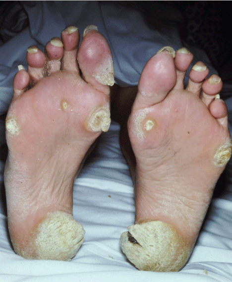
Plantar hyperkeratosis is a thickening of the skin over the metatarsophalangeal joints, secondary to the increased pressure and mechanical stress placed on the feet, in particular the metatarsal heads, due to the weight gain associated with obesity (Figure 3). Studies have shown that nearly 50% of obese people suffer from plantar hyperkeratosis and that the relationship between the development of plantar hyperkeratosis and obesity increases with increasing BMI [13]. While hyperkeratosis is part of the skin's physiologic protective response to increased mechanical stress, it can create a variety of problems including impingement of the plantar nerves, arthritis of the metatarsophalangeal joints, painful ambulation, impaired balance, and difficulty wearing shoes [14]. All of these abnormalities not only disrupt the patient's activities of daily living but put the patient at an increased risk for falls.
.
Figure 3: Plantar hyperkeratosis is a thickening of the skin over the metatarsophalangeal joints, secondary to the increased pressure and mechanical stress placed on the feet, in particular the metatarsal heads, due to the weight gain associated with obesity.
View Figure 3
The best approach to manage plantar hyperkeratosis includes weight loss counseling as this helps to alleviate the primary underlying cause of the lesion [15]. Fortunately, in the interim while the patient is losing weight, there are a number of effective symptomatic treatments. One of the quickest mechanisms of relief is surgical debridement of the lesion. Using a #15 scalpel blade to remove the excess keratin can provide almost immediate relief [15]. It is recommended to follow debridement with the placement of metatarsal padding in the shoe beneath the lesion to help slow the development of further hyperkeratosis and provide symptomatic relief. Silicon toe sleeves are especially effective because not only do they provide the necessary padding, but also they help soften the lesion. Additionally, if the patient is experiencing irritation from the friction between two toes after the debridement, a foam toe spacer can be used [16]. The use of these orthotic devices may require a shoe with a more commodious toe box to avoid the risk of pressure ulcers.
Other novel treatments for plantar hyperkeratosis include lactic acid creams, foot baths, and callus removers. It is important to mention that while pumice stones and other callus removers are used successfully in some patients, the risk of ulceration in the diabetic patient or the patient with peripheral neuropathy outweighs the potential benefit.
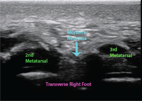
Finally, it is worth addressing common attempts at symptomatic therapy that tend to fail to ameliorate the patients pain and can have unintended consequences. While NSAIDs have many uses, they have not been found to be effective for plantar hyperkeratosis. Additionally, sometimes intralesional steroid injections are attempted. Unfortunately, the resultant atrophy of adipose tissue serves to exacerbate the patient's pain. It should be noted that other causes of foot pain including metatarsal stress fractures, Morton neuroma, intermetatarsal bursitis, and plantar fasciitis may co-exist with plantar hyperkeratosis and contribute to the patient's pain and functional disability (Figure 4).
.
Figure 4: Morton neuroma, intermetatarsal bursitis, and plantar fasciitis may co-exist with plantar hyperkeratosis and contribute to the patient's pain and functional disability. Transverse ultrasound image demonstrating a Morton's neuroma.
View Figure 4
Intertrigo
Intertrigo is an inflammatory skin condition that occurs in body folds as a result of the friction between opposing skin, the localized moisture, and the occlusion fostered by those folds. There is also often an infectious component. These conditions work together to produce an area of intense erythema and maceration that often times is so severe that the lesions weep and crust. Intertrigo occurs most commonly in the groin, the inframammary area, and the axillae [17] (Figure 5). It may also occur between the toes. It is also worth noting that in obese patients intertrigo can occur between folds created by the obesity itself.
.
Figure 5: Intertrigo occurs most commonly in the groin, the inframammary area, and the axillae. This patient is suffering from intertrigo with a candida superinfection.
View Figure 5
Intertrigo has a very strong association with obesity with a direct correlation between the incidence of the disorder and the patient's weight. The reason for this correlation is thought to be three-fold: First, obese persons are found to have increased surface area of body folds thus providing more skin area for friction to occur; Second, obese patients have been found to sweat more into these body folds and to then to retain more moisture within the folds; and Third, the skin of patients with a BMI greater than 25 has been found to be more alkaline than that of patients with normal BMI predisposing overweight and obese patients to superinfection by candida species [18].
How intertrigo presents depends on the location of the lesion. Patients with lesions in body folds often complain of pain, burning, and itching; not to mention emotional distress as a result of the cosmetic morbidity caused by the lesion. It is the experience of the authors that patients with groin intertrigo seem especially prone to emotional distress as a result of their lesions. On the other hand, patients with intertrigo of the toe webs are typically either asymptomatic or complain of a burning sensation.
Evaluating patients with suspected intertrigo involves two steps: First, distinguishing intertrigo from other similar appearing lesions; and Second, ruling out secondary infection of the lesion. Intertrigo can usually be diagnosed clinically based on the characteristic appearance of the lesions. Biopsy of intertrigo is not necessary as there are no defining histologic features specific to intertrigo. With that in mind, it is worth mentioning similar lesions that are often misdiagnosed as intertrigo. Patients with suspected intertrigo that has been refractory to treatment should be carefully evaluated for other possible conditions that may mimic the clinical presentation of intertrigo. Most commonly, inverse psoriasis, seborrheic dermatitis, irritant contact, allergic contact, and atopic dermatitis are confused with intertrigo. Additionally, Hailey-Hailey and Pemphigus Vegetans may also be mistaken for intertrigo.
In order to determine the correct treatment regimen for patients suffering from intertrigo, it is important to rule out concomitant infection as obese patients are especially susceptible to superinfection with candida species. KOH preps are very useful in confirming the presence of candida, with the presence of satellite papules and pustules considered pathognomonic for candida superinfection [19]. Other common secondary infections occurring in obese patients suffering from intertrigo include Streptococcus Pyogenes, Staphylococcus Aureus, Proteus species, Pseudomonas Aeruginosa, Dermatophyes, and Corynebacterium Minitissumum (Etiologic Agent of Cutaneous Erythasma) [19]. In the past it was believed that Malassezia species could cause secondary infections in patients with intertrigo; however, there is currently a debate among dermatologists as to whether these are true secondary infections or whether they are the result of concomitant seborrheic dermatitis. Wood's lamp evaluation will reveal a characteristic green fluorescence with pseudomonas infection and a coral-red fluorescence with infection with corynebacterium minitissumum [20].
Recent evidence points to Group A Streptococcus as an underdiagnosed culprit of secondary infection in obese patients suffering from intertrigo. Clinical signs that should encourage suspicion of infection with Group A Streptococcus include a foul smell, lack of candida's characteristic papular or pustular lesions, and the presence of a much more delineated, fiery red lesion [19]. Bacterial culture can be helpful for determining the specific cause of secondary infection and antibiotic sensitivity test should also be obtained to help guide subsequent therapy.
Several considerations go into determining the appropriate treatment regimen for patients with intertrigo. In the absence of secondary infection, the first line treatment is a short course of topical steroids from either Group VI or VII such as triamcinolone acetonide 0.025% or hydrocortisone 2.5% [6]. It is important to avoid topical steroids from Groups I-V as these stronger preparations will tend to cause atrophy and striae formation due to the markedly increased absorption caused by the occlusive effect of moist body folds. Topical tacrolimus, a macrolide immunosuppressive, can effectively be used at a concentration of 0.1% without concern of development of the aforementioned side effects [20]. It is worth noting that for patients anticipating prolonged duration of treatment, tacrolimus can be used as a solo agent. If intertrigo becomes a recurring problem, topical application of 1% hydrocortisone is rarely associated with striae formation and therefore can be used for long-term treatment. In patients with especially wet or weeping lesions, cool water compresses can be used as an adjunct therapy the first several days to help dry the lesions. Finally, prevention of recurrence of intertrigo can be achieved by the use of barrier creams such as zinc oxide; however, some studies have shown that the use of these creams predisposes patients to develop candida infections [21].
In patients with suspected secondary infection with candida, topical antifungals (e.g. azoles, terbinafine, etc.) and Greer's Goo (combination of nystatin powder, hydrocortisone powder, and zinc oxide paste) have been shown to be effective [19]. In cases resistant to initial topical treatments, oral fluconazole can be used. Catellani's carbolfuchsin paint has been shown to be especially effective for patients suffering from intertrigo of the toe webs [22]. It is important to aggressively treat secondary candida infections to avoid the subsequent development of onchomycosis.
In the same way that patients with secondary infections with candida should undergo specific treatment, those with bacterial infections should as well. As with all bacterial infections, treatment can be started empirically; however, it is important to adjust accordingly based on antibiotic susceptibilities obtained when culturing the lesions. For example, secondary infection with Group A Streptococcus infection can be treated with a combination of oral amoxicillin-clauvlanate 500-875 mg every 12 hours combined with topical application of fusidic acid 20 mg/gram cream pending cultures.
Chronic Venous Insufficiency
Chronic venous insufficiency and its associated dermatologic sequelae are commonly seen in obese patients. It is believed that this relationship is the result of two unique processes: 1) The obesity itself results in increased intra-abdominal pressure which causes increased resistance to venous return flow ultimately culminating in venous valvular insufficiency creating a self-perpetuating cycle of worsening venous insufficiency; and 2) The chronic inflammatory processes triggered by obesity causes on-going damage to the affected veins fostering both insufficiency and a propensity for thromboembolism [23].
As a result of venous insufficiency, the increased hydrostatic pressure within the veins causes fluid, cells, and intracellular components to extravasate into the surrounding tissue. While this process is initially compensated by increased lymphatic drainage from the affected area, the ongoing extravasation of fluid, cells, and intracellular components eventually overwhelm the ability of the lymphatics to compensate resulting in areas of pitting edema, warmth, hyperpigmentation, and scaling of the skin. The warmth and erythema are believed to be the result of an inflammatory response against the breakdown products of extravasated red blood cells (RBC) and intracellular components.
Left untreated, some patients suffering from chronic venous insufficiency will go on to develop stasis dermatitis. While the exact pathogenesis of stasis dermatitis remains unknown, two theories are predominately used to explain the phenomenon: 1) Some authorities believe it is the result of aforementioned inflammatory processes against RBC breakdown products; and 2) Others attribute the condition to an underlying susceptibility of affected skin to physical and chemical assault created by the venous insufficiency [24].
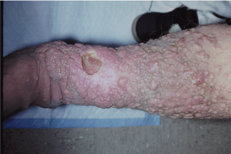
It is important to recognize that not all patients with venous insufficiency will develop stasis dermatitis, but when stasis dermatitis develops, it can present clinically in an acute, subacute, and chronic form. The acute form of stasis dermatitis is characterized by a rapid onset of intense pruritus and its profound redness that may mimic cellulitis [24]. A weeping transudate and crusting may also be present. The subacute form of stasis dermatitis is characterized by the slower development of a brown discoloration of the skin that is the result of the extravasation of hemosiderin from red blood cell breakdown products. Simultaneously, the affected areas of subacute stasis dermatitis will become scaly and dry. Over time, patients with repeated episodes of acute and/or subacute stasis dermatitis can go on to develop chronic stasis dermatitis which is characterized by a thickening of the skin, intractable dark brown discoloration, and presence of cyanotic red plaque (Figure 6). The pathogenesis of chronic stasis dermatitis is thought to be primarily the end result of longstanding inflammation of the affected skin and subcutaneous tissue.
.
Figure 6: Patients with repeated episodes of acute and/or subacute stasis dermatitis can go on to develop chronic stasis dermatitis which is characterized by a thickening of the skin, intractable dark brown discoloration, and presence of cyanotic red plaque.
View Figure 6
Another common sequela of venous insufficiency in some obese patients is varicose veins. Varicose veins are the result of incompetency of venous valves in the legs leading to reflux and dilation of the superficial veins. Clinically, these superficial veins appear as torturous, dilated bluish colored blood vessels that appear to arise from within the skin itself. While varicose veins are often thought of as purely a cosmetic problem, these lower extremity varicosities can cause discomfort of the legs which is aching in nature, with burning and itching often present. The main complication of lower extremity varicosities is venous thrombosis; however, unlike deep vein thrombosis, there is little risk of clinically significant pulmonary embolism [25].
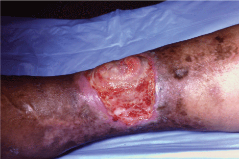
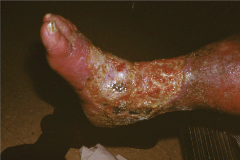
The most serious complication of venous insufficiency in the obese patient is the development of venous stasis ulcers (Figure 7). Once either the perforating or deep venous systems of the leg have become compromised, patients will likely develop ulcerations. These lesions most commonly occur near the medial malleolus, although they can be found anywhere in the distal lower extremities when trauma and/or infection incite the lesions [26]. Once present, these ulcers have the tendency to enlarge, even without the presence of any obvious inciting or exacerbating factor (Figure 8). Patients who develop venous stasis ulcers tend to complain of constant aching pain that is exacerbated by prolonged periods of standing and relieved by elevation of the affected lower extremity. Complicating treatment is the fact that many venous stasis ulcers become infected. This complicates healing and even with early optimal treatment it can take up to several months for venous stasis ulcers to heal. Even with treatment, nearly 50% of ulcers will recur [27]. With this in mind, if a lesion fails to respond to treatment within a reasonable time-frame, it is important to re-evaluate the diagnosis of venous stasis ulcer and determine if another pathologic process such as osteomyelitis, neoplasms, pyoderma gangrenosum, and various types of vasculitis are at play. These pathologic conditions can co-exist with venous insufficiency, complicating the differential diagnosis.
.
Figure 7: The most serious complication of venous insufficiency in the obese patient is the development of venous stasis ulcers.
View Figure 7
.
Figure 8: Once present, venous stasis ulcers have the tendency to enlarge, even without the presence of any obvious inciting or exacerbating factor.
View Figure 8
Treatment for venous insufficiency is in large part driven by the clinical presentation of the disease. For patients with distal lower extremity swelling in the absence of more serious dermatologic issues, compression with elastic stockings or bandages combined with elevation of the affected extremity can provide adequate symptomatic relief; however, it is important to simultaneously promote weight loss as this is an integral part of a rational treatment plan if one hopes to slow the progression of the disease.
Patients suffering from acute stasis dermatitis with lesions characterized by significant weeping will benefit from the initial use of cool water compresses two to four times per day to decrease transepidermal water loss until the lesions began to dry. Topical steroids can be used around the lesions of acute stasis dermatitis to decrease pruritus and rubor, but should not be applied directly into any cutaneous fissures or onto open areas as this can retard the healing process.
For patients with stasis dermatitis in the subacute phase, topical steroids can be used with potency as high as Grade II, e.g. desoximetasone cream 0.25%. Additionally, because of the high likelihood of co-existent bacterial infection it is recommended that the patient concurrently be started on an oral first generation cephalosporin such as cephalexin 500 mg four times a day.
A variety of treatment modalities exist for patients suffering from lower extremity varicosities. Most notably, patients can treat their varicosities with compression, lifestyle modifications, sclerotherapy, surgery, radiofrequency ablation, and laser therapy [27]. While conservative therapy combining the use of compression and elevation has been the mainstay of treatment, studies have shown that it is inferior to sclerotherapy and other aforementioned treatment modalities in terms of both symptom management and cosmetic improvement. In fact, the only symptom which has been shown to be relieved by compression is pain [27]. In patients who fail compression and sclerotherapy or in patients with severe disease, the combination of avulsion and vein stripping (surgery) has been found to successfully treat varicosities and prevent recurrence in the majority of patients. The use of radiofrequency ablation and laser therapy have both been shown to have solid short term results; however, they may not achieve the long term results seen with vein stripping surgery.
Patients suffering from venous stasis ulcers will usually benefit from surgical debridement of the ulcer in conjunction with occlusive gauze dressings impregnated with zinc oxide, white petrolatum, and calamine lotion. Concurrent identification and treatment of infection and treatment of nutritional deficiencies is paramount if healing is to be achieved. Other adjunct therapies, including aspirin and the phosphodiesterase inhibitor pentoxifylline, may also help improve associated vascular insufficiency and enhance healing [28]. Malnourished patients can benefit tremendously from the supplementation of Vitamin E, Zinc, and Vitamin C and correction of any protein deficiencies.
Lymphedema
Lymphedema occurs predominately in the obese and presents serious quality of life ramifications to this special patient population, with over 75% of morbidly obese patients suffering from lymphedema at some time during their life [29]. It is the sheer weight of the patient's soft tissues that cause the obstruction of lymphatic return. This results in the localized accumulation of lymph which causes further mechanical compressive effects in the affected region. The resultant accumulation of lymph causes further vascular compromise, exacerbates the problem, and in turn makes these areas susceptible to bacterial infection. When bacterial infection occurs, it triggers an inflammatory response within the lymphatic system which causes scarring of the lymphatic vessels causing further obstruction and further lymphedema. This process generally starts in the distal lower extremities and works its way proximally. As the resultant vascular compromise and lymphangitis progresses, a characteristic deformity of the affected extremity reminiscent of the elephantiasis caused by filarial disease known as elephantiasis nostra verrucosa occurs.
Studies suggest that there is a BMI threshold at which lower extremity lymphedema occurs and then above that BMI an even higher threshold BMI at which upper extremity lymphedema occurs. Patients with a BMI greater than 30 have a 91% chance of developing lymphedema with the average BMI of patients suffering from elephantiasis nostra verrucosa being 55.8 [30].
In a clinical setting, there are four stages of lymphedema (Stages 0-3), three of which have gross clinical findings. The condition starts off as a subclinical lymphedema (Stage 0). While there are no gross findings in this stage, microscopic examination of affected tissue will demonstrate histologic changes of lymphangiectasia secondary to the initial impedance of lymph transport. From there, the condition can progress to reversible edema (Stage 1) [30]. In this stage pitting edema is present on physical examination; however, it can be resolved by elevation and compression of the affected extremity. Histologically, this stage not only shows lymphangiectasia, but also shows early fibrotic changes and dermal edema. As the disease progresses, patients can develop spontaneously irreversible edema (Stage 2) (Figure 9). Swelling in these patients is so severe that it is non-pitting and feels hard when palpated. As expected, histology of these lesions shows progressive fibrosclerosis as well as adipocyte hyperplasia. Finally, some patients can go on to develop elephantiasis nostra verrucosa (Stage 3) (Figure 10). These patients have significant dermatologic changes characterized by papillomatosis, hyperkeratosis, and wart-like plaque formation in the affected extremity. These lesions can start to crust and weep lymphorrhea making the area a nidus for infectious cellulitis. The resulting deformity is distressing to the patient and family and will often cause the patient to avoid going out in public out of embarrassment. Ultimately, it is the associated pain and the fatigue caused by the huge effort required to move the grossly enlarged extremities that limit the patient's ability to ambulate. Additionally, patients who suffer from recurrent infections can be seriously hampered by the repeated hospitalizations they require for proper treatment.
.
Figure 9: Patients with Stage 2 lymphedema demonstrate early fibrotic changes and dermal edema. As the disease progresses, patients can develop spontaneously irreversible edema.
View Figure 9
.
Figure 10: Patients with Stage 3 lymphedema can go on to develop elephantiasis nostra verrucosa. These patients have significant dermatologic changes characterized by papillomatosis, hyperkeratosis, and wart-like plaque formation in the affected extremity.
View Figure 10
Rare primary forms of lymphedema, secondary to genetic defects in the lymphangiogenesis pathway can occur, but secondary causes of lymphedema including obesity-induced lymphedema are much more common [31]. When evaluating an obese patient with a working diagnosis of lymphedema, it is important to remember that a number of diseases can mimic the clinical presentation of lymphedema. These diseases include venous insufficiency, edema secondary to congestive heart failure, edema secondary to kidney failure, edema secondary to other systemic organ failure, myxedema, edema secondary to radiation therapy and lymphadenectomy, post-traumatic edema, lipedema, and deep venous thrombosis. Given the disastrous sequela of failing to diagnose one of these potentially life threatening diseases, the clinician should always include them in the differential diagnosis of the patient with significant lymphedema, especially when the edema is of acute onset. While both Stage 1 lymphedema and the edema of congestive heart failure can be relieved by elevation of the affected extremities, diuretics will only decrease edema.
In addition to the aforementioned lymphedema secondary to obesity, a rare patient will develop solitary, slow growing lesions called massive localized lymphedema. While these lesions are also derived from the lymphatic drainage obstruction caused by obesity they present in a clinically distinct manner. The lesions associated with massive localized edema are polyp-like masses and are defined by peau d'orange appearance and the presence of non-pitting edema [30]. Many times these lesions are biopsied out of fear that the localized lesion is a liposarcoma. Fortunately, massive localized edema can be easily histologically distinguished from liposarcoma due to the absence of adipocytes and atypical cells commonly associated with liposarcoma. Histologically, the lesions of massive localized lymphedema will be characterized by the presence of lymphangectasia as well as both dermal and epidermal thickening [31].
Prompt, aggressive treatment of diffuse lymphedema, as well as massive localized lymphedema, is mandatory because of the propensity for both of these conditions to develop infection and because of the potential for malignant transformation. This propensity for areas of lymphedema to undergo malignant transformation into a lymphangiosarcoma is known as Stewart-Treves Syndrome and most often seen in patients who have undergone radical mastectomy, but can also be seen in patients with any form of chronic diffuse lymphedema [32].
Unfortunately, treatment options for diffuse lymphedema are limited with goals of therapy directed at symptomatic improvement and prevention of infection. Symptomatic improvement therapy is usually based on elevation and compression with pneumatic compression devices and compression stockings serving as mainstays of treatment. Other modalities include the integration of physical therapy into these approaches.
The recommended treatment for patients suffering from massive localized edema is surgical excision due to the risk of infection and malignant transformation that such lesions pose. In patients who are not amenable to excision, compression, lymph drainage, and weight reduction are the primarily methods of treatment.
Adiposa Dolorosa
Adiposa dolorosa, which is also known as Dercum's disease, is a rare multisystem disease which is most commonly seen in obese, perimenopausal females. The disease has four typical symptoms: 1) generalized obesity, 2) multiple, painful lipoma-like masses 3) psychiatric illness including dementia, depression, confusion, and emotional lability and 4) chronic fatigue [33]. Patients suffering from adiposa dolorosa will typically complain of severe pain which is often out of proportion to what would be expected given the physical findings. While most cases appear spontaneously, the occurrence of the disease in multiple family members has been reported [34]. These patients often are hyperalgesic to palpation of the lipoma-like masses. Additionally, while patients often cannot identify individual masses as painful prior to physical examination, palpation of masses tends to elicit pain in some of them. These masses include the abdomen, thighs, and upper arms; however, lesions have been documented everywhere except for the head. The skin superficial to these masses often is characterized by dilated superficial veins.
Diagnosis of this disease presents a particular challenge as the multiple associated psychiatric symptoms tend to obscure the clinical picture. Additionally, the lack of inflammatory, autoimmune, or general laboratory markers that might aid in diagnosis contribute to the difficulty in accurate diagnosis. It is worth noting that biopsy is likely not of great use in these patients as multiple studies have found that the masses of adiposa dolorosa are indistinguishable from the histologic findings of the common lipoma. Diagnosis is generally made by magnetic resonance imaging (MRI) which will usually reveal the following pathognomonic findings: (1) multiple ill-defined, oval shaped masses that appear to bluish on unenhanced MRI; (2) no enhancement following the administration of gadolinium contrast; (3) decreased T1-weighted signal; and (4) increased water-sensitive sequences [35]. Additionally, some cases are diagnosed via ultrasound by identifying superficial oblong shaped subcutaneous fatty nodules that are hyperechoic and do not produce an increased color Doppler echo.
Treatment of adiposis dolorosa is focused primarily on controlling the pain and psychiatric manifestations of the disease. NSAIDs, oral prednisone, and simple analgesics such as acetaminophen usually fail to provide meaningful pain relief. Additionally, weight loss measurements, even when successful; fail to decrease the pain in most patients suffering from this painful condition. There are some clinical reports that the use of intravenous lidocaine and oral mexilitine may provide a modicum of symptomatic relief [36]. Another approach which has documented success, albeit of varying duration, is the use of intralesional corticosteroid injections. It would seem most logical to use ultrasound guidance for these injections as patients often struggle to successfully localize their pain. Ultimately, the most effective strategy is often surgical excision or liposuction; however, many of the patients are not surgical candidates due to their comorbidities secondary to their obesity [37]. Additionally, even when surgery is successful, recurrence of symptoms is common. Recent anecdotal reports suggest that the injection of interferon alpha-2b may reduce pain symptomatology [38].
Conclusion
The increasing prevalence of obesity needs to be met with an increasing awareness of the dermatologic manifestations that accompany it. The multifaceted impact that obesity has on skin physiology and the diverse pathologic complications that follow serve as a microcosm for the overall effect of obesity on the body. It is especially important not to discount the effect that the mechanical stresses of obesity place on the body. This article serves to outline the many consequences of mechanical stress and will hopefully aid the clinician in diagnosing and treating the mechanical stress-induced dermatologic manifestations of obesity.
Part II of this series will discuss the dermatologic manifestations caused by the hyperandrogenism of obesity.
References
-
Glauser TA, Roepke N, Stevenin B, Dubois AM, Ahn SM (2015) Physician knowledge about and perceptions of obesity management, Obesity Research & Clinical Practice.
-
Al-Himdani S, Ud-Din S, Gilmore S, Bayat A (2014) Striae distensae: a comprehensive review and evidence-based evaluation of prophylaxis and treatment. Br J Dermatol 170: 527-547.
-
Gilmore SJ, Vaughan BL Jr., Madzvamuse A, Maini PK (2012) A mechanochemical model of striae distensae. Math Biosci 240: 141-147.
-
Salter SA, Kimball AB (2006) Striae gravidarum. Clin Dermatol 24: 97-100.
-
Osman H, Rubeiz N, Tamim H, Nassar AH (2007) Risk factors for the development of striae gravidarum. Am J Obstet Gynecol 196: 62.
-
Habif Thomas P (2010) Topical Therapy and Topical Corticosteroids. Clinical Dermatology: A Color Guide to Diagnosis and Therapy. Edinburgh, Mosby 130-135.
-
Soltanipour F, Delaram M, Taavoni S, Haghani H (2014) The effect of olive oil and the Saj® cream in prevention of striae gravidarum: A randomized controlled clinical trial. Complementary Therapies in Medicine 22: 220-225.
-
Hexsel D, Mazzuco R, DaÍForno T (2009) Superficial dermabrasion in the treatment of recent stretch marks (striae rubra). JAAD 60: AB186.
-
Tanzi EL, Lupton JR, Alster TS (2003) Lasers in dermatology: four decades of progress. J Am Acad Dermatol 49: 1-31.
-
Michel JL (2005) Flashlamp pumped dye laser (585 nm, low fluence) for aesthetic and medical indications: Own experience/results and review of literature. Medical Laser Application 20: 77-83.
-
Carniol PJ, Lloyd HW, Zhao AS, Murray K (2010) Laser Treatment for Ethnic Skin. Facial Plastic Surgery Clinics 18: 105-111.
-
Park KY, Kim HK, Kim SE, Kim BJ, Kim MN (2012) Treatment of striae distensae using needling therapy: a pilot study. Dermatol Surg 38: 1823-1828.
-
Spink MJ, Menz HB, Lord SR (2009) Distribution and correlates of plantar hyperkeratotic lesions in older people. J Foot Ankle Res 2: 8.
-
Birtane M, Tuna H (2004) The evaluation of plantar pressure distribution in obese and non-obese adults. Clin Biomech (Bristol, Avon) 19: 1055-1059.
-
Freeman DB (2002) Corns and calluses resulting from mechanical hyperkeratosis. Am Fam Physician 65: 2277-2280.
-
Hatcher RM, Goller WL, Weil LS (1978) Intractable plantar keratoses: a review of surgical corrections. J Am Podiatry Assoc 68: 377-386.
-
Wolf R, Oumeish OY, Parish LC (2011) Intertriginous eruption. Clinics in Dermatology 29: 173-179.
-
Janniger CK, Schwartz RA, Szepietowski JC, Reich A (2005) Intertrigo and common secondary skin infections. Am Fam Physician 72: 833-838.
-
Neri I, Bassi A, Patrizi A (2015) Streptococcal intertrigo. J Pediatr 166: 1318.
-
Chapman M, Brown J, Linowski G (2005) Tacrolimus 0.1% ointment for intertrigo. JAAD 52: P61.
-
Habif TP (2010) Fungal Infections. Clinical Dermatology: A Color Guide to Diagnosis and Therapy. Edinburgh: Mosby 246-289.
-
Shah M (2003) Castellan 's paint. IJDVL 69: 357-358.
-
Heit JA, Rooke TW, Silverstein MD, Mohr DN, Lohse CM, et al. (2001) Trends in the incidence of venous stasis syndrome and venous ulcer: a 25-year population-based study. J Vasc Surg 33: 1022-1027.
-
White J (2013) Non-atopic dermatitis. Medicine 41: 345-346.
-
Tisi PV (2011) Varicose veins. BMJ Clin Evid 2011.
-
Thomas DR (2013) Managing venous stasis disease and ulcers. Clin Geriatr Med 29: 415-424.
-
Willenberg T (2014) Treatment of varicose veins. Reviews in Vascular Medicine 2: 67-72.
-
Jull A, Waters J, Arroll B (2002) Pentoxifylline for treatment of venous leg ulcers: a systematic review. Lancet 359: 1550-1554.
-
Carlson JA (2014) Lymphedema and subclinical lymphostasis (microlymphedema) facilitate cutaneous infection, inflammatory dermatoses, and neoplasia: A locus minoris resistentiae. Clin Dermatol 32: 599-615.
-
Jabbar F, Hammoudeh ZS, Bachusz R, Ledgerwood AM, Lucas CE (2015) The diagnostic and surgical challenges of massive localized lymphedema. The American Journal of Surgery 209: 584-587.
-
Berenji M, Kalani A, Kim J, Kelly K, Wallack MK (2010) Massive localized lymphedema of the thigh in a morbidly obese patient. EJSO 36: 104-106.
-
Sharma A, Schwartz RA (2012) Stewart-Treves syndrome: pathogenesis and management. J Am Acad Dermatol 67: 1342-1348.
-
Schaffer PR, Hale CS, Meehan SA, Shupack JL, Ramachandran S (2014) Adiposis dolorosa. Dermatol Online J 20.
-
Amine B, Leguilchard F, Benhamou CL (2004) Dercum's disease (adiposis dolorosa): a new case-report. Joint Bone Spine 71: 147-149.
-
Tins BJ, Matthews C, Haddaway M, Cassar-Pullicino VN, et al. (2013) Adiposis dolorosa (Dercum's disease): MRI and ultrasound appearances. Clin Radiol 68: 1047-1053.
-
Petersen P, Kastrup J (1987) Dercum's disease (adiposis dolorosa). Treatment of the severe pain with intravenous lidocaine. Pain 28: 77-80.
-
Hansson E, Svensson H, Brorson H (2011) Liposuction may reduce pain in Dercum's disease (adiposis dolorosa). Pain Med 12: 942-952.
-
Gonciarz Z, Mazur W, Hartleb J, Machniak M, Bednarek I, et al. ( 1997) Interferon alfa-2b induced long-term relief of pain in two patients with adiposis dolorosa and chronic hepatitis C. J Hepatol 27: 1141.
