
Trauma Cases and Reviews
Orbital Floor Reconstruction in Facial Asymmetry: A Clinical Case
Michele Maglione1, Davide Sossi1, Paolo Cecchini2 and Roberto Rizzo1*
1Department of Medical, Surgical and Health Sciences, University of Trieste, Italy
2Department of Ophtalmology, University of Trieste, Italy
*Corresponding author: Roberto Rizzo, MD, PhD, Clinica Odontoiatrica e Stomatologica, Piazza dell'Ospitale 2, Italy, Tel: +393385442110, E- mail: r.rizzo@fmc.units.it
Trauma Cases Rev, TCR-1-016, (Volume 1, Issue 3), Case Report; ISSN: 2469-5777
Received: October 17, 2015 | Accepted: November 19, 2015 | Published: November 21, 2015
Citation: Maglione M, Sossi D, Cecchini P, Rizzo R (2015) Orbital Floor Reconstruction in Facial Asymmetry: A Clinical Case. Trauma Cases Rev 1:016. 10.23937/2469-5777/1510016
Copyright: © 2015 Maglione M, et al. This is an open-access article distributed under the terms of the Creative Commons Attribution License, which permits unrestricted use, distribution, and reproduction in any medium, provided the original author and source are credited.
Abstract
Orbital floor blowout fractures may cause severe facial asymmetry, particularly when combined with panfacial fractures with wide bone defects. Surgical repair recommendations depend on multi-disciplinary clinical evaluation and digital imaging to assess the amount of soft tissues herniation, the infraorbital nerve and rectum inferioris muscle entrapments, the ocular bulbus dislocation, and the ocular diplopia. A second surgery intervention may be required particularly when the orbital floor fracture is combined with skull and panfacial fractures in severely traumatized patients when the first surgical stage has been focused more on life saving than on craniofacial reconstruction. Preoperative multiplanar CT scans with 3D image reconstructions permit 3D printing modelling to achieve useful guide datas to combine bone grafts and titanium meshes in an effective and low expensive technique for orbital floor reconstruction when severe facial asymmetry occurs.
Keywords
Blowout fractures, Orbital floor, Bone graft, Titanium mesh, Facial asymmetry, Posttraumatic deformities, Computer assisted surgery
Article Text
Orbital floor can easily be damaged in a craniomaxillofacial trauma due to its weakness. These fractures are mainly located medial to the infraorbital groove and canal, sometimes combined with medial orbital wall fractures due to the reduced bone thickness in this area [1,2]. Diplopia and enophtalmos are relatively common consequences of such fractures whereas other symptoms are unfavourable aesthetics, important facial asymmetry, enophtalmos, persistent diplopia, vertical ocular dystopia and restriction gaze [3,4]. Recommendations for surgical fracture repair depend on a combination of multidisciplinary clinical evaluation and imaging studies to evaluate the inferior rectus and oblique muscles, the infraorbital nerve entrapment and periorbital tissue herniation. Precise repositioning of the fractured bones, particularly of the zygomatic-orbital-maxillary complex if fractured, is the key to ultimately restore original function and aesthetics of the midface [5]. Despite this, posttraumatic deformities could be left such secondary orbital reconstruction is recommended when primary repair has not been performed or when its results have not been satisfying [1-6]. Due to the complex orbital anatomy, thorough reconstruction of orbital traumas remains challenging. Many surgical techniques from osteothomies to inlay bone augmentations are available for reconstructing bone contour and restorating proper orbital volume involving many biomaterials ranging from autogenous or homologous bone grafts to cartilage and fat grafts to titanium meshes, to alloplastic materials. Moreover, lately, computer assisted surgery developed together with virtual planning and intraoperative navigation in helping preoperatory surgical planning and decision making; CT DICOM files can be remotely processed to Standard Triangulation Language (.stl) format by CAD software used for rapid prototyping, 3D printing and computer aided manufacturing such as stereolithographic polymeric models and biomaterial prosthetic parts.
Methods
Case description
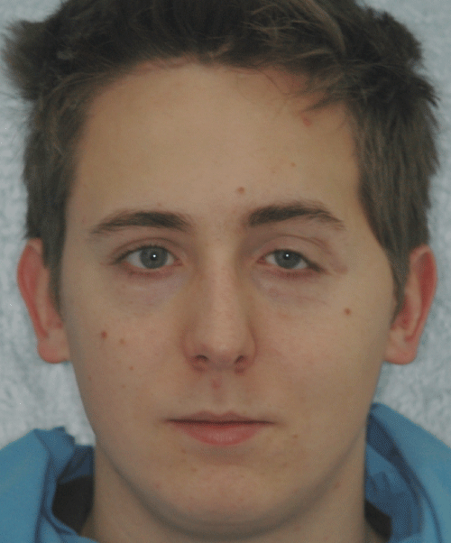
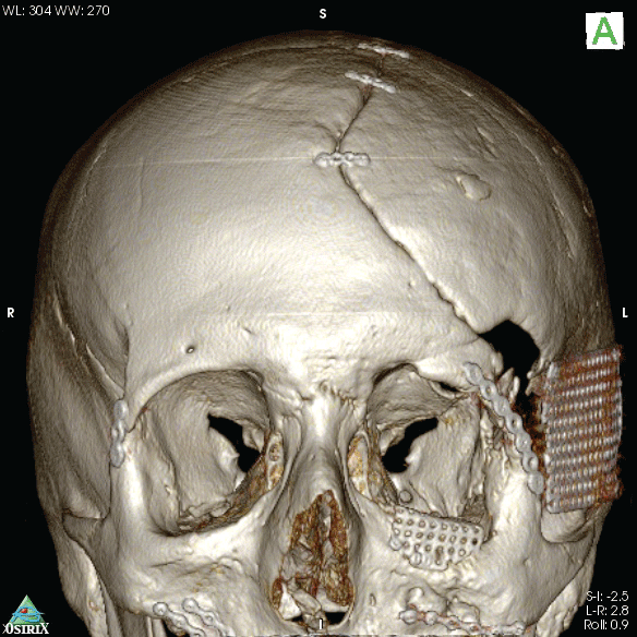
Due to a high-energy motorcycle accident, a male 21 year-old patient suffered frontal anterior cranial vault injuries and skull and pan-facial fractures. He was hospitalized at the local trauma centre and an immediate emergency neurosurgical intervention was performed to reduce the high intracranial pressure level, temporarily removing part of the left parietal and temporal bones and definitively the more comminuted bone fragments from the left orbital rim and roof. When stabilized, a second surgical intervention was performed to practice the cranioplasty, reapplying the previously removed cranial vault bones and to reduce the frontal bone and zygomatic fractures; supraorbital rim comminuted fracture was reconstructed with homologous cadaveric frozen bone; orbital floor defect was sealed with Ethisorb® Dura Patch membrane and the maxillo-zygomatic process was reduced on the left side. Reduction of frontal bone and zygomatic fracture, orbital floor and medial orbital wall reconstruction were also performed on the right side. Despite the treatment, ocular dystopia and severe mid-facial asymmetry with left orbital deformity remained due to the lack of the left orbital floor bone and the presence of still wide missing bone defects (Figure1).
.
Figure 1: Mid-facial asymmetry after the first surgical intervention with ocular dystopia
View Figure 1
At the time, the best corrected visual acuity was 20/20 in both eyes. The ophthalmological examination proved to be unremarkable. Orthotic evaluation was notable for an 8 prism dioptres (PD) left eye esotropia and 4 PD hypertropia. The patient suffered from oblique diplopia for near and distance. A prismatic prescription of 7 PD bases 350 was necessary to obtain binocular single vision in primary position. The vertical squint worsened on downgaze, due to left eye inferior rectus and superior oblique muscles under-action with infraduction impairment. An abnormal head posture with chin depression was observed.
.
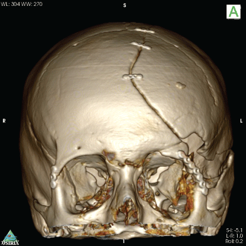
Figure 2: CT 3D rendering after first surgical stage showing the orbital floor defect, mid-facial asymmetry and orbital deformity
View Figure 2
3D Modeling
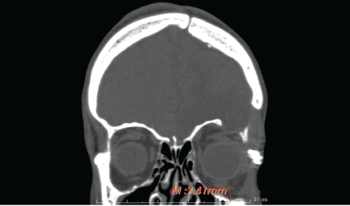
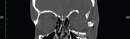
The patient's anatomy was then assessed in multiplanar (axial, coronal, sagittal) and 3D computed tomography via Toshiba Aquilon One Helical CT scanner (Figure 2), gantry = 0, slice = 1mm. The orbital floor malposition on the left side was, as measured through the CT images, 7.4 mm caudal than the contralateral one (Figure 3), causing ocular dystopia. DICOM files were processed by DeVIDE software to a .stl file. Then Netfabb software was used to obtain an ABS polymeric stereolithographic model via HP Designjet 3D printer (Figure 4). A silicon impression material was manually adapted on the stereolithopraphic model defective orbital floor to achieve a steam sterilizable autoclavable mould as a helping device in manual bone graft modelling during the surgery.
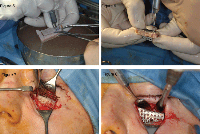
A new surgical intervention was performed 6 months later to solve the left diplopia and the mid-facial asymmetry the patient suffered. Through an infraorbital transcutaneous surgical approach, due to the presence of a scar being left from the previous intervention, the orbicularis muscle was transected, and the dissection along a preseptal orbital plane was performed in order to avoid the orbital fat herniation in the operating field. The inferior orbital rim periosteum was incised through its whole length and raised so that wide access to orbital floor was obtained. A 1.5 cm length and 1, 2 cm deep iliac crest homologous graft bone was hand-shaped by low rpm drilling following the shape of the silicon mould previously adapted on the 3D stereolithographic model and fixed to a titanium mesh plate with titanium screws (Figure 5) just before its fixation to the orbital floor rim (Figure 6, Figure 7, Figure 8). Forced ocular muscles duction test after graft insertion was performed by the ophthalmic surgeon in order to verify the obtained free ocular motility.
.
Figure 5-8: iliac crest homologous bone graft hand shaping and its fixation to the infraorbital rim with titanium mesh
View Figure 5-8
Postoperative CT images showed the resolution of the dystopia and the follow up controls demonstrated good health of the soft tissues and the absence of diplopia. An improvement of the facial symmetry was furthermore noticeable (Figure 9, Figure 10).

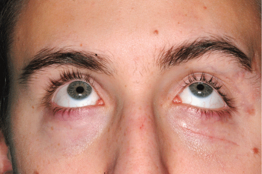
By the ophthalmologist point of view, immediately after surgery, the facial asymmetry was remarkably reduced, and the resolution of diplopia in primary position for distance vision was obtained (Figure 11). Diplopia was still present for near view, due infraduction limitation. The angle of hypertropia was related to the amount of deorsumversion. A 4 PD on 350 axis prescription was useful to avoid diplopia for reading. The post-op CT scan confirmed such good results (Figure 12). Eighteen months after surgery vertical diplopia was present on extreme downgaze only. The patient did not consider that an issue and dismissed the use of any prismatic correction for near and distance vision. On outer examination, the patient showed a limited chin depression to avoid diplopia during downgaze and to achieve binocular single vision.
Discussion
A wide variety of surgical techniques and biomaterials have been proposed for orbital floor reconstruction, including bone grafts, high-density porous polyethylene, titanium mesh, and composites [7]. The ideal material for floor reconstruction and augmentation, should allow easy adapting to an anatomic shape, provide an adequate thickness as showed in our case, be radiopaque (to allow for postoperative radiological confirmation of placement), and be stable over time. Bone grafts are optimally biocompatible and radiopaque, have a smooth surface from which periorbital tissues can be easily dissected in secondary reconstruction, and have no additional costs. Their disadvantages are longer operating time, additional donor site (calvaria, ribs, iliac crest, and mandible) with the attendant morbidity, and chances of resorption. They require additional implants as plates and/or screws to hold them in place too. Titanium mesh has good biocompatibility and is easily adjustable. Because of the mesh structure, connective tissue can grow around and through the implant, preventing its migration. It can be reliably fixed with screws in zones such the infraorbital rim. Titanium mesh has good mechanical strength even in thin sections and produces fewer artifacts on CT scans than other metals [8]. Pre operative stereolithographic model could be useful to plan and achieve an optimal autologous or homologous bone graft adaption and for time saving in the operating theatre; all that, with good clinical results when limited to small areas such the one presented in this case and far less expensive than 3D CAD-CAM alloplastic graft production that could be saved for the treatment of more complex ones, as many authors suggest that preoperative computer design and stereolithographic modelling combined with intraoperative navigation provide a useful guide for and possibly more accurate reconstruction of a variety of complex cranio-maxillofacial deformities [9-12]. An orbito-zygomatic osteothomy with coronal reposition of the fragment that reasonably could have been the first choice in the presented case was preoperatively excluded due to the thinness of the homologous bone grafted during the previous frontozygomatic orbital rim reconstruction and the lack of most of the orbital floor. Our approach to the case of a mid-facial and orbital deformity, due to the bone loss consequent to an high energy panfacial trauma, demonstrates the usefulness of combining traditional clinical assessment with 3D modeling technology for successful facial reconstruction in quite simple cases, in order for time saving in the operatory theater, ultimately with an affordable cost.
Conclusion
Preoperative computer modeling provides useful guide for, and presumably more, accurate reconstruction of complex orbital injuries and posttraumatic orbital defects. The combined use of stereolithographic morphing and modeling with bone grafts and titanium mesh plates could be an effective and cost containing technique for the orbital floor reconstruction in mid-facial asymmetry.
References
-
Schon R, Metzger MC, Zizelmann C, Weyer N, Schmelzeisen R (2006) Individually preformed titanium mesh implants for a true-to-original repair of orbital fractures. Int J Oral Maxillofac Surg 35: 990-995.
-
Tse R, Allen L, Matic D (2007) The white-eyed medial blowout fracture. Plast Reconstr Surg 119: 277-286.
-
Sakakibara S, Hashikawa K, Terashi H, Tahara S (2009) Reconstruction of the orbital floor with sheets of autogenous iliac cancellous bone. J Oral Maxillofac Surg 67: 957-961.
-
Potter JK, Malmquist M, Ellis E 3rd (2012) Biomaterials for reconstruction of the internal orbit. Oral Maxillofac Surg Clin North Am 24: 609-627.
-
Yu H, Shen G, Wang X, Zhang S (2010) Navigation-guided reduction and orbital floor reconstruction in the treatment of zygomatic-orbital-maxillary complex fractures. J Oral Maxillofac Surg 68: 28-34.
-
Banica B, Ene P, Vranceanu D, Ene R (2013) Titanium preformed implants in orbital floor reconstruction - case presentation, review of literature. Maedica (Buchar) 8: 34-39.
-
Shetty P, Senthil Kumar G, Baliga M, Uppal N (2009) Options in orbital floor reconstruction in blowout fractures: a review of ten cases. J Maxillofac Oral Surg 8: 137-140.
-
Gabrielli MF, Monnazzi MS, Passeri LA, Carvalho WR, Gabrielli M, et al. (2011) Orbital wall reconstruction with titanium mesh: retrospective study of 24 patients. Craniomaxillofac Trauma Reconstr 4: 151-156.
-
Klug C, Schicho K, Ploder O, Yerit K, Watzinger F, et al. (2006) Point-to-point computer-assisted navigation for precise transfer of planned zygoma osteotomies from the stereolithographic model into reality. J Oral Maxillofac Surg 64: 550-559.
-
Schicho K, Figl M, Seemann R, Ewers R, Lambrecht JT, et al. (2006) Accuracy of treatment planning based on stereolithography in computer assisted surgery. Med Phys 33: 3408-3417.
-
Gateno J, Xia JJ, Teichgraeber JF, Christensen AM, Lemoine JJ, et al. (2007) Clinical feasibility of computer-aided surgical simulation (CASS) in the treatment of complex cranio-maxillofacial deformities. J Oral Maxillofac Surg 65 :728-734.
-
Bell RB (2010) Computer planning and intraoperative navigation in cranio-maxillofacial surgery. Oral Maxillofac Surg Clin North Am 22: 135-156.
