

Clinical Medical
Reviews and Case Reports
Patellar Tendon Rupture. Repair with Suture Anchors and Percutaneous Reinforcement with Semitendinosus and Gracilis Tendons: Two Cases
Manuel Godino*
Orthopedic surgeon, Hospital Costa del Sol, Spain
*Corresponding author:
Manuel Godino, Orthopedic surgeon, Hospital Costa del Sol, Autovia A-7, Km 187, 29603 Marbella, Málaga, Spain, Tel: 951 976 669, E-mail: manugodino10@gmail.com
Clin Med Rev Case Rep, CMRCR-2-078, (Volume 2, Issue 12), Case Report; ISSN: 2378-3656
Received: October 26, 2015 | Accepted: December 19, 2015 | Published: December 22, 2015
Citation: Godino M (2015) Patellar Tendon Rupture. Repair with Suture Anchors and Percutaneous Reinforcement with Semitendinosus and Gracilis Tendons: Two Cases. Clin Med Rev Case Rep 2:078. 10.23937/2378-3656/1410078
Copyright: © 2015 Godino M. This is an open-access article distributed under the terms of the Creative Commons Attribution License, which permits unrestricted use, distribution, and reproduction in any medium, provided the original author and source are credited.
Keywords
Patellar, Tendon, Repair, Reinforcement, Hamstring, Suture, Anchors
Case Report
Two male patients, of 45 and 33 years of age, presented pain and functional disability for a full flexion and extension of the knee.
.
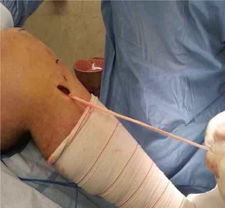
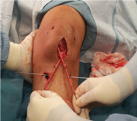
Figure 1: Percutaneous removal of ipsilateral semitendinosus and gracilis tendons.
View Figure 1
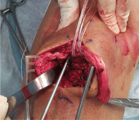
Clinically they presented rupture of the patellar tendon. A knee x-ray was performed to rule out bone lesions. They had emergency surgery, where the tendon injury was repaired with two suture anchors of 5 mm fixed on to the patella. With the sutures the patellar tendon was secured.
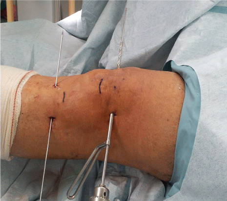
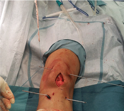
The repair was reinforced with semitendinosus and gracilis tendons, which were fed through percutaneous transosseous tunnels in patella and proximal tibia. We adjusted the patellar high with the knee flexed 30°, and then, in that position, the tendon was attached with an interference screw in the tibia. Postoperatively, the knee was immobilized by a plaster cast set at 20° flexion for two weeks. One week later they started with isometric quadriceps exercise. By the fourth week we proceeded with progressive movement, and by the sixth week they started to put weight on the leg.
.
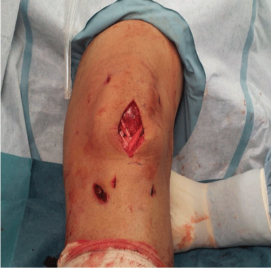
Figure 6: The end result of suture anchors and percutaneous reinforcement with semitendinosus and gracilis tendons.
View Figure 6
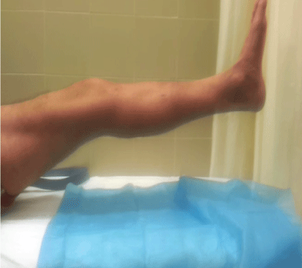
Six months after surgery, Lysholm score was 94 for one patient and 96 for the other. Functional recovery of the extensor unit of the knee was achieved and the patients returned to work and play sports (Figure 1, Figure 2, Figure 3, Figure 4, Figure 5, Figure 6 and Figure 7).
Discussion
Patellar tendon disruptions are relatively uncommon knee injuries compared to fractures, ligaments sprains, or meniscal tears [1]. These tears may be traumatic or they may occur spontaneously in patients with other underlying diseases such as diabetes mellitus, rheumatoid arthritis, steroid medication or kidney failure [2].
MRI can also be useful, by providing additional information such as the location of the rupture. We reported one case of neglected patellar disruption and the other case was traumatic. Several techniques have been used to relocate the patella to its anatomic position and repair the patellar tendon, but there is no widely accepted method [3-5].
Patellar tendon repair with suture anchors yields significantly better biomechanical results than repair with the commonly applied transosseous sutures [6].
We used a free semitendinosus and gracilis tendons [7,8], for reconstruction of the patellar tendon with an interference screw in tibia. Use of a contralateral patellar tendon [9] may result in additional damage to the uninjured leg, whereas the use of allografts [1] increases the risk of bacterial or viral infection. We performed reconstruction and restoration of ruptured patellar tendons using STG tendons without preserved distal insertions, without the use of any synthetic materials. Others authors use semitendinosus and gracilis tendons preserving distal insertions [10].
The treatment goals for ruptured patellar tendons include restoration of the quadriceps mechanism, restoration of the anatomic congruity of the patellofemoral joint to avoid chondral lesions, improved range of motion, and splinting of the patellar tendon to allow early mobilization.
Early surgical treatment can yield optimal clinical results, and repair or reinsertion is used, with or without cerclage reinforcement. The use of reinforcement with semitendinosus and gracilis tendons should be the first therapeutic choice [11].
Conclusion
The use of semitendinosus and gracilis tendons with suture anchors facilitates good functional recovery in patients with patellar tendon rupture.
The technique is relatively simple and feasible. This technique uses an easy to harvest graft which has low donor site morbidity. Additionally, the strength of the graft allows early rehabilitation, and no further surgery for hardware removal is necessary. This method should be the first option for this type of surgical techniques.
References
-
Labib SA, Wilczynski MC, Sweitzer BA (2010) Two-layer repair of a chronic patellar tendon rupture: a novel technique and literature review. Am J Orthop (Belle Mead NJ) 39: 277-282.
-
Takata Y, Nakase J, Numata H, Oshima T, Tsuchiya H (2015) Repair and augmentation of a spontaneous patellar tendon rupture in a patient with Ehlers-Danlos syndrome: a case report. Arch Orthop Trauma Surg 135: 639-644.
-
Greis PE, Holmstrom MC, Lahav A (2005) Surgical treatment options for patella tendon rupture. Part I, Acute. Orthop 28: 672-679.
-
Bushnell BD, Tennant JN, Rubright JH, Creighton RA (2008) Repair of patellar tendon rupture using suture anchors. J Knee Surg 21: 122-129.
-
Capiola D, Re L (2007) Repair of patellar tendon rupture with suture anchors. Arthroscopy 23: 906.
-
Ettinger M, Dratzidis A, Hurschler C, Brand S, et al. (2013) Biomechanical properties of suture anchor repair compared with transosseous sutures in patellar tendon ruptures: a cadaveric study. Am J Sports Med Nov 41: 2540-2544.
-
Van der Bracht H, Verdonk R, Stuyts B (2009) Augmentation of a patellar tendon repair with an autologous semitendinosus graft. Acta Orthop Belg 75: 417-419.
-
Nguene-Nyemb AG, Huten D, Ropars M (2011) Chronic patellar tendon rupture reconstruction with a semitendinosus autograft. Orthop Traumatol Surg Res 97: 447-450.
-
Milankov MZ, Miljkovic N, Stankovic M (2007) Reconstruction of chronic patellar tendon rupture with contralateral BTB autograft: a case report. Knee Surg Sports Traumatol Arthrosc 15: 1445-1448.
-
Takazawa Y, Ikeda H, Ishijima M, Kubota M, Saita Y, et al. (2013) Reconstruction of a ruptured patellar tendon using ipsilateral semitendinosus and gracilis tendons with preserved distal insertions: two case reports. BMC Res Notes 6: 361.
-
Sundararajan SR, Srikanth KP, Rajasekaran S (2013) Neglected patellar tendon ruptures: a simple modified reconstruction using hamstrings tendon graft. Int Orthop 37: 2159-2164.
