

Clinical Medical
Reviews and Case Reports
Is Cock-up Splint the Right Choice for All of the Carpal Tunnel Syndrome Patients? A Case Report
Mahdi Hadidi1, Mohammad-Reza Hadian2,3*, Ali Arab Kheradmand4, Mahmood Farzan5, Hamidreza Monsef6, Maryam-Raheleh Dadras7, Saeed Talebian2 and Gholam Reza Olyaei2
1School of Rehabilitation, Tehran University of Medical Sciences (TUMS), Iran
2Professor, Faculty of Rehabilitation, Tehran University of Medical Sciences (TUMS), Iran
3Professor, Brain and Spinal Research center (BASIR, Imam Hospital), Tehran University of Medical Sciences (TUMS, TUMS-IC), Iran
4Associate Professor, surgeon, Imam Hospital, Tehran University of Medical Sciences (TUMS), Iran
5Professor, orthopedic surgeon, Imam Hospital, Tehran University of Medical Sciences (TUMS), Iran
6Associate Professor, Faculty of Pharmacy, Tehran University of Medical Sciences (TUMS), Iran
7Medical Physics, International Affairs, Iran University of Medical Sciences (IUMS), Iran
*Corresponding author: Dr. Mohammad-Reza Hadian, Professor, Faculty of Rehabilitation, Brain and Spinal Injury Research center (BASIR, Imam Hospital), Tehran University of Medical Sciences (TUMS, TUMS-IC), Tehran, Iran, Tel: +9821-77536134, Fax: +9821-77534133, E-mail: hadianrs@sina.tums.ac.ir, hadian_ras@yahoo.com
Clin Med Rev Case Rep, CMRCR-3-083, (Volume 3, Issue 1), Case Report; ISSN: 2378-3656
Received: December 02, 2015 | Accepted: January 09, 2016 | Published: January 12, 2016
Citation: Hadidi M, Mohammad-Reza H, Kheradmand AA, Farzan M, Monsef H, et al. (2016) Is Cock-up Splint the Right Choice for All of the Carpal Tunnel Syndrome Patients? A Case Report. Clin Med Rev Case Rep 3:083. 10.23937/2378-3656/1410083
Copyright: © 2016 Hadidi M, et al. This is an open-access article distributed under the terms of the Creative Commons Attribution License, which permits unrestricted use, distribution, and reproduction in any medium, provided the original author and source are credited.
Abstract
Introduction: Splinting the hand is a common treatment strategy for the patients with carpal tunnel syndrome.
Case presentation: A fifty-year-old woman was the exceptional one who her clinical signs and symptoms was deteriorated after the administration of splinting. She showed deterioration of clinical and electrophysiological parameters after two weeks of using splint combined with steroid [1]. After removing the splint, the patient showed relief of subsequent signs and symptoms.
Conclusion: It seems that some cautions should be considered in prescribing splints for CTS patients. We have discussed the clinical presentation, possible causes, and management of the patient and a brief review of literature were also presented.
Keywords
Splinting, Carpal Tunnel Syndrome (CTS), Iontophoresis
Introduction
Carpal Tunnel Syndrome (CTS) is the most common entrapment neuropathy of the upper limb [2]. It is more common in women than men and is seen in patients between 40-60 years old [3]. Entrapment of the median nerve within the carpal tunnel is secondary to the compression of the nerve between the transverse carpal ligament (flexor retinaculum) superiorly and the flexor tendons and carpal bones inferiorly [4].
Several treatment options either surgical and or non-surgical have been suggested for relieving the pressure on the nerve. The non-surgical methods have frequently recommended for patients with mild to moderate symptoms and include splinting the wrist in neutral position, physical therapy and medication [2,5]. However, in severe condition of CTS, conservative treatments seem to be less beneficial. Splinting, iontophoresis and phonophoresis are among the non-surgical treatments that have been recommended separately or in combination for CTS patients. Very few researches have focused on the efficacy of combined treatments for CTS patients [1,6-8].
Some previous researches have shown clinical improvements following splinting in spite of non-significant changes in electrophysiological parameters [7,8]. In this report, evidences from one woman who her clinical and electrophysiological signs and symptoms was deteriorated after the application of the splint and iontophoresis will be discussed.
Case Description
A fifty-year-old woman was referred to the Rehabilitation clinic of Tehran University of Medical Science for treatment of idiopathic bilateral CTS. She was out of fourteen women who were referred for CTS treatments in a clinical trial. Diagnosis of CTS was determined based on the clinical signs and symptoms and also electrophysiological findings by surgeons (hand specialist). Exclusion and inclusion Criteria such as Rheumatoid arthritis, previous fracture and dislocation of wrist, diabetes, Hyperthyroidism, myxedema, any wrist operation and severe CTS were considered. In other word, no patient was entered to the main study with the above mentioned pathology and illness and therefore, the same criteria were also regarded for this case.
The onset of signs and symptoms was about 20 years earlier with on and off periods. Her chief complaints were pain, numbness and sensory disturbances of both hands (i.e. positive Phalen & Tinel's signs). The symptoms were awakening her during the night. Furthermore, pain and paranesthesia exacerbated with constant work and gripping activities; and decreased after shaking the hands.
Various parameters of Functional Status Scale during fine tasks such as writing, unscrew the bottle, holding the phone, self-wash and bathing were deteriorated. Furthermore, Symptom Severity Scale parameters such as awakening pain, sleep disorder due to paresthesia and pain, severity of pain during the day time, repletion of pain and sensory disturbances were also increased after the splint administration.
Median nerve sensory and motor action potentials recordings were performed as following
For Median sensory study, recording electrodes were placed in the middle finger and the reference was between the recording electrodes (2.5-3 cm distance). Stimulating electrodes were placed in 14 cm distance from the recording electrodes (i.e. 3 cm above the distal crease). For Median motor distance measurements, recording electrodes were placed on abductor policis brevice and the stimulating electrodes were in 8 cm distance and 3 cm above the elbow for distal and proximal latency measurements respectively [9].
For ulnar sensory study, the recording electrodes were placed in the fifth finger and the reference was in the distal phalanx and the reference was between the recording electrodes (2.5-3 cm distance). Stimulating electrodes were placed in 14 cm distance from the recording electrodes. For motor measurements, recording electrodes were placed on hypothenar eminence and the stimulating electrodes were in 8 cm distance in wrist and 4 cm distal to medial epicondyle for distal and proximal motor latencies, respectively [9].
F response could be recorded following supramaximal stimulation of motor branch for nerve conduction study. In Median and Ulnar nerves, the F response (wave) could easily be recorded by stimulation of the distal parts of the nerves; the parameters of recording similar to motor nerve study with exception of sweep speed and amplitude (i.e. sweep speed, more than 10 m. seconds; sensitivity, μV200 respectively).
Electrophysiological parameters such as sensory and motor latencies were compatible with the clinical signs & symptoms and suggested the mild and moderate involvements of left and right Median nerves respectively (Table 1).
![]()
Table 1: Nerve conduction values of the subject before, in the middle of treatment, after treatment and follow up.
View Table 1
The patient was provided with Cock-up splints and recommended to wear them during the sleep time and whenever possible during the day time (i.e. four weeks from the beginning of treatment). Iontophoresis of dexamethasone sodium phosphate was also administered during the first two weeks of treatment for ten sessions. Clinical and electrophysiological assessments were performed four times (before treatment, after fifth treatment session, after tenth treatment session and after two weeks from the last session as follow up).
All of the Nerve Conduction Study (NCS) procedures were performed in accordance with guidelines for measurement, precautions and electrode placement [9].
Besides, clinical evaluation of signs and symptoms were performed with Levin and Simmon's carpal tunnel (median nerve) function disability form [3]. It consists of two separate parts:
1. Symptom Severity Scale (SSS) comprise of eleven questions.
2. Functional Status Scale (FSS) comprise of eight questions. The overall score for each scale was calculated as the mean.
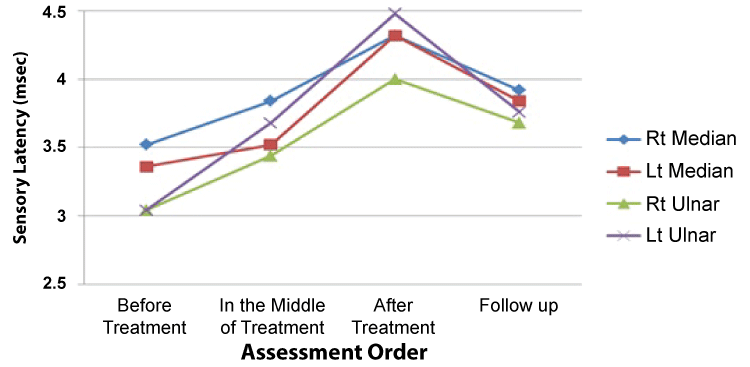
One week after the beginning of treatment, clinical signs and symptoms and electrophysiological tests were worsened. At this stage, the splint was replaced with a more comfortable one based on the assumption that the prior one was inappropriate. After second week, she showed deterioration of signs and symptoms again. At this stage, we decided to stop using the splint and planned to follow up the patient for two more weeks. The modification of treatment was fruitful and helped the patient to return to the initial state (Figure 1).
.
Figure 1: Changes of Median and Ulnar distal sensory latencies during four assessment sessions for both hands.
View Figure 1
Conclusion
Splinting is known as one of the most effective non-surgical treatment means for CTS patients [7]. Application of splints to immobilize the wrist in neutral position often helps to relieve the signs and symptoms by keeping the pressure in the carpal canal at the lowest level [5,8].
Although, research had suggested that the therapeutic effects of wrist splinting is due to keep the carpal tunnel pressure on the median nerve at the minimum level [8]. However a report by Rempel et al. had suggested higher carpal tunnel pressure with splint application at rest and also during repetitive hand activities, perhaps suggesting some external compression. They offered some cautions when applying splints for these patients and suggested removing the splint as soon as the inflammation and pain were reduced [10]. In addition, a review had suggested that if a reduction in symptoms was not occurred following the initial splinting, further evaluation should be conducted and splinting should be discontinued [5].
CTS is a classic example of chronic compression-ischemic neuropathy [4,11]. According to Sunderland, the two major categories of compression nerve injury are the acute injury with immediate effects and the chronic injury with delayed and gradual effects on the compressed nerve. Both of them can be due to some external force from outside the body and/or from a source within the body. When the nerve is gradually compressed, as in the development of chronic lesions, a vascular component is inevitably introduced due to the impairment of the blood flow to the nerve [11]. From the clinical point of view, the rapid relief of symptoms which frequently occurs after carpal tunnel release suggests an ischemic component [2,4]. A common feature of pain in this compression lesion is the nocturnal nature of the attacks. It would appear that muscular inactivity during sleep results in venous congestion around the nerve. As a result, the nerve fibers respond at a lower threshold to ischemia and causes pain. Movement and exercise of the limb improves venous return, decreases the pressure and relieves the pain [11]. Correspondingly, Luchetti et al. (1994) showed that intra-carpal tunnel pressure of the patients always exceeded the critical pressure of 30 mmHg at night and reached to the highest values at 6 a.m. slightly lower pressures were found when the wrist was splinted, but the difference was not significant, nor were critical pressure levels prevented by splinting [12].
The present case became worse clinically and electrophysiologically after application of the splints. This event may be due to the increase of carpal canal pressure with immobilization. She was an artist and used to paint a lot. As the patient herself reported, the onset of problem was about 20 years ago. It was hypothesized that she had thickened synovial lining of tendons that put mechanical pressures within the carpal tunnel [12]. Immobilizing the wrist with the splint might have disturbed the blood circulation by restricting hand movements. Furthermore, we found that both Ulnar and Median nerves electrophysiological parameters deteriorated bilaterally (almost equally). If the source of problem was just an increase of internal pressure within the carpal canal, then it was only expected the increased latency of Median nerve. Therefore, an external factor might be responsible for conduction blocks of both nerves. Considering motor conduction velocity measurements, an existence of external pressure due to tight straps or inappropriate splints might be suggested as the cause of deterioration of symptoms. At this stage, we decided to replace the splints with a more convenient pair and instructed the patient not to extremely tighten the straps. After a week, we tested the nerves again, but unfortunately the exacerbation continued, thus, we stopped using the splints. Patient was followed after two weeks and it was observed that she was gradually returning to the initial state.
Continuation of splints application resulted in the subsequent worsening of signs and symptoms in both median nerves. This might suggest the role of increased intrafunicular pressure, edema and to some extent obstruction of the arterial flow.
Nowadays, many CTS patients take the benefits of application of splints; however, there are some conditions that require more attention and caution. Perhaps the splint is not the right choice for all of the patients and some prerequisites should be defined (e.g. duration of symptoms). Therefore, in addition to prescribing the splint, providing the patients with necessary instructions has to be a part of the treatment protocol.
Acknowledgment
This project was supported by a grant from the Postgraduate Studies and Research Program, Tehran University of Medical Sciences, Tehran, Iran. The authors would like to acknowledge the generous assistance of the staff of clinic in the Faculty of Rehabilitation and Imam hospital (Tehran University of Medical Sciences, TUMS).
References
-
Aygul R, Ulvi H, Karatay S, Deniz O, Varoglu AO (2005) Determination of sensitive electrophysiologic parameters at follow-up of different steroid treatments of carpal tunnel syndrome. J Clin Neurophysiol 22: 222-230.
-
Donatelli R, Wooden MJ (2001) Orthopaedic physical therapy. Churchill Livingstone, Philadelphia.
-
Magee DJ (2013) Orthopedic physical assessment. Elsevier Health Sciences.
-
Werner RA, Andary M (2002) Carpal tunnel syndrome: pathophysiology and clinical neurophysiology. Clin Neurophysiol 113: 1373-1381.
-
Zimmerman GR (1994) Carpal tunnel syndrome. J Athl Train 29: 22-30.
-
Gurcay E, Unlu E, Gurcay AG, Tuncay R, Cakci A (2012) Assessment of phonophoresis and iontophoresis in the treatment of carpal tunnel syndrome: a randomized controlled trial. Rheumatol Int 32: 717-722.
-
Papez BJ, Turk Z (2004) Clinical versus electrodiagnostic effectiveness of splinting in the conservative treatment of carpal-tunnel syndrome. Wien Klin Wochenschr 2: 24-27.
-
Walker WC, Metzler M, Cifu DX, Swartz Z (2000) Neutral wrist splinting in carpal tunnel syndrome: a comparison of night-only versus full-time wear instructions. Arch Phys Med Rehabil 81: 424-429.
-
Oh SJ (2003) Clinical electromyography: nerve conduction studies. Lippincott Williams & Wilkins.
-
Rempel D, Manojlovic R, Levinsohn DG, Bloom T, Gordon L (1994) The effect of wearing a flexible wrist splint on carpal tunnel pressure during repetitive hand activity. J Hand Surg Am 19: 106-110.
-
Sunderland S (1991) Nerve injuries and their repair. Churchill Livingstone. New York.
-
Luchetti R, Schoenhuber R, Alfarano M, Deluca S, De Cicco G, et al. (1994) Serial overnight recordings of intracarpal canal pressure in carpal tunnel syndrome patients with and without wrist splinting. J Hand Surg Br 19: 35-37.
