

Clinical Medical
Reviews and Case Reports
Coronary Artery Imaging in Patients with Congenital Heart Disease: Improved Image Quality Using an Intravascular Contrast Agent and Specific Magnetic Resonance Sequence Design
Nathalie Dedieu1#*, Dirk Lossnitzer2#, Marcus R Makowski3, Miguel Silva Nodgueira Vieira1, Tarique Hussain1, James Wong1, Reza Razavi1, Rene M Botnar1 and Gerald F Greil1
1Division of Imaging Sciences and Biomedical Engineering, King’s College London, United Kingdom
2Department of Cardiology, University Hospital Heidelberg, Heidelberg, Germany
3Department of Radiology, Charite - Universitätsmedizin, Chariteplatz, Germany
#The first two authors contributed equally to this manuscript
*Corresponding author: Nathalie Dedieu, MD, Division of Imaging Sciences & Biomedical Engineering, The Rayne Institute, King's College London, 4th Floor, Lambeth Wing St. Thomas' Hospital, London SE1 7EH, United Kingdom, Tel: +44-0-20 7188 5444, Fax: +44-0-20 718 85442, E-mail: nathaliededieu@hotmail.com
Clin Med Rev Case Rep, CMRCR-3-092, (Volume 3, Issue 2), Original Research; ISSN: 2378-3656
Received: January 05, 2016 | Accepted: February 27, 2016 | Published: February 29, 2016
Citation: Dedieu N, Lossnitzer D, Makowski MR, Vieira MSN, Hussain T, et al. (2016) Coronary Artery Imaging in Patients with Congenital Heart Disease: Improved Image Quality Using an Intravascular Contrast Agent and Specific Magnetic Resonance Sequence Design. Clin Med Rev Case Rep 3:092. 10.23937/2378-3656/1410092
Copyright: © 2016 Dedieu N, et al. This is an open-access article distributed under the terms of the Creative Commons Attribution License, which permits unrestricted use, distribution, and reproduction in any medium, provided the original author and source are credited.
Abstract
Background: In patients with congenital heart disease (CHD) imaging of coronary artery origin and course can be crucial for preoperative planning. A novel intravascular contrast agent Gadofosveset trisodium (GdT) has demonstrated to be superior for angiography due to improved intravascular contrast compared to the currently used extravascular contrast agent Gadopentetate dimeglumine (GdD).
The aim of this study was to compare conventional post-contrast coronary magnetic resonance angiography (CMRA) using GdT or GdD to an optimized imaging sequence combined using only the novel agent, GdD.
Methods: Ten patients with CHD (age range 22 to 40 years; mean 31 years) were scanned at a 1.5 T clinical MR scanner (Achieva, Philips Healthcare, Best, The Netherlands) using a 32-element cardiac coil. An extravascular contrast agent (GdD) was administrated first using a standard T2-prepared SSFP sequence. Within 72 hours patients were re-scanned using an intravascular contrast agent (GdT) and IR SSFP additional to the standard T2-prepared SSFP MR Sequence. Image quality was graded form 0 (non-diagnostic) to 4 (best image quality). The left anterior descending (LAD), the left circumflex (LCX) and the right coronary artery system (RCA) were assessed separately. Contrast-to-noise ratio (CNR), Signal-to-noise ratio (SNR), blood-to-myocardial contrast (BMC) and image quality were analyzed for the three techniques. A p-value ≤ 0.05 indicated statistical significance.
Results: The intravascular contrast agent GdT showed significantly improved CNR and BMC when combined with an optimized IR SSFP sequence. There was a trend towards being able to image longer segments of the LAD and RCA and towards improved vessel sharpness but the level of significance was not reached. As expected, coronary artery diameter and image quality, were not significantly different since we imaged the same individuals.
Conclusion: The combination of a novel intravascular contrast agent (GdT) and the addition of an adapted inversion recovery whole heart sequence design seems to provide improved contrast of the coronary artery system compared to currently available extravascular contrast agents and conventional sequence design.
Keywords
Congenital heart disease, Coronary artery angiography, Whole heart imaging, Intravascular contrast agent, Inversion recovery steady-state-free-precession
Introduction
Major improvements in the last decades in CHD diagnosis and treatment have led to an increased patient survival up to approximately 85% to adulthood [1,2]. Consequently, with this continuously growing and aging population, physicians are facing new diagnostic challenges. Knowledge about coronary anatomy can be essential for long-term follow-up of these patients. Examples are patients after surgical repair of transposition of the great arteries [3,4] and patients requiring a pulmonary homograft or transcatheter valve implantation to exclude abnormal course of the coronaries [1,5-8]. Imaging of coronary arteries with echocardiography is challenging in young children and becomes extremely difficult in older patients due to poorer acoustic windows. Catheter-based angiography is invasive, costly and entails radiation, which carries the risk of cancer especially in patients who need regular follow-up exams including cardiac imaging [3,4,9]. Therefore, MRI as a radiation free non-invasive diagnostics tool represents a great advantage for these patients. Classically the standard MR angiography (MRA) for assessment of arterial and venous anomalies in congenital heart disease is performed using a 3-dimensional (3D) single- or multiphase first-pass MRA sequence with an extracellular contrast agent without cardiac gating during at least one breath hold. Unfortunately this entails limited spatial resolution related to the limited capacity of breath holding of sick patients or limited compliance in young children or infants. Moreover, this approach generally does not include ECG gated sequences or respiratory motion compensation and is therefore quite prone to artifacts.
Whole heart coronary MR angiography (CMRA) has been previously used in adults and children with congenital heart disease [2,7,10]. This free-breathing high spatial resolution three-dimensional sequence allows acquisition of extensive cardiac data via a navigator-gated ECG-triggered T2-prepared SSFP sequence. Good diagnostic accuracy (up to 84% according to Tangcharoen et al.) [7,11] and extensive details on cardiac anatomy, volumes as well as function assessment, via a single and easy to apply sequence have been demonstrated [12-16].
The use of a conventional extravascular contrast agent like Gadopentetate dimeglumine (GdD) has been widely described in MR angiography [13,17]. However, first pass techniques carry limitations due to the fast diffusion of GdD into the extravascular space. For visualization of small structures such as coronary arteries, this technique would benefit from increased and longer blood pool contrast compared to the extravascular tissue or fluid [12,13,18].
The novel intravascular contrast agent, Gadofosveset trisodium (GdT) has the potential to increase intravascular contrast to improve image quality and therefore diagnostic performance for the coronary arteries [19].
Furthermore IR SSFP provides enhancement of contrast between tissues with different T1-relaxation times through the addition of a 180° inversion pre-pulse [20], with a determined tissue signal being specifically nulled by selecting an optimal inversion time. Originally this technique was developed and used to detect myocardial infarction [21,22] but it has since then been extended to other cardiac diagnosis i.e. cardiomyopathies, myocarditis, amyloidosis, Anderson-Fabrydisease etc.
The aim of this study was to compare conventional post-contrast CMRA using GdT or GdD to an optimized imaging sequence combined using only the novel agent, GdD.
Materials and Methods
The study was approved by the local ethics committee (Guy’s and St. Thomas' NHS Research Ethics Committee, London, England) and was registered with the United Kingdom Medicines and Healthcare Products Regulatory Agency.
Ten patients with CHD agreed to take part in the study (eight men, two women, age range 22 to 40 years, mean 31 years, table 1). Written consent was obtained and medical history was assessed in all patients. Only patients with a history of CHD who were scheduled for a routine MR imaging examination were included in this study. Patients with significant renal dysfunction were excluded (defined as estimated glomerular filtration rate [eGFR] < 30 ml/min/1.73 m2). All patients had follow up clinical appointments after the scan and at 1year post MRI.
![]()
Table 1: Patients characteristics.
View Table 1
MRI technique
Patients were scanned on 2 occasions at a 1.5 T clinical MR scanner (Achieva, Philips Healthcare, Best, The Netherlands) using a 32-element cardiac coil. During the first scan, gadopentetate dimeglumine (Magnevist®; Bayer Schering Pharma, Berlin, Germany) was administered at a dose of 0.2 mmol per kilogram of body weight (maximum volume, 40 mL) and patients were scanned using a standard commercially available respiratory navigator–gated and ECG-triggered T2-prepared SSFP sequence. During the second scan (within 72 h), a dose of 0.03 mmolper kilogram of body weight gadofosveset trisodium (Vasovist®; Bayer Schering Pharma, Berlin, Germany) was injected and all patients were re-scanned using a IR preparation- pre-pulse SSFP sequence additional to the standard T2-prepared SSFP MR sequence.
The optimal inversion time to suppress extravascular tissue was determined by a Look-Locker sequence [1,2,23]. In this study, it was used to determine the optimal inversion time (approximately 260-280 msec) to minimize signal from extravascular tissue (Table 2).
![]()
Table 2: MR imaging parameters for contrast-enhanced 3D sequences.
View Table 2
Images analysis
Image processing, reformatting and analysis were performed with commercially available software (View Forum, Philips Healthcare, Best, The Netherlands), as well as a specialised custom-made coronary analysis software ("Soap bubble") [1-8,10]. Two experienced investigators analysed the images independently (DL with 8 years and ND with more than 2 years’ experience in cardiac MRI). Consensus reading was performed in case of disagreement.
Signal to noise and contrast to noise
For quantitative image analysis, regions of interest (ROIs) were defined in areas of myocardium, the intra-aortic blood-pool close to coronary ostia, and in a region anterior to the chest wall, where no respiration-induced motion artifacts were visually identified. Signal-to-noise (SNR) was defined as the mean signal intensity found in a ROI divided by the standard deviation found in the ROI anterior to the chest wall [3,4,9] (Equation 1).
SNR was evaluated using the following formula:
SMean in [1] denotes the mean signal intensity in the user defined region of interest and SDEVMean relates to the standard deviation of the mean of the signal intensity anterior to the chest (parallel imaging was used: sensitivity encoding for fast MR imaging [Philips specific]) [2,5-8,10].
At the same anatomical level, SNR was also quantified in the blood-pool of ascending aorta. SNR of the muscle signal was determined in the muscle of the LV anterolateral wall at the level of proximal RCA.
Contrast to noise ratio (CNR) was defined as the difference of the mean signal intensities in two user specified ROIs divided by the standard deviation found in the ROI anterior to the chest wall [4,7,9,11] (Equation 2). The CNR between blood and muscle was defined as
Blood to myocardium contrast: BMC
BMC was defined as the mean signal intensity of the blood divided by the mean intensity of the myocardium in the previously described chosen ROIs.
Vessel sharpness, length and diameter measurements
For the quantitative image analysis of coronaries vessel wall sharpness, length and area a custom-made analyzing tool ("Soap Bubble") was used [2,7,10,12-14]. As previously described by Botnar et al. [10,13,17,24] vessel sharpness can be obtain using a Deriche algorithm [7,11-13,18]. Briefly, this algorithm allows the calculation of an edge image by using a first-order derivative of the input image; the local value in a Deriche image, which represents the magnitude of local change in signal intensity. A vessel sharpness of 100% refers to a maximum signal intensity change at the vessel border. A lower edge value is consistent with inferior vessel sharpness. For the identification of the vessel edges along the path, a semiautomatic vessel tracking algorithm is used.
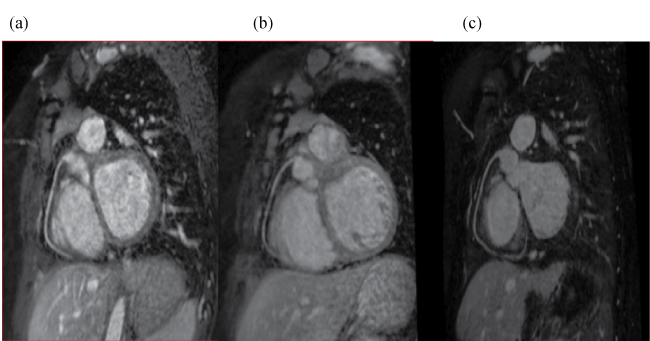
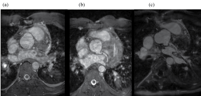
Each one of the main coronary arteries (LAD, LCX and RCA) were analysed for the use of the extracellular (T2-prepared SSFP) contrast agent and intracellular (T2-prepared SSFP and IR-SSFP) contrast agent (Figure S1 and Figure S2).
.
Figure S1: Representative reformatted images of the RCA in the same patient using GdD with a T2-prepared SSFP sequence (a), GdT with a T2-prepared SSFP (b) and GdT with IRSSFP (c).
View Figure S1
.
Figure S2: Representative reformatted images of the LCX in the same patient using GdD with a T2-prepared SSFP sequence (a), GdT with a T2-prepared SSFP (b) and GdT with IRSSFP (c).
View Figure S2
Visual scoring of the quality of the coronary images
For qualitative image analysis, after coronary reformat using Soap Bubble software, image quality was graded form 0 (non-diagnostic) to 4 (best image quality) [12-14,19] for each vessel of the main coronary system using GdD (T2-prepared SSFP) and GdT (T2-prepared SSFP and IR-SSFP) -image quality assessment (from McConnell et al.) [13,17,20] (Table 3).
![]()
Table 3: Image Quality Assessment (from McConnell [10,13]).
View Table 3
Statistical analysis
For statistical comparison, non-parametric methods using statistics software (IBM SPSS Statistics Version 20) were applied. In light of the small sample size we checked the preconditions for parametric testing, using Kolmogorov-Smirnov tests. Since variables were not normally distributed, we conducted the non-parametric Wilcoxon tests for dependent variables. A p-value ≤ 0.05 was considered to indicate statistical significance.
Results
Ten patients completed the study protocol (Table 1: patient characteristics) without any complications. All indications for MRI were part of the clinical follow-up, and all patients agreed to undergo a second scan for research purposes. One of these patients had an abnormal origin of the LCX. None of them experienced any side effects of the MRI scan as well as contrast agent administration.
Quantitative analysis
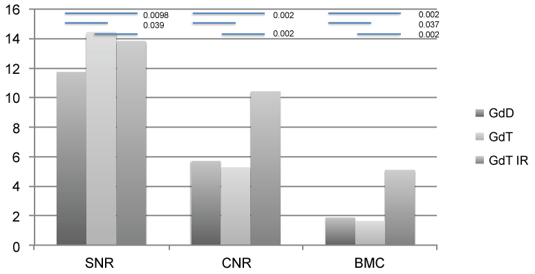
SNR, CNR and BMC: The use of IR SSFP with GdT resulted in significantly improved CNR and blood to myocardial contrast (BMC) when compared with the conventional T2-prepared SSFP sequence using GdT (p = 0.002 for CNR and BMC) as well as GdD (p = 0.002 for CNR and BMC). Furthermore GdT improved SNR significantly using T2-preparedSSFP (p = 0.039) and IR SSFP (p = 0.0098) when compared to GdD (Figure 1).
.
Figure 1: Graph showing qualitative analysis of the images with SNR, CNR and BMC displayed for the different sequences (T2-prepared SSFP for GdD, GdT and IR SSFP GdT), p-values are indicated above the column graphs .p-values only mentioned when significant.
View Figure 1
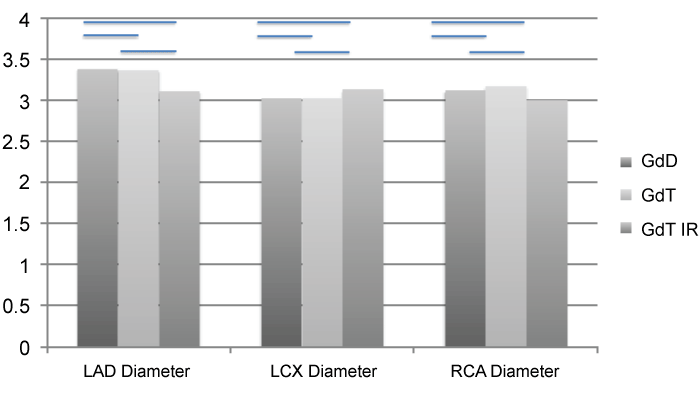
Vessel length and vessel diameter: We did not find any statistically significant difference in vessel diameter when comparing the two contrast agents and three different imaging protocols. This indicates that, as expected, neither the contrast agent nor the sequences let to a difference in vessel diameter within each of the scanned individuals (Figure 2).
.
Figure 2: Graph showing vessel diameter for each of the coronary artery and each of the contrast agent and sequence combination.
View Figure 2
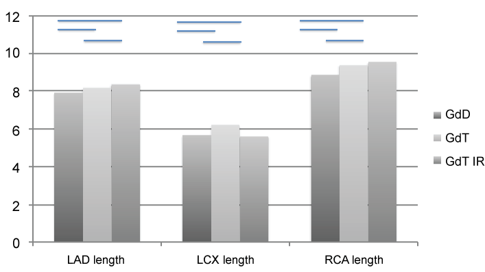
Despite not reaching statistical significance, there was a trend towards increased length of LAD or RCA imaged when using IR SSFP with GdT (Figure 3).
.
Figure 3: Graph showing vessel length for each of the coronary arteries and each of the contrast agent and sequence combination.
View Figure 3
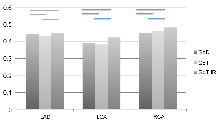
Vessel sharpness: Mean vessel sharpness was measured (GdD T2-prepared SSFP, GdT T2-prepared SSFP, GdT IRSSFP respectively) for LAD (0.44, 0.43 and 0.45), LCX (0.39, 0.42 and 0.48) and RCA (0.46, 0.46 and 0.48). Once again these results seem to indicate a trend in favour of IRSSFP with GdT for the LAD and RCA compared to the conventional T2-prepared SSFP sequence but did not reach statistical significance (Figure 4).
.
Figure 4: Graph showing vessel sharpness for each of the coronary artery and each of the contrast agent and sequence combination.
View Figure 4
Qualitative image analysis
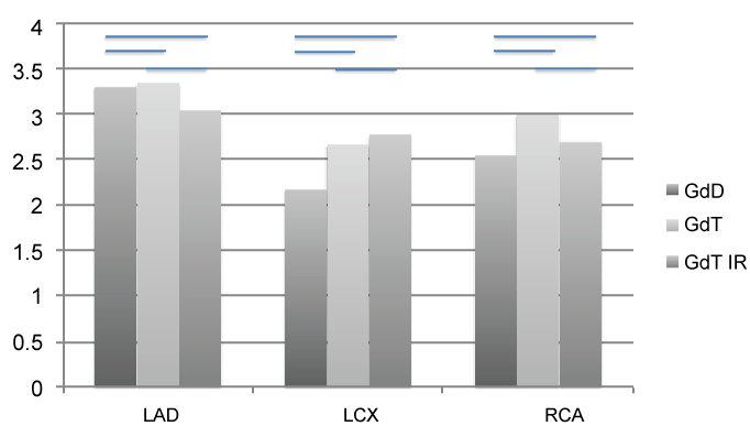
Image quality was scored for the RCA, left main, LAD, and LCX artery according to McConnell et al. [12,13,18,21,22] as stated in table 3.Mean image scoring (GdD T2 prepared SSFP, GdT T2-prepared SSFP, GdT IR SSFP respectively) was for LAD (3.3, 3.35 and 3.05) for LCX (1.95, 3.0 and 2.7) and for RCA (2.3, 3.2 and 2.7) respectively and did not reach statistical significance (Figure 5).
.
Figure 5: Image rating score for each of the coronary artery and each of the contrast agent and sequence combination in accordance with table 3.
View Figure 5
Discussion
The present study evaluated the impact of an intravascular contrast agent on whole-heart MR angiography for characterization of origin and course of coronary arteries as well as the identification of possible coronary abnormalities. Additionally IR SSFP in combination with GdT was evaluated. Whole heart MR angiography enables visualization of the majority of cardiovascular structures of the thorax, assessing simultaneously venous and arterial anomalies including the coronary artery system with a free breathing acquisition [1,12,19]. If the data are acquired isotropic, the complete coronary artery system can be reformatted from a single whole heart dataset by post-processing software applications. Currently, a whole heart T2-prepared SSFP sequence is the most commonly used approach [3,20,25-28].
Unfortunately, some of the disadvantages of the whole-heart CMRA are the relatively long acquisition time; the dependence of an adequate navigator efficiency to compensate for breathing motion artifacts and the reduced contrast between vascular structures and surrounding tissues or fluids [1,5,7,8,12,21,22]. In that context, intravascular contrast agents may provide a significant advantage compared to extravascular contrast agents. Since GdD is an extravascular contrast agent its binding capacity to albumin is relatively low. On the contrary, GdT has a non-covalent, transient and reversible but much stronger binding to albumin, that results in a prolonged intravascular enhancement [3,9,23,29-32].
Thus, GdT’s non-covalent binding to albumin not only contributes to slower renal excretion but also increases T1 relaxivity up to 6 to 10 times when compared to GdD [7,10,32-35]. The intravascular contrast staying in the blood pool for a longer amount of time allows characterization of all cardiac segments and structures, including arterial and venous phases, with a reduced requirement for precise timing of the contrast bolus. Recently published data has shown that intravascular agents are superior in defining anatomical structures in patients with congenital heart disease [24,36].
Although, Raman et al. demonstrated in a recent study that GdT was associated with an increase in CNR (24%) between arteries and myocardium as well as an improvement in image quality when compared to GdD, the differences between the 2 groups were not as significant [12,14,37]. It is worth mentioning that this study was conducted on a 3T scanner while our study was performed on a 1.5T scanner. However, these results are similar to the results published by Wagner et al. [38] where GdT whole heart MRA showed a slightly better image quality.
In our study CNR and BMC were significantly better for GdT using the IR SSFP sequence that allows a suppression of fluids and extravascular tissue in the presence of the short T1 values of the blood. As GdT provides short T1 values of the blood for several hours, CNR and BMC values increased significantly when combining GdT and IR-SSFP (Figure 1).
We would have also expected a higher image quality in terms of image rating for IR SSFP when compared to T2-prepared SSFP sequences [23,36]. In our study the LCX seemed to benefit most from higher intravascular contrast and the IR SSFP sequence (Figure 5). As image quality is mainly influenced by respiratory and cardiac motion as well as heart rate variability, but not intravascular contrast, techniques to suppress respiratory and cardiac motion will predominantly influence image quality. Respiratory motion remains a significant impediment in image quality and speed. The standard1D navigator used assumes a constant linear relationship between cardiac and diaphragmatic motion, which is incorrect and not only increases scan time but, on occasion, leads to residual motion artefacts. The recent introduction of 2D and 3D self-navigating result in an improved coronary artery imaging and shorter scanning times [12,18-20,39,40]. These new navigator techniques in combination with GdT and IR-SSFP might therefore offer a significant improvement in imaging the coronary artery system and need to be evaluated in future studies.
Regarding vessel length, we did not find a statistically significant difference between the two contrast agents. However when using GdT in combination with both IR SSFP and T2-preparedSSFP, the trend is favourable for the intravascular agent for the LAD and RCA. As expected, vessel diameter did not differ between the three groups since we compared the coronary arteries in the same individuals.
Vessel sharpness did not significantly improve with GdT. However, there was a tendency for better vessel wall sharpness of the GdT- IR-SSFP combination (Figure 4). Similar results were reported by Makowski et al. [21,22,36] with better identification and definition of the anatomy of the great vessel using this technique. It is very likely that we could not reach statistical significance due to the small sample size as well as the much smaller vessel size.
Conclusion
A single injection of an intravascular contrast agent in combination with an IR SSFP sequence showed significantly improved CNR and BMC. Although there was a trend towards being able to image longer segments of the LAD and RCA and towards improved vessel sharpness but the level of significance was not reached. Image quality was not significantly different, which could be addressed in the future with novel motion compensation techniques for cardiac and respiratory motion. Using this technique, also more vessel length of the LAD and RCA could be imaged with improved vessel sharpness for all coronaries, but level of significance was not reached.
Acknowledgements
This study has received funding by the Department of Health through the National Institute for Health Research (NIHR) comprehensive Biomedical Research Centre award to Guy's & St Thomas' NHS Foundation Trust in partnership with King's College London and King’s College Hospital NHS Foundation Trust. The Division of Imaging Sciences receives also support as the Centre of Excellence in Medical Engineering (funded by the Wellcome Trust and EPSRC; grant number (WT 088641/Z/09/Z) as well as the BHF Centre of Excellence (British Heart Foundation award RE/08/03). N. Dedieu is funded through a BHF grant (PG/12/5/29350).The magnetic resonance imaging scanner is partly supported by Philips Healthcare. The study was partly supported by Bayer Schering Pharma GmbH, Berlin, Germany. Otherwise there are no financial or other relations that could lead to a conflict of interest.
References
-
Warnes CA (2005) The adult with congenital heart disease: born to be bad? J Am Coll Cardiol 46: 1-8.
-
Etienne A, Botnar RM, Van Muiswinkel AM, Boesiger P, Manning WJ, et al. (2002) "Soap-Bubble" visualization and quantitative analysis of 3D coronary magnetic resonance angiograms. Magn Reson Med 48: 658-666.
-
Taylor AM, Dymarkowski S, Hamaekers P, Razavi R, Gewillig M, et al. (2005) MR Coronary Angiography and Late-Enhancement Myocardial MR in Children Who Underwent Arterial Switch Surgery for Transposition of Great Arteries1. Radiology 234: 542-547.
-
Arai AE, Epstein FH, Bove KE, Wolff SD (1999) Visualization of aortic valve leaflets using black blood MRI. J Magn Reson Imaging 10: 771-777.
-
Tangcharoen T, Jahnke C, Koehler U, Schnackenburg B, Klein C, et al. (2008) Impact of heart rate variability in patients with normal sinus rhythm on image quality in coronary magnetic angiography. J Magn Reson Imaging 28: 74-79.
-
Pruessmann KP, Weiger M, Scheidegger MB, Boesiger P (1999) SENSE: sensitivity encoding for fast MRI. Magn Reson Med 42: 952-962.
-
Tangcharoen T, Bell A, Hegde S, Hussain T, Beerbaum P, et al. (2011) Detection of Coronary Artery Anomalies in Infants and Young Children with Congenital Heart Disease by Using MR Imaging. Radiology 259: 240-247.
-
Khambadkone S, Coats L, Taylor A, Boudjemline Y, Derrick G, et al. (2005) Percutaneous pulmonary valve implantation in humans: results in 59 consecutive patients. Circulation 112: 1189-1197.
-
Coles DR, Smail MA, Negus IS, Wilde P, Oberhoff M, et al. (2006) Comparison of Radiation Doses From Multislice Computed Tomography Coronary Angiography and Conventional Diagnostic Angiography. Journal of the American College of Cardiology 47: 1840-1845.
-
Botnar RM, Stuber M, Danias PG, Kissinger KV, Manning WJ (1999) Improved coronary artery definition with T2-weighted, free-breathing, three-dimensional coronary MRA. Circulation 99: 3139-3148.
-
IEEE: 00028260. 2004: 1-5.
-
Sørensen TS, Körperich H, Greil GF, Eichhorn J, Barth P, et al. (2004) Operator-independent isotropic three-dimensional magnetic resonance imaging for morphology in congenital heart disease: a validation study. Circulation 110: 163-169.
-
McConnell MV, Khasgiwala VC, Savord BJ, Chen MH, Chuang ML, et al. (1997) Comparison of respiratory suppression methods and navigator locations for MR coronary angiography. AJR Am J Roentgenol 168: 1369-1375.
-
Beerbaum P, Sarikouch S, Laser K-T, Greil G, Burchert W, et al. (2009) Coronary anomalies assessed by whole-heart isotropic 3D magnetic resonance imaging for cardiac morphology in congenital heart disease. J Magn Reson Imaging 29: 320-327.
-
Uribe S, Hussain T, Valverde I, Tejos C, Irarrazaval P, et al. (2011) Congenital heart disease in children: coronary MR angiography during systole and diastole with dual cardiac phase whole-heart imaging. Radiology 260: 232-240.
-
Delgado JA, Abad P, Rascovsky S, Calvo V, Castrillon G, et al. (2014) Assessment of cardiac volumes using an isotropic whole-heart dual cardiac phase sequence in pediatric patients. J Magn Reson Imaging 39: 708-716.
-
Boxt LM, Rozenshtein A (2003) MR imaging of congenital heart disease. Magn Reson Imaging Clin N Am 11: 27-48.
-
Weber OM, Martin AJ, Higgins CB (2003) Whole-heart steady-state free precession coronary artery magnetic resonance angiography. Magn Reson Med 50: 1223-1228.
-
Fink C, Goyen M, Lotz J (2007) Magnetic resonance angiography with blood-pool contrast agents: future applications. Eur Radiol 17 Suppl 2: B38-44.
-
Simonetti OP, Kim RJ, Fieno DS, Hillenbrand HB, Wu E, et al. (2001) An improved MR imaging technique for the visualization of myocardial infarction. Radiology 218: 215-223.
-
Kim RJ, Wu E, Rafael A, Chen EL, Parker MA, et al. (2000) The use of contrast-enhanced magnetic resonance imaging to identify reversible myocardial dysfunction. N Engl J Med 343: 1445-1453.
-
Wagner A, Mahrholdt H, Holly TA, Elliott MD, Regenfus M, et al. (2003) Contrast-enhanced MRI and routine single photon emission computed tomography (SPECT) perfusion imaging for detection of subendocardial myocardial infarcts: an imaging study. Lancet 361: 374-379.
-
Look DC, Locker DR (1970) Time Saving in Measurement of NMR and EPR Relaxation Times. Rev Sci Instrum 41: 250.
-
Bunce NH, Lorenz CH, Keegan J, Lesser J, Reyes EM, et al. (2003) Coronary artery anomalies: assessment with free-breathing three-dimensional coronary MR angiography. Radiology 227: 201-208.
-
Deshpande VS, Shea SM, Laub G, Simonetti OP, Finn JP, et al. (2001) 3D magnetization-prepared true-FISP: a new technique for imaging coronary arteries. Magn Reson Med 46: 494-502.
-
Shea SM, Deshpande VS, Chung YC, Li D (2002) Three-dimensional true-FISP imaging of the coronary arteries: improved contrast with T2-preparation. J Magn Reson Imaging 15: 597-602.
-
Maintz D, Aepfelbacher FC, Kissinger KV, Botnar RM, Danias PG, et al. (2004) Coronary MR angiography: comparison of quantitative and qualitative data from four techniques. AJR Am J Roentgenol 182: 515-521.
-
Spuentrup E, Börnert P, Botnar RM, Groen JP, Manning WJ, et al. (2002) Navigator-gated free-breathing three-dimensional balanced fast field echo (TrueFISP) coronary magnetic resonance angiography. Invest Radiol 37: 637-642.
-
Bi X, Carr JC, Li D (2007) Whole-heart coronary magnetic resonance angiography at 3 Tesla in 5 minutes with slow infusion of Gd-BOPTA, a high-relaxivity clinical contrast agent. Magn Reson Med 58: 1-7.
-
Rohrer M, Bauer H, Mintorovitch J, Requardt M, Weinmann HJ (2005) Comparison of magnetic properties of MRI contrast media solutions at different magnetic field strengths. Invest Radiol 40: 715-724.
-
Bock M, Schulz J, Ueltzhoeffer S, Giesel F, Voth M, et al. (2008) Intravascular contrast agent T1 shortening: fast T1 relaxometry in a carotid volunteer study. MAGMA 21: 363-368.
-
Lauffer RB, Parmelee DJ, Dunham SU, Ouellet HS, Dolan RP, et al. (1998) MS-325: albumin-targeted contrast agent for MR angiography. Radiology 207: 529-538.
-
Caravan P (2009) Protein-targeted gadolinium-based magnetic resonance imaging (MRI) contrast agents: design and mechanism of action. Acc Chem Res 42: 851-862.
-
Nassenstein K, Waltering KU, Kelle S, Schlosser T, Breuckmann F, et al. (2008) Magnetic resonance coronary angiography with Vasovist: in-vivo T1 estimation to improve image quality of navigator and breath-hold techniques. Eur Radiol 18: 103-109.
-
Kelle S, Thouet T, Tangcharoen T, Nassenstein K, Chiribiri A, et al. (2007) Whole-heart coronary magnetic resonance angiography with MS-325 (Gadofosveset). Med Sci Monit 13: 469-474.
-
Makowski MR, Wiethoff AJ, Uribe S, Parish V, Botnar RM, et al. (2011) Congenital heart disease: cardiovascular MR imaging by using an intravascular blood pool contrast agent. Radiology 260: 680-688.
-
Raman FS, Nacif MS, Cater G, Gai N, Jones J, et al. (2013) 3.0-T whole-heart coronary magnetic resonance angiography: comparison of gadobenate dimeglumine and gadofosveset trisodium. Int J Cardiovasc Imaging 29: 1085-1094.
-
Wagner M, Rösler R, Lembcke A, Butler C, Dewey M, et al. (2011) Whole-heart coronary magnetic resonance angiography at 1.5 Tesla: does a blood-pool contrast agent improve diagnostic accuracy? Invest Radiol 46: 152-159.
-
Henningsson M, Koken P, Stehning C, Razavi R, Prieto C, et al. (2012) Whole-heart coronary MR angiography with 2D self-navigated image reconstruction. Magn Reson Med 67: 437-445.
-
Henningsson M, Prieto C, Chiribiri A, Vaillant G, Razavi R, et al. (2014) Whole-heart coronary MRA with 3D affine motion correction using 3D image-based navigation. Magn Reson Med 71: 173-181.
