

Clinical Medical
Reviews and Case Reports
The Use of the Gallbladder Wall for Reconstruction of the Extrahepatic Bile Ducts in a Case of Agenesis of the Common Bile Duct: A Case Report
Maher N Ghabriel*
Department of Surgery, St. Peter's Hospital, Heliopolis, Cairo, Egypt
*Corresponding author: Dr. Maher N Ghabriel, Consultant Surgeon, Department of Surgery, St. Peter’s Hospital, Heliopolis, Cairo, Egypt, Tel: +01223564812, E-mail: maherghabriel@hotmail.com
Clin Med Rev Case Rep, CMRCR-3-106, (Volume 3, Issue 5), Case Report; ISSN: 2378-3656
Received: April 11, 2016 | Accepted: May 14, 2016 | Published: May 16, 2016
Citation: Ghabriel MN (2016) The Use of the Gallbladder Wall for Reconstruction of the Extrahepatic Bile Ducts in a Case of Agenesis of the Common Bile Duct: A Case Report. Clin Med Rev Case Rep 3:106. 10.23937/2378-3656/1410106
Copyright: © 2016 Ghabriel MN. This is an open-access article distributed under the terms of the Creative Commons Attribution License, which permits unrestricted use, distribution, and reproduction in any medium, provided the original author and source are credited.
Abstract
Unexpected anomalies of the extrahepatic biliary ducts were found intraoperatively during open cholecystectomy in a 49-year-old male, with calcular obstructive jaundice. The patient had congenital absence of the common bile duct that appears to have been replaced by the cystic duct, which opened directly into the duodenum. The right and left hepatic ducts drained into the posterior wall of the gallbladder by a short common hepatic duct. Intraoperatively the gallbladder wall (with uncompromised nerve and blood supply) was used as a vital flap to reconstruct a tubular bridge across the defect in the biliary ducts, while retaining a functional sphincter of Oddi at the end of the original cystic duct. The patient showed uneventful recovery and had no further complaints at subsequent follow-up for nine years after surgery. Knowledge of such an anomaly is essential for hepatobiliary surgeons. Intraoperative assessment of the biliary ducts should be performed prior to excision of the gallbladder in order to allow repair of any anomalies in the duct system using the gallbladder wall while maintaining its cystic arterial supply.
Keywords
Choledochoplasty, Common bile duct agenesis, Hepaticocystic duct, Cholecystectomy, Cholangiography, Biliary surgery
Introduction
Biliary channels anomalies are well recognized [1]. Many such anomalies are often discovered during surgery and may become further complicated by the pathological changes in biliary diseases. The biliary system develops from a primary hepatic bud at the distal end of the embryonic foregut region. Further development of the hepatic bud into the ventral mesentery and septum transversum leads to the formation of the endoderm-derived liver cells and the cells lining the biliary system, while the mesoderm of the septum transversum provides the supporting walls of the biliary ducts [2]. Varying degrees of ductal abnormalities in the biliary system may arise during the successive division of the hepatic bud, and the pars cystica (which will form the gallbladder) as it buds off the pars hepatica. Thus familiarity with and anticipation of anomalies of this system are essential for successful surgical outcome. The most common anomalies are found in the cystic duct termination and in the intrahepatic ducts from the eight hepatic segments [3]. However, absence of the typical common bile duct (CBD) is considered to be very rare [1,4] and was not reported among biliary variations in a magnetic resonance cholangiographic study [3]. In the current case, a transversely positioned gallbladder, short common hepatic duct draining the left and right hepatic ducts opening in the posterior wall of the gallbladder, and an absence of the CBD are reported. The surgical procedure performed, using the gallbladder wall to reconstruct a bridge across the defect in the extrahepatic biliary tract is described.
Case Report
A 49-year-old male was admitted to hospital diagnosed with calcular obstructive jaundice. Abdominal ultrasound examination showed a 1.7 cm gallstone at the neck of a contracted gallbladder. The liver was of normal size and had a smooth surface with no focal lesions, but showed dilated intra-hepatic radicals suggestive of obstruction of the biliry ducts. Ultrasound also reported what appeared to be a CBD of normal diameter. Liver function tests showed raised alkaline phosphatase and direct serum bilirubin. Endoscopic retrograde cholangio-pancreatography (ERCP) and Magnetic resonance cholangio-pancreatography (MRCP) were not available at that time, and the patient was prepared for open cholecystectomy. Intraoperatively, the gallbladder was found to lie transversely, adherent to the liver at its fundus. Exploration revealed absence of the typical CBD, and that the cystic duct opened in the duodenum, replacing the typical CBD. During dissection of the gallbladder from the liver bed a hepatic duct that opened into the back of the gallbladder was inadvertently injured thus was temporarily loosely ligated and secured on a sling. Further exploration showed that the injured duct was a short common hepatic duct formed from a right and left hepatic ducts, and opened into the posterior wall of the gallbladder near its fundus. The gallbladder was opened to explore its cavity. A large gallstone was found and removed. The cystic duct patency was assessed through its full length to the duodenum. The gallbladder wall was fashioned to form a flap while retaining its continuity with the cystic duct and arterial and neural supply. A buttonhole opening was made into that flap and the long limb of a T-drainage tube was threaded through it. One of the short arms of the T-tube was inserted into the cystic duct while the other short arm inserted into the common hepatic duct after releasing the slinging suture. The gallbladder flap was wrapped round the transverse arm of the T-tube, sutured to fashion a new common bile duct and its proximal end was anastomosed to the common hepatic duct (Figure 1). The abdominal incision was closed with a peritoneal tubal drain and the T-tube was left in situ for four weeks. Cholangiography through the T-tube performed 26 days post operatively displayed patency of the refashioned biliary tract (Figure 2). The patient had an uneventful postoperative course with no further complaints when followed up for nine years post surgery.
.
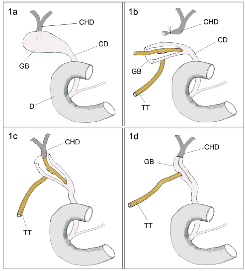
Figure 1: (a) Diagram showing the organisation of the biliary system in the current case, where the common hepatic duct (CHD) formed of right and left hepatic ducts terminated in the posterior wall of the gall bladder (GB) near the fundus, and the cystic duct (CD) opened directly in the second part of the duodenum (D) thus replacing a congenitally absent common bile duct;
(b) The common hepatic duct (CHD), inadvertently cut during dissection of the gall bladder (GB) from the liver bed, was loosely ligated and put on a sling. The long arm of drainage T-tube (TT) was inserted through a buttonhole made in the wall of the gall bladder (GB) while one of the short arms of the T-tube was threaded through the cystic duct (CD);
(c) The other short arm of the T-tube (TT) was threaded through the common hepatic duct (CHD);
(d) The wall of the gall bladder (GB) was refashioned around the transverse arm of the T-tube (TT) to create a duct, the proximal end of which was anastomosed to the common hepatic duct (CHD).
View Figure 1
.
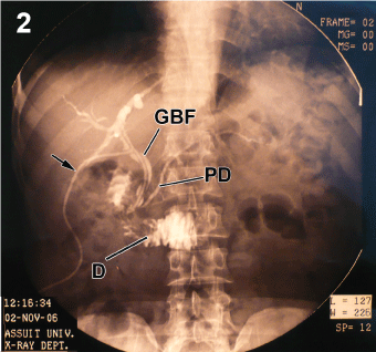
Figure 2: Retrograde trans-tubal cholangiogram (arrow) performed 26 days post surgery showing the gall bladder flap (GBF) fashioned around the transverse arm of the T-tube. The dye entered the duodenum (D) and the pancreatic duct (PD) indicating patency of the refashioned common bile duct without any leaks outside the biliary tree.
View Figure 2
Discussion
The current report is perhaps the first to document common bile duct (CBD) agenesis in the Egyptian population. Although considered very rare [1,4], a few cases of agenesis of the CPD, have been reported in Libyan [5], Turkish [6], Eastern Mediterranean [7,8], Eastern European [9,10], Indian [11] and Asian [12] populations. In the current case, the cystic duct opened directly into the duodenum and replaced the typical CBD, while a short common hepatic duct opened in the posterior wall of the gallbladder [9,12]. During surgery the wall of the gallbladder was used successfully as a flap to reconstruct the biliary ducts while retaining the intact sphincter of Oddi. A previous experimental study in pigs used a gallbladder flap to reconstruct the CBD [13] and concluded that the wall of the gallbladder is suitable for such procedure. Histologically the gallbladder wall closely resembles that of the common bile duct, including a muscular layer that is capable of contraction. In the experimental study in pigs, two animals that had tubal reconstruction from gallbladder flaps showed common bile duct stenosis ten days postoperatively [13]. However, in the current case retrograde cholangiography 26 days post surgery showed patency of the biliary system. In addition follow-up of the patient clinically for nine years post surgery showed no signs of hepatobiliary complications. In humans, gallbladder flaps have been used in surgical treatment of cases of Mirizzi syndrome by forming cholecysto-choledocho-jejunostomy [14]. However, the intraoperative decision taken in the current case was necessary to repair the inadvertently injured common hepatic duct, and it also retained the functional sphincter of Oddi.
In two reported cases of hepaticocystic duct the common hepatic duct opened close to the neck of the gallbladder thus it was possible to excise the gallbladder keeping the hepatic ducts' continuity with the cystic duct while suturing-closed the neck of the bladder [12,15]. These cases were considered as Type IIIB anomaly [4]. In a third case of hepaticocystic duct [16] a right and left hepatic ducts opened into the superior part of the gallbladder and cholecystectomy was achieved leaving that portion of the gallbladder wall that received the natural openings of the hepatic ducts to maintain biliary drainage. The natural opening of hepatic ducts into the gallbladder was also preserved in another case [8] where the hepatic ducts opened near the neck of the gallbladder. However, in the current case a short common hepatic duct opened in the posterior aspect of the body of the gallbladder near the fundus, and according to Losanoff et al. classification is considered as Type IIIC [4]. As stated above, the common hepatic duct was inadvertently severed during surgery, and the gallbladder flap was reconstructed into a tube and anastomosed to the end of the common hepatic duct to bridge the defect in the biliary ducts. As the flap was continuous with the neck of the gallbladder and cystic duct, the reconstituted flap retained its nerve supply, arterial supply and venous drainage. To the author's knowledge the surgical procedure applied in the current case has not been reported previously.
Hepatobiliary surgeons should be acquainted with the wide range of extrahepatic biliary system anomalies. Knowledge of such variations is essential for optimal surgical outcome. Preoperative identification of the anatomy may not be enough, and further anomalies may be encountered intraoperatively as seen in the current case. Intraoperative assessment of the biliary ducts should be done prior to excision of the gallbladder in order to identify any anomalies in the duct system may then be repaired using the gallbladder wall whilst maintaining its neurovascular supply.
The author declares that he has no competing interest.
References
-
Lamah M, Karanjia ND, Dickson GH (2001) Anatomical variations of the extrahepatic biliary tree: review of the world literature. Clin Anat 14: 167-172.
-
Hamilton WJ, Boyd JD, Mossman HW (1972) Hamilton, Boyd and Mossman's human embryology: prenatal development of form and function. (4th edn), Heffer, Cambridge, 646.
-
Mortele KJ, Ros PR (2001) Anatomic variants of the biliary tree: MR cholangiographic findings and clinical applications. AJR American journal of roentgenology. 177: 389-394.
-
Losanoff JE, Jones JW, Richman BW, Rangnekar NJ (2002) Hepaticocystic duct: a rare anomaly of the extrahepatic biliary system. Clin Anat 15: 314-315.
-
Elhamel A (1989) A rare extrahepatic biliary anomaly. HPB Surg 1: 353-358.
-
Ozaslan E, Dagli N, Balkanci F, Bayraktar Y (2002) Absence of the common bile duct and junction of the cystic duct with the left hepatic duct in a patient with chronic portal vein thrombosis. J Clin Gastroenterol 34: 280-281.
-
Olsha O, Steiner A, Rivkin LA, Sheinfeld A (1987) Congenital absence of the anatomic common bile duct. Case report. Acta Chir Scand 153: 387-390.
-
Hashmonai M, Kopelman D (1995) An anomaly of the extrahepatic biliary system. Arch Surg 130: 673-675.
-
Losanoff JE, Kjossev KT, Katrov E (1996) Hepaticocystic duct--a case report. Surg Radiol Anat 18: 339-341.
-
Nikolov K, Chobanov G, Belchev B, Donev S (1994) A rare case of gallbladder interposition in the extrahepatic bile ducts. Khirurgiia 47: 52-53.
-
Phani Krishna R, Behari A (2011) Interposition of gallbladder - a rare extrahepatic biliary anomaly. Indian J Surg 73: 453-454.
-
Harada N, Sugawara Y, Ishizawa T, Kaneko J, Sakamoto Y, et al. (2013) Resection of hepaticocystic duct which is a rare anomaly of the extrahepatic biliary system: a case report. J Med Case Rep 7: 279.
-
Mortensen FV, Ishibashi T, Hojo N, Yasuda Y (2004) A gall bladder flap for reconstruction of the common bile duct. An experimental study on pigs. J Hepatobiliary Pancreat Surg 11: 112-115.
-
Safioleas M, Stamatakos M, Revenas C, Chatziconstantinou C, Safioleas C, et al. (2006) An alternative surgical approach to a difficult case of Mirizzi syndrome: a case report and review of the literature. World J Gastroenterol 12: 5579-5581.
-
Shah O (2007) The missing common bile duct (hepaticocystic duct). Surgery 142: 424-425.
-
Jackson Jb, Kelly Tr (1964) Cholecystohepatic Ducts: Case Report. Ann Surg 159: 581-584.
