

International Journal of Neurology and Neurotherapy
Developmental Venous Anomaly Adjacent to Aqueduct of Sylvius
Kenichi Nishiyama* and Akira Hasegawa
Department of Neurosurgery, Niigata Medical Center, Niigata, Japan
*Corresponding author:
Kenichi Nishiyama, Department of Neurosurgery, Niigata Medical Center, Niigata, Japan, Tel: +81 25 232 0111, Fax: +81 25 231 3431, E-mail: nishiken@d4.dion.ne.jp
Int J Neurol Neurother, IJNN-3-048, (Volume 3, Issue 3), Case Report; ISSN: 2378-3001
Received: February 17, 2016 | Accepted: May 11, 2016 | Published: May 13, 2016
Citation: Nishiyama K, Hasegawa A (2016) Developmental Venous Anomaly Adjacent to Aqueduct of Sylvius. Int J Neurol Neurother 3:048. 10.23937/2378-3001/3/3/1048
Copyright: © 2016 Nishiyama K, et al. This is an open-access article distributed under the terms of the Creative Commons Attribution License, which permits unrestricted use, distribution, and reproduction in any medium, provided the original author and source are credited.
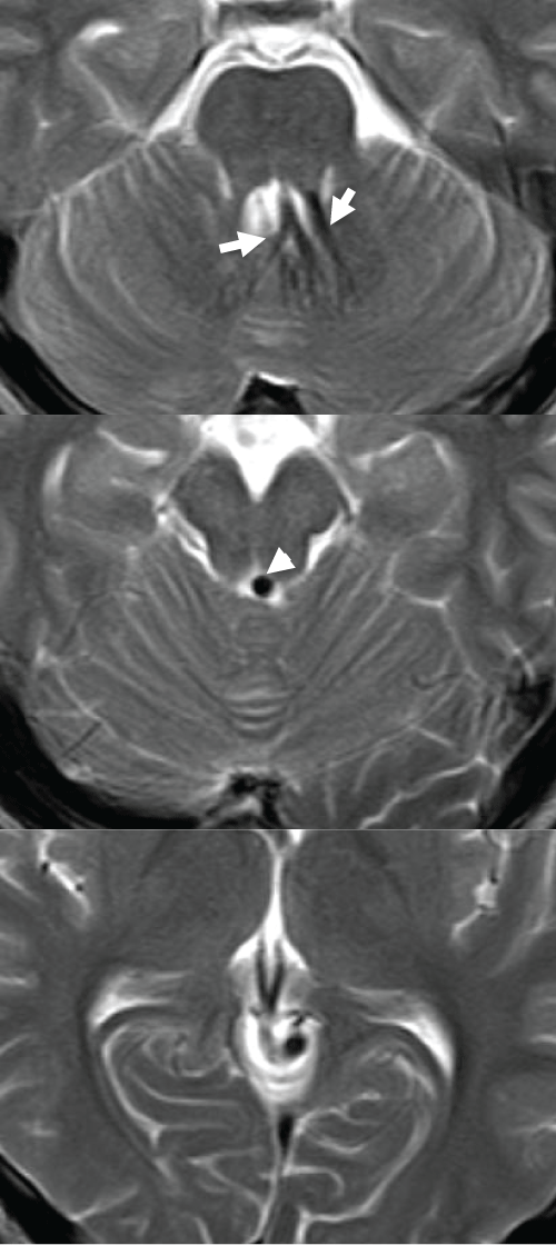
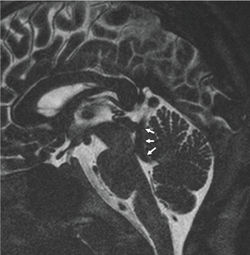
A 15-year-old boy presented to our hospital with headache after head trauma caused by a traffic accident. T2-weighted axial MR-images demonstrated the deep medullary veins of the cerebellum converging on the roof of the fourth ventricle. These veins drained into the subependymal vein, which then drained into the precentral cerebellar vein (Figure 1). MR-cisternography with 3D Fourier transformation constructive interference in steady state (CISS), which can depict the venous anatomy when it is surrounded by CSF, demonstrated a dilated precentral cerebellar vein located in the fourth ventricle. This dilated precentral cerebellar veinran adjacent to exit area of the aqueduct of Sylvius, penetrated the superior medullary velum and finally drained into the vein of Galen (Figure 2). The lesion was diagnosed as a developmental venous anomaly (DVA). MR-images did not show any hemorrhage or ventricular dilatation. This young man did not require any specific medical treatment for his venous anomaly.
.
Figure 1: T2-weighted axial MR-image showing posterior DVA collector veins (arrow) draining in to the precentral cerebellar vein (arrowhead).
View Figure 1
.
Figure 2: MR-cisternography with CISS showing the dilated precentral cerebellar vein (arrow) located adjacent to the exit of the aqueduct and penetrating the superior medullary velum.
View Figure 2
Developmental venous anomalies (DVAs) are known as congenital intracranial vascular lesion. Most DVAs are asymptomatic; however, they may occasionally cause obstructive hydrocephalus and should be considered in cases of chronic or intermittent headaches [1-3]. Paulson et al. reported a case with hydrocephalus caused by the DVA passing through the orifice of the aqueduct and referred 10 previously reported cases which had presented with hydrocephalus [2]. Their conclusion was that no surgical treatment was necessary for the DVAs for itself, but CSF diversion might be necessary in some cases. Although MR-images demonstrated a dilated precentral cerebellar vein adjacent to the aqueduct in our case which presented with headache, an aqueduct stenosis resulting in hydrocephalus was not present and surgical intervention was not considered necessary.
Disclosure
The authors report no conflicts of interest concerning the materials or methods in this paper. None of the authors received any financial or material support.
References
-
Lee C, Pennington MA, Kenney CM (1996) MR evaluation of developmental venous anomalies: medullary venous anatomy of venous angiomas. AJNR Am J Neuroradiol 17: 61-70.
-
Paulson D, Hwang SW, Whitehead WE, Curry DJ, Luerssen TG, et al. (2012) Aqueductal developmentalvenousanomaly as an unusual cause of congenital hydrocephalus: a case report and review of the literature. J Med Case Rep 6:7.
-
Giannetti AV, Rodrigues RB, Trivelato FP (2008) Venous lesions as a cause of sylvianaqueductal obstruction: case report. Neurosurgery 62: E1167-1168.
