

International Journal of Ophthalmology and Clinical Research
Management of Descemet Detachment following Crescentic Lamellar Excision with Autolamellar Dissection for Pellucid Marginal Corneal Degeneration
Radwan Almousa1,2* and Sheraz M. Daya1,3
1Corneoplastic Unit, Queen Victoria Hospital, East Grinstead, UK
2Department of Ophthalmology, University Hospitals Coventry and Warwickshire, UK
3Private clinic, Centre for Sight, East Grinstead, UK
*Corresponding author: Radwan Almousa, Department of Ophthalmology, University Hospitals Coventry and Warwickshire NHS Trust, Coventry CV2 2DX, UK, Tel: +447799641635, E-mail: radomousa@hotmail.com
Int J Ophthalmol Clin Res, IJOCR-2-015, (Volume 2, Issue 1), Case Report; ISSN: 2378-346X
Received: September 03, 2014 | Accepted: February 23, 2015 | Published: February 26, 2015
Citation: Almousa R, Daya SM (2015) Management of Descemet Detachment following Crescentic Lamellar Excision with Autolamellar Dissection for Pellucid Marginal Corneal Degeneration. Int J Ophthalmol Clin Res 2:015. 10.23937/2378-346X/1410015
Copyright: © 2015 Almousa R, et al. This is an open-access article distributed under the terms of the Creative Commons Attribution License, which permits unrestricted use, distribution, and reproduction in any medium, provided the original author and source are credited.
Abstract
A 35 year-old male patient with pellucid marginal corneal degeneration underwent crescentic lamellar excision with autolamellar dissection on his left eye. One month later, the patient developed localized corneal edema overlying the area of the crescentic resection due to Descemet membrane detachment. The Snellen corrected visual acuity reduced from 6/15 one week postoperatively to 2/60 following the development of corneal edema. The fluid that collected in the interface was removed with a blunt 30-gauge cannula, and air was injected into the anterior chamber to oppose the two corneal layers.
Corrected visual acuity improved to 6/12 over 1 month, and to 6/9 seven months later. The corneal edema resolved over 2 months.
Crescentic lamellar excision with autolamellar dissection could be associated with Descemet membrane detachment and corneal edema; however, this complication has a very good outcome when managed with a minimally invasive procedure.
Keywords
Cornea, Descemet detachment, Lamellar dissection, Crescentic
Introduction
Pellucid Marginal Corneal Degeneration (PMCD) is a progressive, noninflammatory peripheral corneal thinning disorder characterized by a peripheral band of thinning, mostly of the inferior cornea, from the 4-o�clock to the 8-o�clock position accompanied by 1 to 2 mm of normal cornea between the limbus and the area of thinning [1]. The clinical picture of PMCD results in flattening in the vertical meridian and the appearance of marked against-the-rule (ATR) astigmatism [2].
Several surgical methods have been advocated for visual rehabilitation in PMCD [3-10]; however, none of the present modalities of treatment reliably achieves an optimum visual result. This is mainly due to the progression of ATR astigmatism, as in the case of crescentic wedge resection, or due to the inherent suboptimal visual results associated with penetrating keratoplasty [3,4].
Crescentic lamellar excision with auto lamellar dissection (CLEAD) was developed by one of the authors (SD) [5] to reduce the chance of ATR astigmatism progression postoperatively. This is a new procedure that could be associated with new side effects.
Case Presentation
Patient informed consent form was obtained. A 35 year-old male of Indian origin had been treated for seasonal allergic conjunctivitis at the age of 11, and he used to rub his eyelids excessively. The seasonal allergic conjunctivitis settled down as he grew into adulthood, and had ceased by the time he was 21. At the age of 24 he began to experience reduced visual acuity and started wearing glasses at the age of 25. His vision continued to deteriorate rapidly; he was subsequently diagnosed with a possible keratoconus and started on scleral Contact Lenses (CL) by his local ophthalmologist. By the age of 28, the patient could no longer tolerate the scleral CL and he was started on soft toric CL. These failed to improve his sight adequately, so he was referred to a corneal tertiary referral centre in the south east of England. On examination, the Snellen Best Spectacle Corrected Visual Acuity (BSCVA) was 6/16 in the right eye and 6/12 in the left eye. Both corneas showed inferior thinning extending from 4 to 8 o�clock, sparing the peripheral 1mm of the cornea. There was minimal peripheral corneal neovascularization, reaching the inferior edge of the thin area, without scarring or lipid deposition. The thin area straddled over 1mm, with corneal ectasia above it. The condition was progressive giving the continuous deterioration of vision since the age of 24. At this stage the patient was diagnosed with a bilateral Pellucid Marginal Corneal Degeneration (PMCD), which was confirmed with Pentacam corneal tomography (Pentacam�, Oculus Optikger�te GmbH, Wetzlar, Germany) that showed bilateral inferior, butterfly shaped steepness and against-the-rule astigmatism. After discussing options for surgical management with the patient, we decided to treat the left eye with CLAED, which involved resecting an inferior lamellar crescenting corneal wedge to remove the thin area. Opposite the resected area, we made a partial thickness peripheral corneal incision at 12 o�clock, through which we carried out a lamellar dissection inferiorly until reaching the resected area. The final result was a lamellar dissection of the normal cornea with excision of the thinned area. This facilitates the wound edge opposition and reduces the chance of Descemet protrusion into the anterior chamber (AC), which usually appears with lamellar crescentic resection alone. Our hypothesis is that lamellar dissection would release the tension on the resected wound edges, which can cause progression of ATR astigmatism following conventional wedge resection. We used 10/0 mersilene suture to close the wound, and injected an air bubble into the AC to press Descemet membrane against the stroma. The patient was started on topical dexamethazone 0.1% and levofloxacine 0.3 % four times a day on the left eye.
One day postoperatively, both corneal flaps were attached and the left eye BSCVA was 6/15. The patient came back 1 month later with corneal edema overlying the area of the crescentic resection. Anterior segment ocular coherent tomography (Visante;Carl Zeiss Meditec, Dublin, CA) showed Descemet detachment underlying the excised corneal wedge (Figure 1). The cornea surface was irregular (Figure 2) and the visual acuity dropped to 2/60.
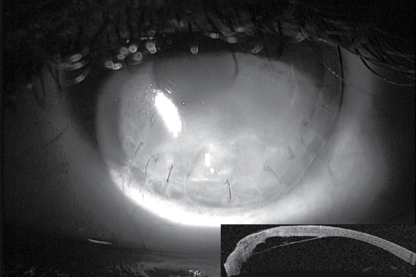
Figure 1: Corneal edema involving the inferior third of the cornea, one
month following crescentic lamellar excision with autolamellar dissection for
Pellucid Marginal Corneal Degeneration. Inset shows Descemet membrane
detachment underlying the corneal edema.
View Figure 1
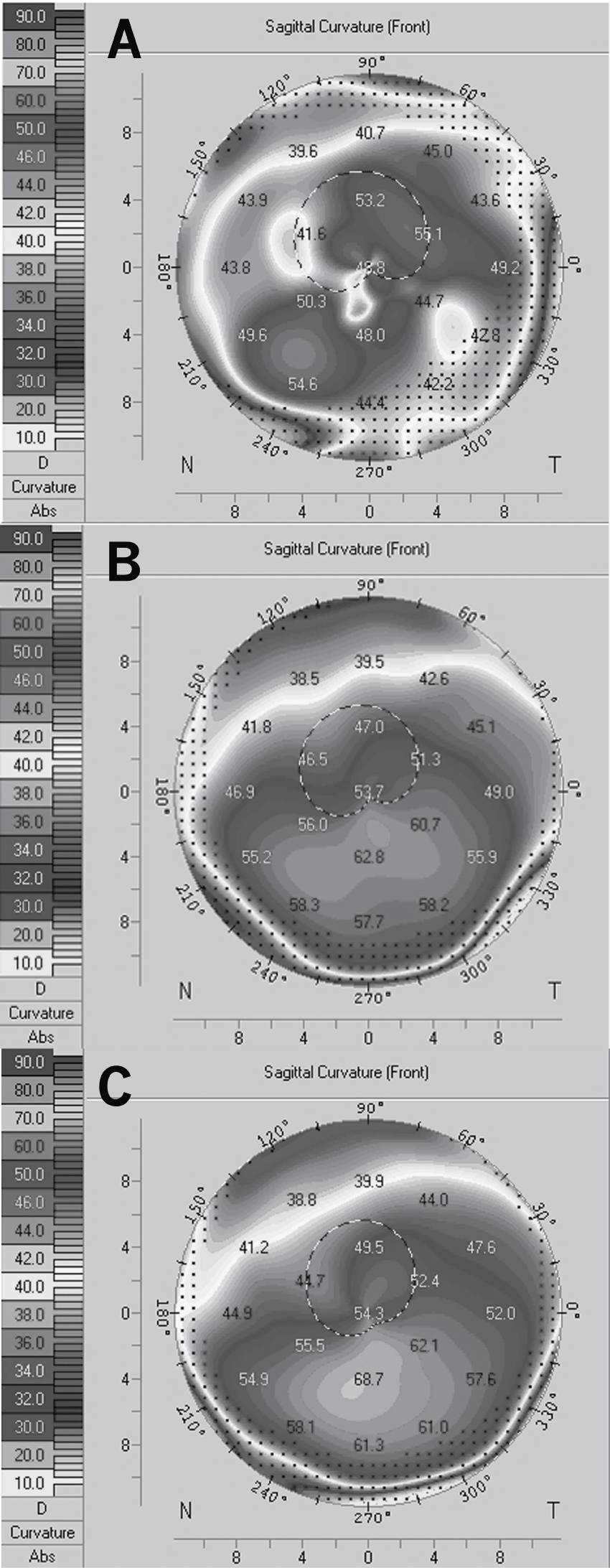
Figure 2: Corneal topography. A. Corneal surface irregularity Following
Descemet membrane detachment and corneal edema. B. Three months
following treatment with interface fluid removal and air injection in the anterior
chamber. The cornea surface is regular with with-the-rule astigmatism. C. The
last visit the corneal still shows regular with-the-rule astigmatism.
View Figure 2
A week later, the fluid was removed from the interface with a blunt 30-gauge cannula with air injected into the AC. Next day both corneal flaps were attached and over the next two months the corneal edema settled down, leaving behind corneal scarring inferior to the visual axis (Figure 3). Pentacam corneal tomography showed improvement in corneal surface irregularity and the mean K reading reduced from 57.2 preoperatively to 54.7 at the final follow-up with highest K reading at 88� (with-the-rule astigmatism) (Figure 2). BSCVA was 6/12 after one month and 6/9.5 after three months. At the final follow-up visit 7 months later, BSCVA was 6/9. Unaided visual acuity with the operated eye has improved from 3/60 preoperatively to 6/60 at the final follow-up. Manifest refraction spherical equivalent (MRSE) reduced from -11.5 (-9.25-4.5�100) preoperatively to -10 (-7.5-5�135) at the final follow-up.
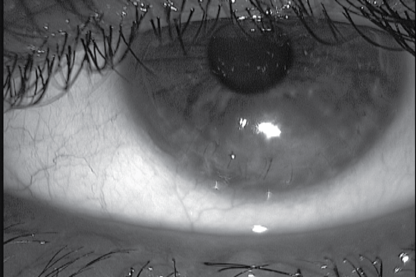
Figure 3: Two months following treatment with interface fluid removal and
air injection inside the anterior chamber, the corneal edema resolved with
residual corneal scarring.
View Figure 3
Discussion
The correction of irregular astigmatism caused by a primary corneal ectasia, such as the PMCD, remains a challenge for eye care practitioners. Spectacles and rigid gas permeable contact lenses might be an adequate choice for mild cases; however, as the topographic changes progress, these conservative measures are no longer adequate. Patients with early and moderate PMCD who are intolerant to contact lenses and whose peripheral corneal thickness is more than 450 micron could be treated with intracorneal rings.6 Various surgical procedures have been tried, such as crescenitc wedge resection, crescentic lamellar keratoplasty, C-shaped lamellar keratoplasty [3,4,7]. These procedures remove the pathological thin cornea; however, they were all reported to result in progressive ATR astigmatism over a mean of 9.5 months [3,4,7]. Oversized central penetrating keratoplasty (PK) or decentered PK are prone to rejection because of the proximity of the graft to the limbus and its blood vessels. Furthermore, grafts that are deliberately decentered inferiorly are associated with large degrees of astigmatism [8]. Two staged procedures, lamellar graft followed by smaller penetrating graft, has evolved with the idea to avoid the complication of a large graft, and to achieve a topographically acceptable refracting corneal surface [9] however, 2 corneal grafts are needed for these procedures and the patient have to return back to the operating theatre for the second stage surgery and this is difficult to apply in many eye departments. Collagen cross-linking is a treatment for progressive corneal ectasia, albeit corneal thinning below 400 micron is a relative contraindication due to the potential damage to corneal endothelium [10]. Our patient was relatively young without any signs of cataract, hence clear lens exchange with toric IOL was not an option.
Crescentic lamellar excision with autolamellar dissection was developed to reduce the chance of ATR astigmatism progression without the risk of inducing corneal rejection. As there is misalignment of the wound and complete separation between the posterior and anterior corneal lamella, there is increased chance of posterior corneal flap separation; therefore the procedure is usually ended with air tamponade in the AC to keep both flaps in opposition. In the case we have described, the patient had Descemet separation underlying the area of crescentic lamellar excision, causing overlying corneal stromal edema and disturbed vision. Removal of the fluid from the interface with repeat air injection into the AC resulted in the fast resolution of the corneal edema with reattachment of Descemet membrane.
Patient with PMCD who decide to have surgical treatment for visual improvement should be made aware of the various procedures and their possible side effects.
CLEAD is a less invasive surgical procedure compared to full thickness or lamellar large corneal graft. It is associated with side effects; however, these are relatively easy to manage. The visual outcomes of CLEAD in terms of ATR astigmatism progression and stability of vision postoperatively need further assessment with a large cohort of patients and longer follow-ups.
Acknowledgments
Helga Perry, Electronic Systems & Resources Librarian at University Hospitals Coventry & Warwickshire NHS Trust assisted with proof-reading and preparation of this paper.
The Authors has no proprietary or commercial interest in any materials discussed in this article.
Ethical Statement
Patient informed consent form was taken and patient anonymity was preserved.
None of the authors have any financial or proprietary interest in any product or procedure discussed in this study.
References
-
Krachmer JH (1978) Pellucid marginal corneal degeneration. Arch Ophthalmol 96: 1271-1221.
-
Rao SK, Fogla R, Padmanabhan P, Sitalakshmi G (1999) Corneal topography in atypical pellucid marginal degeneration. Cornea 18: 265-272.
-
Sridhar MS, Mahesh S, Bansal AK, Nutheti R, Rao GN (2004) Pellucid marginal corneal degeneration. Ophthalmology 111: 1102-1107.
-
Cheng CL, Theng JT, Tan DT (2005) Compressive C-shaped lamellar keratoplasty: a surgical alternative for the management of severe astigmatism from peripheral corneal degeneration. Ophthalmology 112: 425-430.
-
Maccheron LJ, Daya SM (2012) Wedge resection and lamellar dissection for pellucid marginal degeneration. Cornea 31: 708-715.
-
Mularoni A, Torreggiani A, di Biase A, Laffi GL, Tassinari G (2005) Conservative treatment of early and moderate pellucid marginal degeneration: a new refractive approach with intracorneal rings. Ophthalmology 112: 660-666.
-
Javadi MA, Karimian F, Hosseinzadeh A, Noroozizadeh HM, Sa'eedifar MR, et al. (2004) Lamellar crescentic resection for pellucid marginal corneal degeneration. J Refract Surg 20: 162-165.
-
Rasheed K, Rabinowitz YS (2000) Surgical treatment of advanced pellucid marginal degeneration. Ophthalmology 107: 1836-1840.
-
Kremer I, Sperber LT, Laibson PR (1993) Pellucid marginal degeneration treated by lamellar and penetrating keratoplasty. Arch Ophthalmol 111: 169-170.
-
Wittig-Silva C, Chan E, Islam FM, Wu T, Whiting M, et al. (2014) A randomized, controlled trial of corneal collagen cross-linking in progressive keratoconus: three-year results. Ophthalmology 121: 812-821.
