

International Journal of Ophthalmology and Clinical Research
Relation of Pupil Size and Cataract Surgery using PupilX
Annekatrin Rickmann1*, Maria Waizel1, Peter Szurman1,2 and Karl T Boden1
1Knappschaft Eye Clinic Sulzbach, Knappschaft Hospital Saar, Sulzbach/Saar, Germany
2Centre for Ophthalmology, University Eye Clinic Tuebingen, Tuebingen, Germany
*Corresponding author: Dr. Annekatrin Rickmann, Knappschaft Eye Clinic Sulzbach, Knappschaft Hospital Saar, An der Klinik 10, 66280 Sulzbach/Saar, Germany, Tel: 06897574-1119, Fax: 06897574-2139, E-mail: annekatrinrick@gmail.com
Int J Ophthalmol Clin Res, IJOCR-3-059, (Volume 3, Issue 3), Original Article; ISSN: 2378-346X
Received: May 26, 2016 | Accepted: July 06, 2016 | Published: July 08, 2016
Citation: Rickmann A, Waizel M, Szurman P, Boden KT (2016) Relation of Pupil Size and Cataract Surgery using PupilX. Int J Ophthalmol Clin Res 3:059. 10.23937/2378-346X/1410059
Copyright: © 2016 Rickmann A, et al. This is an open-access article distributed under the terms of the Creative Commons Attribution License, which permits unrestricted use, distribution, and reproduction in any medium, provided the original author and source are credited.
Abstract
Background: The aim of this study was to evaluate the influence of cataract surgery on pupil size at well-defined measurement conditions with a novel pupillometer (PupilX).
Methods: Pupil size of healthy study participants was measured with infrared-video PupilX pupillometer (MEye Tech GmbH) at 5 different illumination levels (0, 0.5, 4, 32 and 250 lux) before and after cataract surgery. Measurements were performed by the same investigator. 90 images were recorded during a measurement period of 3 seconds. The absolute linear camera resolution was approximately 20 pixels per mm.
Results: This prospective study analysed the pupil size of 35 patients (mean age 73.2 ± 8.6 years, range 50-87 years). Beside the gradually decreasing pupil size with an increasing illumination level there is a significant decreased pupil size after cataract surgery at all illumination levels. One week after cataract surgery mean pupil diameter for all patients decreased significantly by 13.1% at 0 Lux, 13.6% at 0.5 Lux, 8.4% at 4 Lux, 7.3% at 32 Lux and 5.1% at 250 Lux. Four weeks after cataract surgery mean pupil diameter increased back to comparably preoperative pupil size.
Conclusion: In conclusion, this study showed a significant decrease of pupil size after cataract surgery in a normal population under scotopic, mesopic and photopic static illuminance conditions. After 4 weeks postoperatively pupil size returned to approximately preoperative levels. The variability of illuminance conditions of PupilX allows the possibility of investigations of pupil size in relation to refractive surgery.
Keywords
Pupillometer, PupilX, Pupil size, Digital pupillometer, Cataract surgery
Introduction
The pupil size and its relation to visual outcome in cataract and refractive surgery is an important element of optical quality. So far, there are controversial studies about a large scotopic pupil size as a potential risk factor for night vision complaints including halos, glare and vision deterioration [1]. Earlier studies reported influence of cataract surgery on pupil size [2]. Kanellopoulos could show with a Scheimpflug-system that a cataract extraction surgery reduced photopic pupil size and induced a more circular shaped pupil, appearing more significant with increasing age [3]. A retreating pupil size with age has been reported already [4]. Previously, many studies compared various pupillometers and demonstrated a disparity of the pupil size among different devices [5-7]. The majority of previous pupil size studies were performed under scotopic conditions in healthy eyes [8,9]. In contrast, studies under well-defined mesopic and photopic illumination conditions are rare [10,11]. In clinical routine, precise and easy measurement of pupil size is important for planning and outcome of refractive surgery [6,12,13]. Furthermore, multiple binocular measurements gain a precise description of pupil behaviour before refractive surgery [14]. A recently developed pupillometer PupilX (MEye Tech GmbH) enables to measure bilateral pupil sizes with indirect infrared illuminance conditions simultaneously under constant and adjustable illumination levels, facilitating comparable measurements under scotopic, mesopic and photopic conditions [5]. Schilde et al. reported on most precise measurements with PupilX [5]. This device offers high comparability and standardisation for research purposes.
Therefore, the aim of this study was to evaluate the pupil size of healthy participants at different well defined illumination levels before and after cataract surgery. We used a commonly implantated intraocular lens Tecnis One (Abbott Medical Optics, Illinois, USA). Tecnis One is an aspheric hydrophobic acryl-intraocular lens which affects a less slight spheric aberration than others [15]. Furthermore it does not block blue light with a wavelength of 450-500 nm, so that there is no influencing factor of light irradiation at scotopic conditions [16].
Methods
The pupil size of healthy study participants was measured using the PupilX pupillometer (MEye Tech GmbH). This study was approved by the local Ethical Committee (Landesärztekammer Saarland) with a positive vote for prospective investigations.
Patients
This prospective study analysed 35 patients, 20 male and 15 female. Exclusion criteria were any history of eye disease, trauma or interfering topical or systemic medication. An additional slit lamp examination excluded other morphological abnormalities of the anterior eye segment in addition to senile cataract. All eyes underwent a cataract surgery performed with topical anesthesia (lidocaine gel), clear-corneal incision, phacoemulsification and in-the-bag intraocular implantation. No flexible iris retractor was used during the cataract surgery. The postoperative regimen consisted of a combination of tobramycin and dexamethasone drops for 3 weeks.
Technical procedure
Each patient's eyes were measured at 5 illumination levels with 0, 0.5, 4, 32 and 250 lux before, one and four weeks after cataract surgery. No subject was exposed to bright light directly before the measurement. Furthermore, all patients underwent a 2 minute retinal adaptation phase in the same room under low mesopic conditions (darkened room where the only light was from the device). This was monitored by the same investigator. The participants saw a white uniform LED field with adjustable brightness, which could be switched off for scotopic conditions. By using light-tight rubber eyecups, the PupilX pupillometer allows for measurements under scotopic conditions without the need to completely darken the examination room (Figure 1). We performed the measurements at 0.5, 4, 32 and 250 lux, allowing 5-10 seconds for participants to adapt to the illuminance level in advance. The protocol order proceeded from the dimmest to the brightest condition. All measurements were performed and observed via display by the same investigator (AR). The subjects were requested to look straight ahaid and keep their eyes open for the whole measurement period. At each continuous level of illumination, 90 images were acquired, 30 images per second over a measurement period of 3 seconds.
.
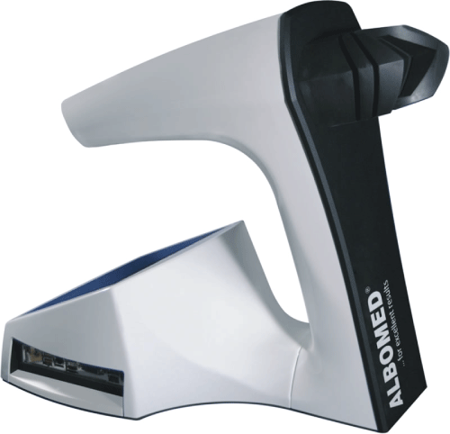
Figure 1:Picture of the device, PupilX. The participant needs to look into theblack light-tight rubber eyecups. The investigator could monitor all data at the screen sinistral at this picture [www.albomed.de].
View Figure 1
Data analysis
The absolute linear camera resolution was approximately 20 pixels per mm. The resolution of measurements of the entire system is +/- 0.1 mm for pupils < 3.3 mm and +/- 3% for pupils > 3.3 mm. Pupil tracking circles were automatically fitted to image data indicating the pupil borders. This could be observed on the monitor. Recordings with blink artefacts were automatically discarded and repeated. Both eyes were recorded simultaneously. The software analyzed average pupil size with standard deviation (i.e., short term variability within the measurement period) and anisocoria over the measurement period of 3 seconds. Data could also be selected for all 90 single measurements taken during the measurement period. The accepted thresholds for the CIE (international Commission on Illumination) curve for vision are: scotopic = illuminance levels below 0.05 lux, photopic = illuminance levels above 50 lux (National Physical Laboratory, London, United Kingdom).
.
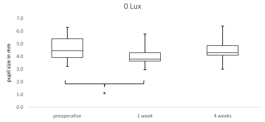
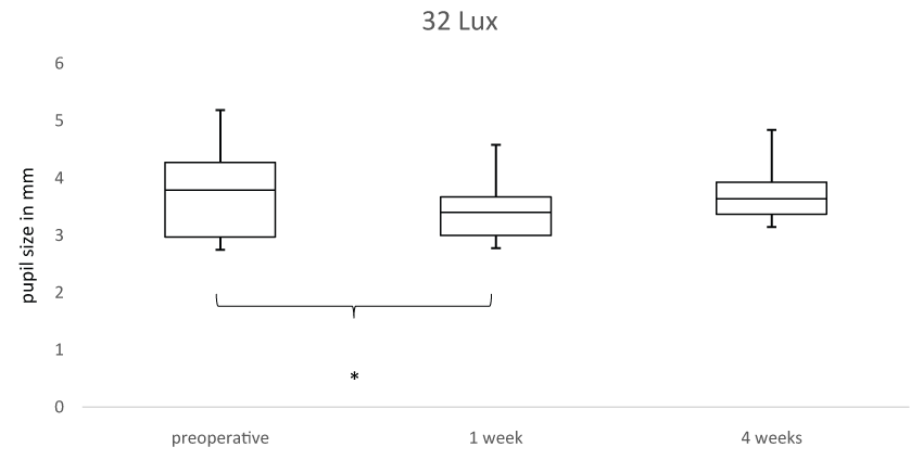
Figure 2: Box plot analysis presents the pupil size in mm for all subjects before, one week and 4 weeks after cataract surgery at (A) 0 lux. Statistically significant results (paired Wilcoxon test, p < 0.05) are marked with an asterisk.
View Figure 2
.
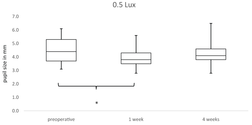
Figure 3: Box plot analysis presents the pupil size in mm for all subjects before, one week and 4 weeks after cataract surgery at (A) 0.5 lux. Statistically significant results (paired Wilcoxon test, p < 0.05) are marked with an asterisk.
View Figure 3
Statistical methods
For statistical evaluation ANOVA and a post-hoc paired Wilcoxon rank sum test was performed, p < 0.05 was defined as statistically significant. Statistical analysis was performed with Matlab® R2014b (The Math Works, Version 8.4.0).
.
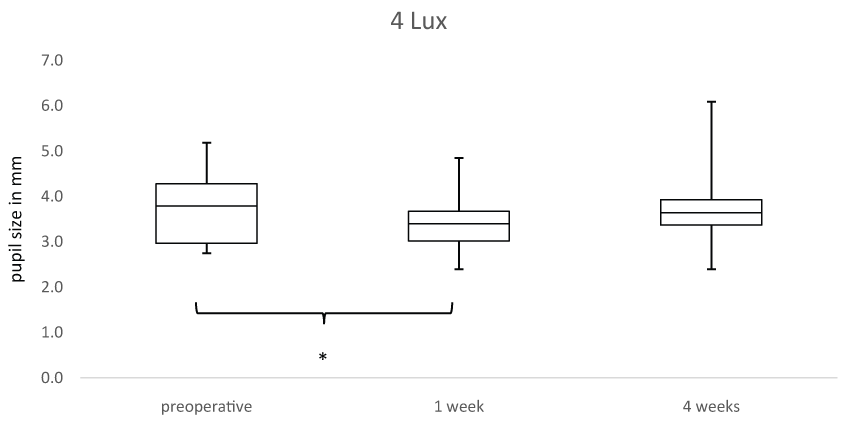
Figure 4: Box plot analysis presents the pupil size in mm for all subjects before, one week and 4 weeks after cataract surgery at (A) 4 lux. Statistically significant results (paired Wilcoxon test, p < 0.05) are marked with an asterisk.
View Figure 4
Results
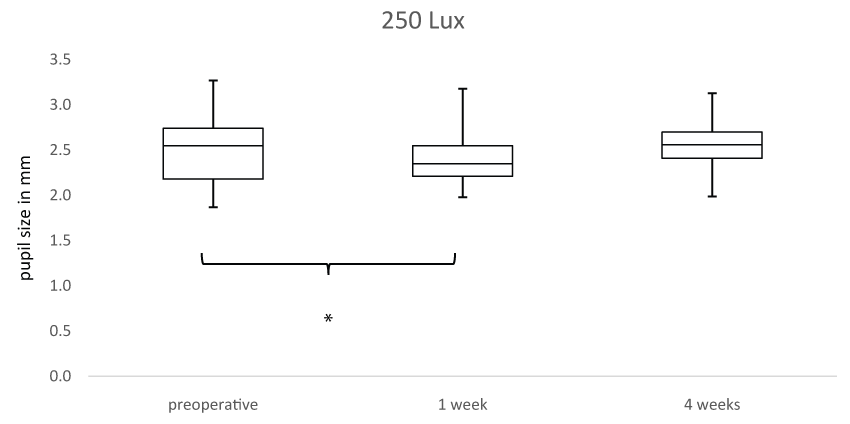
This prospective study analysed 35 patients, 20 male and 15 female. The mean age was 73.2 ± 8.6 years; participants ranged in age from 50-87 years. The best index of the true value of pupil size is the mean of 90 measurements during the 3 second period. The mean pupil diameter for all patients at 0 lux was preoperatively 4.69 ± 0.85 mm and decreased significantly one week after cataract surgery to 4.08 ± 0.035 mm (-13.1%, p < 0.001) and increased 4 weeks after cataract surgery to 4.45 ± 0.89 mm (p = 0.56) (Figure 2). At 0.5 lux the mean pupil diameter was preoperatively 4.49 ± 0.84 mm and decreased significantly one week after cataract surgery to 3.88 ± 0.63 mm (-13.6%, p < 0.001) and increased 4 weeks after cataract surgery to 4.29 ± 0.93 mm (p = 0.52) (Figure 3). At 4 lux the mean pupil diameter was preoperatively 3.69 ± 0.74 mm and decreased significantly one week after cataract surgery to 3.38 ± 0.49 mm (-8.4%, p = 0.021) and increased 4 weeks after cataract surgery to 3.76 ± 0.83 mm (p = 0.86) (Figure 4). At 32 lux the mean pupil diameter was preoperatively 3.04 ± 0.52 mm and decreased significantly one week after cataract surgery to 2.82 ± 0.38 mm (-7.3%, p = 0.0015) and increased 4 weeks after cataract surgery to 3.18 ± 0.57 mm (p = 0.47) (Figure 5). At 250 lux the mean pupil diameter was preoperatively 2.51 ± 0.39 mm and decreased significantly one week after cataract surgery to 2.38 ± 0.25 (-5.1%, p = 0.0305) and increased 4 weeks after cataract surgery to 2.67 ± 0.26 mm (p = 0.73) (Figure 6) (paired Wilcoxon test). Our data show a statistically significant difference in pupil size after cataract surgery at all illumination levels. After 4 weeks postoperatively pupil size returned to approximately preoperative levels. Differences in pupil size in general were less with increasing illumination level. Just two of 35 patients had the same pupil size after cataract surgery. No patient had a mydriatic effect.
.
Figure 5: Box plot analysis presents the pupil size in mm for all subjects before, one week and 4 weeks after cataract surgery at (A) 32 lux. Statistically significant results (paired Wilcoxon test, p < 0.05) are marked with an asterisk.
View Figure 5
.
Figure 6: Box plot analysis presents the pupil size in mm for all subjects before, one week and 4 weeks after cataract surgery at (A) 250 lux. Statistically significant results (paired Wilcoxon test, p < 0.05) are marked with an asterisk.
View Figure 6
Discussion
This study provides data of pupil size before and after cataract surgery at scotopic, mesopic and photopic illuminance conditions. There was a significant decrease of pupil size at all illumination levels after cataract surgery. We could show that the decreased pupil size returned to approximately preoperative levels after one month postoperatively.
Previous studies reported an influence of cataract surgery to the pupil size [2], especially with increasing age [3]. It is well-known that the pupil size and its relation to visual outcome in cataract and refractive surgery is an important element of optical quality. The pupil size determines the quality of the retinal image via optimizing diffraction, ocular aberrations and depth of focus. Particularly, mesopic and scotopic pupil sizes are important for corneal ablation zone and multifocal intraocular lens in refractive surgery in order to prevent optical refraction and diffused light [14]. The highest differences of pupil size before and after cataract surgery were shown in this study at scotopic conditions. So far, there are controversial studies about a large scotopic pupil size as a potential risk factor for night vision complaints like halos, glare and vision deterioration [1]. In addition, data of mesopic pupil sizes are necessary for current studies about accommodative intraocular lenses [10]. Fliedner et al. suggested already that the pupil feasibly could control an artificial lens throughout the pupillary near reflex arc by increasing the depth of focus [17].
Our results provide information on pupil size after cataract surgery in a healthy population. The postoperative miosis after one week returned to approximately preoperative levels after one month. This is on accordance with other studies [18,19]. A possible explanation of the current results are the mediation of inflammatory reaction after cataract surgery via cyclooxygenase-1 (COX-1) and COX-2 enzymes and prostaglandins. This inflammation cascade with prostaglandin synthesis leads to decreased pupil size [20]. Normalisation of pupil diameter after one month is therefore also comprehensible by terminated inflammation reaction. On the contrary, Kanellopoulos et al. demonstrated a reduced mesopic and photopic pupil size after one month of cataract surgery [2,3]. However, the experimental set up is different as Kanellopoulos et al. imaged the pupil size and corneal vertex location via determination of the Cartesian coordinates of the first Purkinje reflection point to the pupil geometric center. Furthermore, Peters et al. recorded pupillary responses up to 20 weeks after surgery and did not find an evidence of dilation lag or segmental palsy in the operated eyes. The latency of constriction was also not prolonged [21].
A possible limitation of this study is the variable length of time needed for full dark adaptation of the retina in different individuals. In this study 2 minutes were allowed for each participant. We choose this retinal adaptation phase, because it is practicable in clinical routine and is also comparable to daily life illuminance conditions. Furthermore, the pupil reaction is the fast part of retinal adaptation and takes only few seconds to a minute [22]. The normal experimental set-up for the following mesopic and photopic luminance conditions took into account that light adaptation takes only seconds. Therefore possible differences might be acceptable. Factors influencing pupil size including fatigue or emotional stress can hardly be excluded [23]. In addition, accommodation cannot be completely excluded and could lead to smaller pupil sizes [19]. However, it is unlikely that accommodation plays a major role, as the device uses a telecentric system and does not offer a near target.
The intraoperative usage of topical lidocaine gel is known to have an additional mydriatic effect during cataract surgery [24]. In contrary, dexamethasone eyedrops act at the level of phospholipase A with the resultant inhibition of prostaglandine release [25]. Therefore, real pupil size after one week might be even smaller without postoperative treatment with topical corticosteroids. In our experience, the advantages of PupilX were easy handling and good measurements even for participants with dark irises and darkened eyelashes. However this study analysed white subjects only. Recordings with blink artefacts were automatically discarded. In cases of obvious artefacts individual measurement data could be excluded for experimental set-ups. However, the whole measurement should be observed via display to minimize artefacts caused by inobservance, eye-movements, pendant eyelids or long darkened eyelashes.
In conclusion, this study showed a significant decrease of pupil size after cataract surgery with normalisation of pupil size after one month in a normal population under scotopic, mesopic and photopic static illuminance conditions. The variability of illuminance conditions of PupilX allows the possibility of investigations on pupil size in relation to refractive surgery. Due to easy handling and comparative experimental set-up PupilX might be interesting for further researches.
Acknowledgements
We thank Axel von Wallfeld (MEye Tech GmbH) for material and technical support.
Disclosure
Rickmann A (none), Waizel M (none), Szurman P (none), Boden KT (none).
Competing Interests
None.
References
-
Salz JJ, Trattler W (2006) Pupil size and corneal laser surgery. Curr Opin Ophthalmol 17: 373-379.
-
Kanellopoulos AJ, Asimellis G, Georgiadou S (2015) Digital pupillometry and centroid shift changes after cataract surgery. J Cataract Refract Surg 41: 408-414.
-
Kanellopoulos AJ, Asimellis G (2014) Clear-cornea cataract surgery: pupil size and shape changes, along with anterior chamber volume ad depth changes. A Scheimpflug imaging study. Clin Ophthalmol 8: 2141-2150.
-
Winn B, Whitaker D, Elliott DB, Phillips NJ (1994) Factors affecting light-adapted pupil size in normal human subjects. Invest Ophthalmol Vis Sci 35: 1132-1137.
-
Schilde T, Bende T, Matallana M, Kohlhaas M (2013) Messung des Pupillendurchmessers. Der Augenspiegel 06: 32-34.
-
Kohnen T, Terzi E, Bühren J, Kohnen EM (2003) Comparison of a digital and a handheld infrared pupillometer for determining scotopic pupil diameter. J Cataract Refract Surg 29: 112-117.
-
Schmitz S, Krummenauer F, Henn S, Dick HB (2003) Comparison of three different technologies for pupil diameter measurement. Graefes Arch Clin Exp Ophthalmol 241: 472-477.
-
Kohnen EM, Zubcov AA, Kohnen T (2004) Scotopic pupil size in a normal pediatric population using infrared pupillometry. Graefes Arch Clin Exp Ophthalmol 242: 18-23.
-
Schnitzler EM, Baumeister M, Kohnen T (2000) Scotopic measurement of normal pupils: Colvard versus Video Vision Analyzer infrared pupillometer. J Cataract Refract Surg 26: 859-866.
-
Heine C, Yazdani F, Wilhelm H (2013) Pupillary diameter in everyday situations. Klin Monbl Augenheilkd 230: 1114-1118.
-
Witting MD, Goyal D (2003) Normal pupillary size in fluorescent and bright light. Ann Emerg Med 41: 247-250.
-
Freedman KA, Brown SM, Mathews SM, Young RS (2003) Pupil size and the ablation zone in laser refractive surgery: considerations based on geometric optics. J Cataract Refract Surg 29: 1924-1931.
-
Wickremasinghe SS, Smith GT, Stevens JD (2005) Comparison of dynamic digital pupillometry and static measurements of pupil size in determining scotopic pupil size before refractive surgery. J Cataract Refract Surg 31: 1171-1176.
-
Rosen ES, Gore CL, Taylor D, Chitkara D, Howes F, et al. (2002) Use of a digital infrared pupillometer to assess patient suitability for refractive surgery. J Cataract Refract Surg 28: 1433-1438.
-
Oshika T, Klyce SD, Applegate RA, Howland HC (1999) Changes in corneal wavefront aberrations with aging. Invest Ophthalmol Vis Sci 40: 1351-1355.
-
Mainster MA (2006) Violet and blue light blocking intraocular lenses: photoprotection versus photoreception. Br J Ophthalmol 90: 784-792.
-
Fliedner J, Heine C, Bretthauer G, Wilhelm H (2014) The pupil can control an artificial lens intuitively. Invest Ophthalmol Vis Sci 55: 759-766.
-
Rocha KM, Soriano ES, Chamon W, Chalita MR, Nosé W (2007) Spherical aberration and depth of focus in eyes implanted with aspheric and spherical intraocular lenses: a prospective randomized study. Ophthalmology 114: 2050-2054.
-
Hayashi K, Hayashi H (2004) Pupil size before and after phacoemulsification in nondiabetic and diabetic patients. J Cataract Refract Surg 30: 2543-2550
-
Srinivasan BD, Kulkarni PS (1989) Inhibitors of the arachidonic acid cascade in the management of ocular inflammation. Prog Clin Biol Res 312: 229-249.
-
Peters DR, Tychsen L (1989) Recovery of pupillomotor function after cataract surgery. Aviat Space Environ Med 60: 586-588.
-
Campbell FW, Rushton WA (1955) Measurement of the scotopic pigment in the living human eye. J Physiol 130: 131-147.
-
Bradley MM, Miccoli L, Escrig MA, Lang PJ (2008) The pupil as a measure of emotional arousal and autonomic activation. Psychophysology 45: 602-607.
-
Behndig A (2007) Effects on pupil size and accommodation from topical lidocaine hydrochloride and tetracaine hydrochloride. J Ocul Pharmacol Ther 23: 591-598.
-
Hirneiss C, Neubauer AS, Kampik A, Schönfeld CL (2005) Comparison of prednisolone 1%, rimexolone 1% and ketorolac tromethamine 0.5% after cataract extraction: a prospective, randomized, double-masked study. Graefes Arch Clin Exp Ophthalmol 243: 768-773.
