

International Journal of Radiology and Imaging Technology
Extranodal Rosai-Dorfman Disease of the Pediatric Female External Genitalia: A Case Report
Adebunmi O Adeyiga* and Anjum N Bandarkar
Division of Diagnostic Imaging & Radiology, Children's National Health System, USA
*Corresponding author:
Adebunmi O Adeyiga, M.D., Division of Diagnostic Imaging & Radiology, Children's National Health System, 111 Michigan Ave, N.W., Washington, DC 20010, USA, Tel: 202-476-5714, Fax: 202-476-3644, E-mail: aadeyiga@childrensnational.org
Int J Radiol Imaging Technol, IJRIT-2-018, (Volume 2, Issue 2), Case Report
Received: August 16, 2016 | Accepted: November 21, 2016 | Published: November 24, 2016
Citation: Adeyiga AO, Bandarkar AN (2016) Extranodal Rosai-Dorfman Disease of the Pediatric Female External Genitalia: A Case Report. Int J Radiol Imaging Technol 2:018.
Copyright: © 2016 Adeyiga AO, et al. This is an open-access article distributed under the terms of the Creative Commons Attribution License, which permits unrestricted use, distribution, and reproduction in any medium, provided the original author and source are credited.
Abstract
Rosai-Dorfman disease (RDD), also referred to as sinus histiocytosis with massive lymphadenopathy (SHML), is a rareproliferative disorder of phagocytic histiocytes of unknown etiology. Systemic RDD most typically presents as painless cervical lymphadenopathy with constitutional symptoms. Extranodal involvement by RDD has been reported in nearly half of all cases, including skin, head/neck and upper respiratory tract. Genitourinary involvement has rarely been described. We report a case of a 6-year-old girl who presented with a large firm vulvar mass with greenish discoloration of the overlying skin. Partial surgical excision was performed at the request of the patient's parents, and biopsy results were consistent with extranodal RDD.We present this case in order to increase awareness of the diagnosis and variable presentations of Rosai-Dorfman disease.
Keywords
Rosai-Dorfman, Histiocytosis, Genitourinary, External genitalia
Introduction
Rosai-Dorfman disease (RDD) is a rare, benign proliferative disorder of phagocytic histiocytes of unknown etiology [1,2]. Systemic RDD most typically presents as painless cervical lymphadenopathy with constitutional symptoms and elevated inflammatory markers [3-5]. Extranodal involvement by RDD has previously been reported to involve multiple organ systems; however genitourinary involvement has rarely been described [6,7]. The imaging features of extranodal RDD are quite nonspecific, and thus can be difficult to distinguish from other differential diagnostic considerations including infection, vascular malformation, and malignancy. As such, the diagnosis of RDD must be confirmed with pathology demonstrating diffuse histiocytosis with emperipolesis, which is defined as the presences of intact lymphocytes within the cytoplasm of another cell [8]. We report a case of a 6-year-old girl found to have extranodal RDD presenting as a mass of her external genitalia in order to expand awareness of this diagnosis among radiologists. The imaging features, differential diagnosis, and pathologic correlation are reviewed.
Case Report
A 6-year-old Spanish-speaking girl was referred to the pediatric surgery department at our institution for evaluation of an enlarging mass of her external genitalia. Her parents reported first noticing the mass around age 1, and observed gradual enlargement since the initial observation. She had no other symptoms. Her past medical history was significant only for Trisomy 21. Physical exam showed a large firm, predominantly vulvar mass with greenish discoloration of the overlying skin. Laboratory work-up was unremarkable.
MR imaging of the mass was accomplished utilizing a 1.5 Tesla GE MR scanner.A large, diffuse infiltrating mass was identified involving the external genitalia. The lesion also extended along the lower abdominal wall, as well as the subcutaneous tissues, muscles and myofascial planes of the proximal thighs bilaterally (Figure 1 and Figure 2). On T1-weighted images, the lesion appeared predominantly hypointense, but less so than skeletal muscle signal intensity. There were scattered T1-hyperintese foci throughout the lesions which were consistent with interspersed fat. On T2-weighted images, the lesion demonstrated heterogeneous but predominantly hyperintense signal. Normal flow voids were present in the iliac and femoral vessels, and non-enlarged bilateral inguinal lymph nodes were present.
.
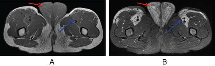
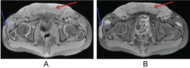
Figure 1: T1-weighted (A) and fat-suppressed T2-weighted; (B) axial MR images of the perineum demonstrate predominantly T1-hypointense and T2 hyperintense mass enlarging the labia majora (red arrows). There is extension along the subcutaneous tissues, muscle and myofascial planes of the proximal thighs (blue arrows).
View Figure 1
.
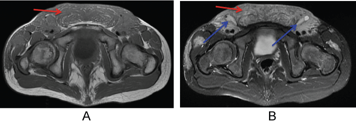
Figure 2: T1-weighted (A) and fat-suppressed T2-weighted; (B) axial MR images of the pelvis demonstrate predominantly T1-hypointense and T2 hyperintense mass enlarging the mons pubis (red arrows). Non-enlarged inguinal lymph nodes are noted bilaterally (blue arrows).
View Figure 2
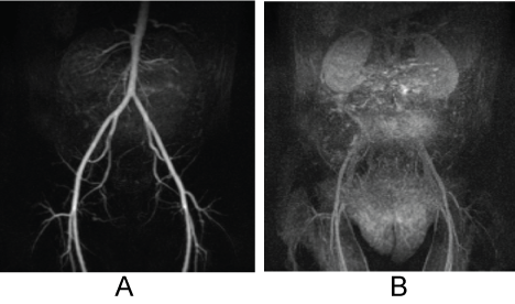
Contrast-enhanced MRA was performed, and showed no evidence of a dominant arterial feeding vessel (Figure 3A and Figure 3B). Standard fat-suppressed T1-weighted post contrast sequences were also obtained, and demonstrated diffusely heterogeneous enhancement throughout the lesion (Figure 4, Figure 5 and Figure 6).
.
Figure 3: Coronal MRA subtraction images of the lower abdomen and pelvis. Early image (A) demonstrates no evidence of a dominant arterial feeding vessel. Late image; (B) demonstrates diffuse parenchymal enhancement throughout the lesion.
View Figure 3
.
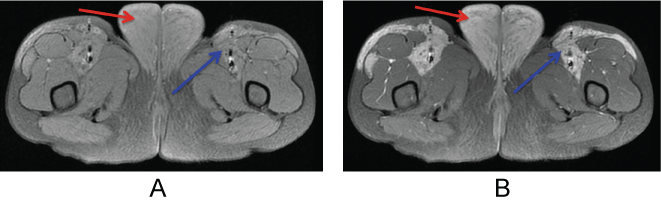
Figure 4: Pre-contrast (A) and post-contrast; (B) fat-suppressed T1-weighted axial MR images of the perineum demonstrate heterogeneous enhancement of the large mass of the labia majora (red arrows) and of the infiltrative components extending along the proximal thighs (blue arrows).
View Figure 4
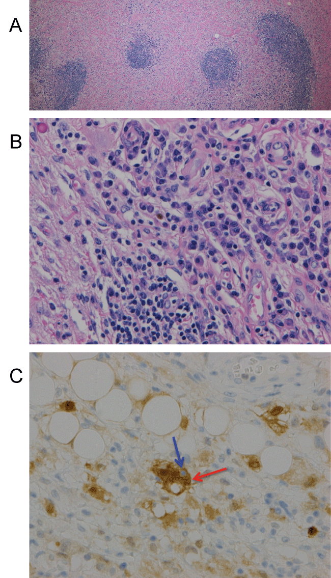
The patient's parents opted for staged resection of the mass, which was initially believed to represent a form of vascular malformation. Surgical pathology was notable for lymphoplasmacytic infiltrate (Figure 7A and Figure 7B) and S100 stain highlighting macrophages with rare emperipolesis (Figure 7C), a term used to describe the presence of an intact cell within the cytoplasm of another cell. The histopathologic findings were consistent with RDD.
.
Figure 5: Pre-contrast (A) and post-contrast; (B) fat-suppressed T1-weighted axial MR images of the pelvis demonstrate heterogeneous enhancement of the large mass of the mons pubis (red arrows) and of the infiltrative components extending along the proximal thighs (blue arrows).
View Figure 5
.
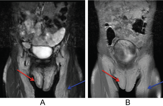
Figure 6: FSEIR (A) and post-contrast fat-suppressed T1-weighted; (B) coronal MR images of the pelvis demonstrate large enhancing mass of the external genitalia (red arrows) extending along the proximal thighs (blue arrows).
View Figure 6
Discussion
Rosai-Dorfman disease (RDD) also referred to as sinus histiocytosis with massive lymphadenopathy (SHML), is a rare, benign proliferative histiocytic disorder first described in 1969 by Rosai and Dorfman [1,2]. Systemic and cutaneous forms of the disease have been described, the systemic form occurring more commonly in the pediatric population and with a slight male predominance. Systemic RDD most typically presents as painless cervical lymphadenopathy with constitutional symptoms, including fever, night sweats, weight loss and anemia. Extranodal involvement by RDD has previously been reported in nearly half of all cases, including skin, head/neck and upper respiratory tract [3-5].
.
Figure 7: (A) Low power microscopic H&E image demonstrating lymphoid aggregates in a fibrogenic background; (B)High power microscopic H&E image showing lymphoplasmacytic infiltrate; (C) S100 stain highlighting macrophages with rare emperipolesis (blue ghost cell (blue arrow) within the central brown cell (red arrow)).
View Figure 7
Genitourinary involvement of extranodal RDD is a rarely described entity. Leiva, et al. described a case of an adult female with a retroperitoneal pelvic mass causing urinary obstruction [6]. Yamaguchi, et al. described a case of extranodal RDD involving the brain who subsequently developed bilateral ovarian involvement due to implantation following ventriculoperitoneal shunt placement [7]. In our case, extranodal RDD manifested as a diffuse genital mass without lymphadenopathy. Additionally of note, our patient was otherwise asymptomatic, and exhibited none of the clinical symptoms or laboratory derangements typically seen with systemic RDD such as fever, leukocytosis, and elevated erythrocyte sedimentation rate (ESR) [8].
Our patient's past medical history was significant only for Trisomy 21. While there are several known associations between Trisomy 21 and blood cell disorders such as transient myeloproliferative disorder, acute lymphoid leukemia, and acute myeloid leukemia [9], there are no reported links between Trisomy 21 and RDD. This may be a potential topic for future investigations.
The MR imaging features of the lesion were fairly non-specific. Due to the diffuse infiltrative nature of the lesion, as well as the clinical observations of skin discoloration and gradual growth of the lesion with growth of the child, the primary differential diagnostic considerations included venolymphatic vascular malformation, infiltrative vascular anomaly, soft tissue infection, and malignancy [10]. Given this wide range of considerations, imaging assessment alone cannot establish a diagnosis of RDD. The definitive diagnosis of RDD could only be made with tissue sampling and microscopic analysis. Pathologic specimens in RDD typically reveal diffuse histiocytosis associated with positive immunohistochemical stainingfor CD68 and S-100 proteins, but negative staining for CD1a. High power views of specimens demonstrate emperipolesis, the classic feature of RDD whereby intact lymphocytes are contained within the cytoplasm of many of the histiocytes [4,8,11]. In our case, the presence of lymphoid aggregates in a fibrogenic background and positive immunohistochemical stainingfor S100 protein demonstrating emperipolesis were consistent with RDD.
Manifestations of extranodal RDD have been treated with surgical resection, medical/chemotherapy and radiation therapy; however surgical therapy has been most successful [6]. In our case, staged surgical resection yielded modest results.
Conclusion
Primary genitourinary involvement of extranodal RDD is quite rare. We highlight the clinical presentation of a young girl with a genital mass, and MR imaging features with pathologic correlation in order to increase awareness of the diagnosis of extranodal RDD in the pediatric population.
References
-
Rosai J, Dorfman RF (1969) Sinus histiocytosis with massive lymphadenopathy. A newly recognized benign clinicopathological entity. Arch Pathol 87: 63-70.
-
Rosai J, Dorfman RF (1972) Sinus histiocytosis with massive lymphadenopathy: a pseudolymphomatous benign disorder. Analysis of 34 cases. Cancer 30: 1174-1188.
-
Woodcock RJ Jr, Mandell JW, Lipper MH (1999) Sinus histiocytosis (Rosai-Dorfman disease) of the suprasellar region: MR imaging findings-a case report. Radiology 213: 808-810.
-
Najafi-Sani M, Saneian H, Mahjoub F (2011) Rosai-Dorfman disease with nodal and extranodal involvements: A case report. J Res Med Sci 16: 1251-1256.
-
Baruah D, Guleria S, Chandra T, Kumar R, Jain KS (2007) Rosai-Dorfman disease with extensive extra nodal involvement. Eur J Radiol Extra 62: 31-33.
-
Leiva BJ, Bofill RM, Sevin BU, Geiger SJ (2006) Extranodal Rosai-Dorfman disease as a pelvic mass. Int J Gynecol Cancer 16: 312-315.
-
Yamaguchi M, Yahata T, Fujita K, Sakurada J, Hasegawa G, et al. (2009) Extranodal Rosai-Dorfman disease involving bilateral ovaries in a patient with a ventriculoperitoneal shunt. J Obstet Gynaecol Res 35: 1000-1003.
-
Mantilla JG, Goldberg-Stein S, Wang Y (2016) Extranodal Rosai-Dorfman disease: Clinicopathologic series of 10 patients with radiologic correlation and review of the literature. Am J Clin Pathol 145: 211-221.
-
Choi JK (2008) Hematopoietic disorders in Down Syndrome. Int J Clin Exp Pathol 1: 387-395.
-
Siegel MJ, Hoffer FA (2002) Magnetic resonance imaging of nongynecologic pelvic masses in children. Magn Reson Imaging Clin N Am 10: 325-344.
-
Lou X, Chen ZY, Wang FL, Ma L (2012) MR findings of Rosai-Dorfman disease in sellar and suprasellar region. Eur J Radiol 81: 1231-1237.
