

International Journal of Stem cell Research and Therapy
Development of Better Treatments for Retinal Disease Using Stem Cell Therapies
Rachel Gater1, Dan Nguyen1,2, Alicia J El Haj1 and Ying Yang1*
1Institute for Science and Technology in Medicine, School of Medicine, Keele University, UK
2Mid Cheshire Hospitals NHS Foundation Trust, Crewe, UK
*Corresponding author: Professor Ying Yang, Institute for Science and Technology in Medicine, School of Medicine
Keele University, Stoke-on-Trent, ST4 7QB, UK, Tel: +44 1782 674386, Fax: +44 1782 674467, E-mail: y.yang@keele.ac.uk
Int J Stem Cell Res Ther, IJSCRT-3-032, (Volume 3, Issue 1), Short Review; ISSN: 2469-570X
Received: April 24, 2016 | Accepted: May 24, 2016 | Published: May 28, 2016
Citation: Gater R, Nguyen D, Haj AJE, Yang Y (2016) Development of Better Treatments for Retinal Disease Using Stem Cell Therapies. Int J Stem Cell Res Ther 3:032. 10.23937/2469-570X/1410032
Copyright: © 2016 Gater R,et al. This is an open-access article distributed under the terms of the Creative Commons Attribution License, which permits unrestricted use, distribution, and reproduction in any medium, provided the original author and source are credited.
Abstract
The retina is a complex, light sensitive tissue layer on the inner surface of the eye, which functions to translate light stimuli into nerve impulses which travel to the brain via the optic nerve. Retinal diseases such as glaucoma, macular degeneration and retinitis pigmentosa can lead to significant vision problems and even blindness. Although there are some existing treatments for retinal disease, current treatments are invasive, need to be regularly repeated and do not have the capability of regenerating the damaged retinal architecture. The demand for more effective treatments for retinal disease is therefore high. Stem cell therapies and tissue engineering techniques for ocular disease have advanced significantly over recent years with promising results. However, challenges still remain with aspects such as; the delivery and integration of regenerative materials to the eye, overcoming the possibility of immune rejection and the guidance of neural growth to establish functional connections. This review describes the main approaches developed for retinal repair using regenerative medicine to date, including stimulating possible endogenous repair processes and cell replacement therapies. Subsequently, we report some of the newer and emerging strategies for retinal tissue engineering using biomaterials, which attempt to address the current challenges associated with retinal cell replacement therapy.
Keywords
Stem cell therapies, Retinal tissue engineering, Scaffold materials, Macular degeneration, Glaucoma, Retinitis pigmentosa
Introduction
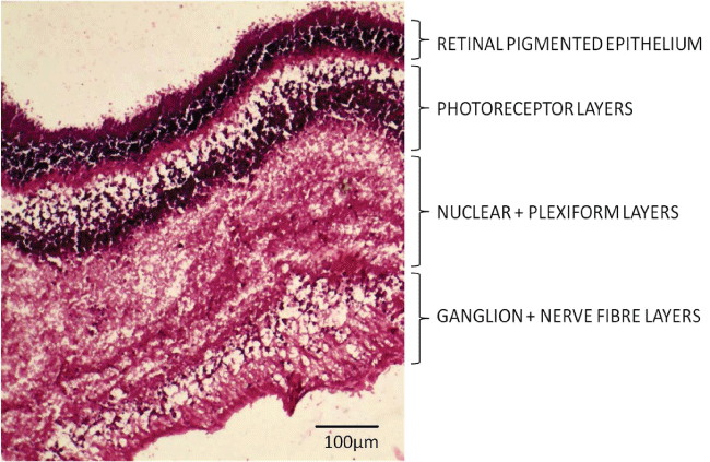
Retina is a vital tissue determining human vision. It lines the back of the eye and converts focused light from the lens into neural signals, and then sends these signals on to the brain for visual recognition [1]. The tissue is made up of several neuronal layers consisting of at least eight different cell types, as shown in figure 1. Degenerative diseases of the retina include glaucoma, macular degeneration, diabetic retinopathy and retinitis pigmentosa. According to the World Health Organisation, glaucoma and diabetic retinopathy account for approximately 17% of world blindness. Furthermore, age-related macular degeneration is ranked the third leading cause of world blindness, after cataract and glaucoma [2]. Macular degeneration and retinitis pigmentosa are typically characterised by the degeneration of the photoreceptors and retinal pigmented epithelium (RPE), a monolayer of cells which aligns a basement membrane, known as the Bruch's membrane, situated between the neural retina and the choroid [3]. Glaucoma is characterised by the degeneration of retinal ganglion cells (RGCs). These conditions can lead to significant vision problems and blindness if left untreated [4].
.
Figure 1: Optical image of a cross section of pig retinal tissue stained with hematoxylin and eosin, showing the multiple layered structure.
View Figure 1
Existing treatments for glaucoma include a surgical procedure named trabeculectomy, for the treatment of advanced glaucoma which can no longer be controlled with medical therapy. This procedure involves surgically making an incision in the conjunctiva and sclera so that fluid can egress into the sub-conjunctiva of the eye and thereby reduce intraocular pressure (IOP). However, the control of IOP after trabeculectomy deteriorates over time, furthermore surgery is an invasive procedure and can lead to inflammation [5] and infection. Alternatives to trabeculectomy such as a tube shunt procedure can still lead to complications such as bleeding inside the eye, infection and a buildup of fluid behind the retina [6]. Other retinal diseases such as macular degeneration or diabetic maculopathy can be treated by intravitreal injections containing ocular drug treatments to target the retina, which can be effective in restoring some level of vision. However, the injections are invasive, require repeated treatments and can lead to significant adverse events such as endophthalmitis, a potentially blinding infection [7]. There are currently no commercially available treatments for retinitis pigmentosa.
There is therefore need for more effective treatments for retinal diseases. Stem cell based therapies and tissue engineering for retinal disease has advanced significantly over recent years with promising results. However, challenges still remain with aspects such as; the delivery and integration of regenerative materials to the eye, overcoming the possibility of immune rejection and the guidance of axonal growth to establish useful connections [8]. This review describes the main approaches developed for retinal repair using regenerative medicine to date, including possible endogenous repair processes and cell replacement therapies. Subsequently, we report some of the newer and emerging strategies for retinal tissue engineering using biomaterials, which attempt to address the current challenges associated with retinal cell replacement therapy.
Endogenous Repair Processes
One approach to improve the treatment of retinal disease is the possibility of inducing endogenous repair processes to regenerate lost retinal neurons and damaged RPE. Studying the regenerative capability of organisms which already naturally possess the ability of retinal regeneration can give researchers an insight into the possibility of inducing endogenous regeneration in the human retina [9]. Potential sources of endogenous retinal regeneration include the RPE and Müller glia [10].
RPE cells
RPE cells of various species have demonstrated their differentiation potential into photoreceptor, amacrine, glial and ganglion cells [11-13]. Remarkably, it is known that certain species of amphibian, such as the newt, are capable of endogenous regeneration of the entire retina via RPE cells [14]. In an attempt to enhance the endogenous repair potential of mammalian RPE, some groups have investigated the regenerative potential of the mouse retina via RPE. One study found that intravenous administration of a low dose of sodium iodate in mice enhances RPE cell proliferation and migration by selectively injuring the RPE and subsequently triggering a sequence of pathophysiological effects which support endogenous regeneration of retinal tissue following injury [15]. However, further studies have also shown higher doses of sodium iodate to be toxic to RPE cells and retina [16]. Therefore, further experiments to investigate the correct dosage of sodium iodate would be encouraged to ensure its safety as a potential therapeutic. The existence of RPE stem cells in the human retina was reported recently in 2012 [17]. The regenerative potential of human RPE is still relatively unknown; however the possibility of RPE replacement for retinal repair in humans appears to be promising. This will be discussed further in the review.
Müller glia
Another potential source of endogenous regeneration is the Müller glia. It is known that organisms such as the Zebrafish are capable of endogenous retinal regeneration via the müller glia, as their müller glia de-differentiates and develops into proliferating neurogenic progenitor cells after injury [18]. Some in vitro studies have found that Müller glia from the primate retina can also develop into progenitor-like cells, but the capability to regenerate retinal neurons remains unclear [19]. Retinal regeneration via Müller glia-derived progenitor cells (MGPCs) requires five cellular actions, driven by signals emitted from the damaged or dying neurons. The first action is de-differentiation, where cells acquire progenitor phenotype and cease to function as glia. The second action is proliferation, which is required to produce a greater quantity of cells, including progeny to differentiate into new neurons and replace the Müller glia. However, it is believed that much of the proliferation which takes place in the mammalian retina is part of scar formation, rather than neuronal replacement [20]. The third action is the migration of dividing progenitor cells, in order to replace neurons within the various retinal layers. Following migration is the action of neural differentiation, to allow Müller glia derived cells to entirely differentiate into retinal neurons. The final action then involves integration into the retinal circuitry, where newly-generated neurons create synaptic connections in order to contribute in retinal function. Currently, this integration process also appears to be lacking in the bird and mammalian retina [21]. Initial studies in the human retina include Jayaram et al. (2014), who found that human MGPCs can differentiate into photoreceptors when cultured on basement membrane protein in the presence of taurine, fibroblast growth factor 2, insulin-like growth factor 1 and retinoic acid. Furthermore, they found that MGPCs had the potential to recover retinal function in a disease model of retinitis pigmentosa [22]. However, difficulty to culture primary human Müller glial cells means that current research is limited.
Cell Replacement Therapies
A more widely considered approach to improve the treatment of retinal disease is the possibility of retinal repair using cell replacement therapies. Therapies include embryonic stem cells (ESCs), induced pluripotent stem cells (iPSCs), mesenchymal stem cells (MSCs), adipose stem cells (ASCs) and retinal progenitor cells (RPCs), as well as RPE replacement. Many groups have successfully transplanted these cells into animal models and some therapies are even commencing into Phase I/II clinical trials.
Embryonic stem cells
ESCs are capable of self-renewal indefinitely when provided with the correct culture conditions, whilst still maintaining pluripotency [23]. Various research groups have found the potential to guide ESCs toward lineages such as RPE cells and photoreceptors [24], as well as RGCs [25], for transplantation into the retina. Some research groups have even found transplanted ESC derived photoreceptors to restore some level of visual function in Crx-deficient mice [26]. However, challenges with ESC derived cell transplantation include the ability to obtain stable cell sources that integrate safely into the diseased retina. There is concern that the plasticity of ESCs may be a problem, raising the risk of inappropriate progeny such as tumours [27]. A second challenge is the ability to develop a realistic up-scaling strategy in order to generate the high amount of cells required for transplantation into patients [28]. A third hurdle is the possibility of immuno rejection. The extensive remodelling that occurs following retinal degeneration can result in upregulation of inflammatory proteins and alteration of the blood-retinal barrier, which is likely to increase the chance of immuno rejection. Therefore, a better understanding of the immune status of a degenerated retinal environment is required [29]. A fourth major hurdle for the transplantation of stem cell derived retinal cells, such as photoreceptors and RGCs, is that very few cells manage to integrate successfully into the retina [30]. This has also been found for photoreceptors and RGCs derived from other types of stem cell.
Induced pluripotent stem cells
iPSCs are derived from reprogrammed peripheral cells (e.g. fibroblasts and lymphoblasts) into a pluripotent state. These cells can be directly generated from adult cells and therefore carry less ethical concerns than ESCs [31]. Most cell types in the retina (including RPE and photoreceptors) have been differentiated successfully from iPSCs [32]. Retinal cells derived from patient-specific iPSCs could be used for autologous transplantation, which has a reduced risk of immune rejection as it uses the patient's own cells. However, personalised cell therapy is time-consuming and expensive; furthermore iPSCs may require gene correction before transplantation. Human leukocyte antigen (HLA) matched allogeneic iPSCs, may also provide an alternative cell therapy in certain cases [33]. Information of current clinical trials using iPSCs can be found in table 1. Although ongoing clinical trials appear promising, previous trials such as an age-related macular degeneration treatment developed by the Masayo Takahashi group in Japan recently had to be halted due to a risk of tumour development [34]. Therefore, iPSC safety as a cell therapy is unknown until current and ongoing clinical trials have ran successfully and released their results.
![]()
Table 1: A summary of cell replacement therapies currently in development for retinal repair, including the main challenges and clinical trial information (adapted from http://clinicaltrials.gov).
View Table 1
Mesenchymal stem cells
Mesenchymal stem cells (MSCs) are multipotent stem cells derived from mesenchymal tissues such as bone marrow, adipose tissue, umbilical cord and placenta [35]. MSCs have been found to differentiate into photoreceptor-like cells, as well as RGC-like cells, making cell replacement therapy a future possibility using these cells. MSCs have also been found to provide neuroprotection for degenerating retinal cells via expression of a variety of neurotrophins which are beneficial for retinal cell survival [36]. Whilst retinal cell replacement therapy using MSCs remains problematic at the present time, some studies using MSCs for their neuroprotective mechanism are promising. For example, one study which involved the injection MSCs intravitreally into a mouse model of acute retinal injury, found that transplanted cells survived for at least 3 months and significantly protected damaged retinal cells long term [37].
Adipose stem cells
ASCs are mesenchymal stem cells which, like other stem cells, have the capacity to differentiate into multiple cell types. However, these cells are derived from the more easily accessible adipose tissue, rather than bone marrow [38]. The administration of ASCs into animal models of retinal degeneration is currently being investigated for the potential treatment of diseases such as diabetic retinopathy [39], glaucoma [40] and macular degeneration [41]. As autologous transplantation is also possible with ASCs, the risk of immune rejection is reduced. A transplantation study involving injection of human ASCs into a rat vitreous cavity found that cells survived and integrated with the eye relatively successfully. However, there was concern that some cells appeared to migrate across the blood-retina barrier and towards non-targeted regions, which could be considered a risk for transplantation to patients [42]. Therefore, more experiments to fully understand ASC cell mechanisms during transplantation will be required before an optimised treatment can be developed.
Retinal progenitor cells
RPCs are typically more predisposed towards a cell fate in comparison to stem cells because they are obtained from donated foetal tissue, rather than embryonic tissue [43]. Some groups have successfully transplanted this cell type into animal models of retinal degeneration [44] and some have even shown evidence of improved visual acuity in rats, due to photoreceptor preservation via transplanted RPCs [45]. Companies such as ‘ReNeuron' are currently commencing this therapy into Phase I/II clinical trials as a potential treatment for patients with retinitis pigmentosa [46,47]. If RPCs can be successfully transplanted into patients, one of the most likely challenges for this treatment will be whether cells are able to integrate into the complex human retinal circuitry. Subsequently, if evidence for photoreceptor preservation is observed, the ultimate challenge will then be whether the photoreceptors can function well enough to improve visual acuity. Furthermore, like ESCs donor tissue is used to obtain RPCs, meaning there could be a chance of immuno rejection.
RPE replacement
Another possible cell replacement therapy is replacement of the RPE. The ability to generate large amounts of functional RPE from ESCs and iPSCs makes this cell type an ideal candidate for transplantation [48]. However, RPE transplantation in suspension and RPE/choroid patch translocation has previously faced several challenges. Allogeneic RPE transplants have shown eventual rejection and/or a lack of functional recovery, plus autologous RPE transplants have also shown problems [49]. The aging process and degenerative disease causes the RPE to accumulate lipofuscin, which diminishes cellular structures and causes a slowing of metabolic capacity. The Bruch's membrane also accumulates debris, making nutrient transport less efficient and the survival/function of transplanted RPE challenging on such an altered substrate [50]. The future potential of each cell therapy will be unknown until current clinical study results are revealed. One notable clinical study was undertaken in 2012, where ESC derived RPE was transplanted into patients with stargardt's macular dystrophy and Macular degeneration. Results found transplanted RPE to have no apparent rejection after 4 months. Therefore, with further development this therapy could overcome the challenge of immune rejection and potentially be used for photoreceptor and central visual rescue [51]. A summary of all cell replacement therapies discussed, including current clinical trial information, can be found in table 1.
Retinal Tissue Engineering Approaches
Whilst cell replacement therapies appear promising, low cell survival and limited integration remain major challenges for this method of retinal repair. Furthermore it can be difficult to retain injected cells at specifically targeted regions of the retina, such as underneath the fovea [67]. Current tissue engineering approaches attempt to overcome these hurdles by supporting cell survival, delivery and integration. These include the development of 2 or 3-dimensional biomaterial scaffolds, as well as development of improved retinal disease models for the testing of neuroregenerative materials.
Scaffolds for retinal progenitor cell grafting
Many biomaterial scaffolds are currently in development for the grafting of RPCs. For example, Neeley et al. (2008) developed a microfabricated thin, porous poly (glycerol-sebacate) scaffold for RPC grafting. The reported scaffold possesses improved mechanical properties which are more closely similar to the retina in comparison to other scaffolds. Results indicate that RPCs adhered strongly to this scaffold and expressed a combination of immature and mature markers, suggesting a tendency towards cell differentiation [68]. In continuation of this work, the same research group has since transplanted the RPC grafted scaffold into mice. Transplantation proved relatively successful with long term RPC survival and expression of mature markers in the retinal tissue [69]. More recently, Yao et al. (2015) developed a biodegradable, thin-film polycaprolactone scaffold. Results indicate that the nano-topographies of the scaffold can interact with RPCs and have the potential to direct these cells towards photoreceptor cell fate before transplantation. The therefore scaffold provides an improved biodegradable platform to guide RPC differentiation and allowed for better organisation and delivery of RPCs to retinal tissue [70]. Although polycaprolactone is known to have improved biodegradability and good ability to drive hRPC differentiation into photoreceptors, poor adhesive qualities limit its use. Therefore, another research group has made a surface modification to a polycaprolactone scaffold by incorporating vitronectin-mimicking oligopeptide, forming a hybrid. Results showed improved cell adhesion of human RPCs and, despite no evidence of functional rescue, the RPCs showed improved migration, integration and survival when transplanted into a degenerated mouse retina on the hybrid scaffold [71].
Scaffolds for RPE transplantation
Many biomaterial scaffolds are also in development for the improvement of RPE transplantation. For example, Liu et al. (2014) developed a supporting scaffold on which RPE would be pre-cultured and supported to mature into a functional monolayer. The scaffold was tested in vitro using human foetal RPE cells and transplanted subretinally into rabbits. Results found that RPE culture was enhanced with the scaffold and showed improved subretinal biocompatibility with scaffolds of 200 nm fibre topography. Therefore, the use of biomaterial scaffolds can be considered a future direction for improving cell-based therapies to treat retinal disease [72]. However, challenges continue to remain in ensuring that scaffolds do not cause further damage to the retina, whilst still allowing for sufficient interaction between the retina and other cell layers. Therefore the surface topography, thickness, mechanical properties and degradation characteristics must be considered in scaffold design to ensure that they are suitable and beneficial for the treatment of ocular disease.
Biomaterials for potential RGC repair
At the moment, few biomaterial scaffolds have been developed for the treatment of RGC degenerative diseases such as glaucoma and optic nerve stroke. In diseases such as glaucoma RGCs are permanently damaged, leading to cell death and interruption of retinal signalling to the brain [73]. Due to their highly delicate and disparate axonal projection patterns, RGC replacement therapy for diseases such as glaucoma is highly challenging. Many more experiments to fully understand the complex in vivo circuitry and how replacement cells could integrate are required before a potential RGC replacement therapy can be developed [74]. Over the next few years however, we may see the development of biomaterial scaffolds and peripheral nerve bridges which attempt to repair injured endogenous ganglion cell axons, rather than replace them. Research into smart biodegradable implants aiding axonal regeneration following spinal cord injury has already been reported, which aim to stimulate repair by ‘bridging the gap' between damaged axon sites. This approach has significant potential to be used in the visual system and appears to be more promising than RGC replacement therapy at the present stage [75].
Development of in vitro or ex vivo retinal tissue models
Another tissue engineering strategy which will be hugely beneficial for improving the therapeutic potential of cell replacement therapies is the development of improved in vitro or ex vivo retinal tissue models, for the testing of neuroregenerative materials such as cell replacement therapies or drug intervention. Multicellular retinal tissue models with preserved cellular interactions are preferable in comparison to in vivo study using live animal models, as there are less ethical implications and the tissue environment can be better controlled and manipulated [76]. Organotypic slice culture involves the in vitro growth of complex tissues, such as the retina, whilst still preserving a large amount of in vivo function and physiology [77]. Many groups have successfully cultured rat retina using this method [78,79].
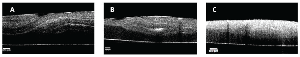
As the neuronal structure of the porcine retina is significantly similar to that of the human retina [80], it is preferable to explore organotypic explant culture of the porcine retina. One notable organotypic retinal tissue system developed recently preserves all layers of full thickness porcine retinal tissue for approximately 7 days. As the photoreceptor layers faces upwards, oxygen supply to the photoreceptors and RPE layer is increased and therefore improves the viability of cultured tissue [81]. However, the filter paper substrate used in this model appears to be very basic and is likely to dehydrate the retinal tissue. Another model currently in development attempts to address this issue further by culturing full thickness porcine retinal tissue on a more biocompatible substrate; collagen hydrogel. The collagen hydrogel mimics a vitreous-like substrate which better supports the integrity of full thickness retinal tissue explants with minimal distortion, as well as improving tissue viability for up to 2 weeks [82]. Figure 2 is an optical coherence tomography image showing a pig retinal explant cultured over 4 days on collagen hydrogel with delineated layered structure. With further development, it can be anticipated that the testing of potential cell replacement therapies and drug interventions on improved retinal tissue models such as this one will advance the progress of these cells into clinical trials and therapeutic applications significantly.
.
Figure 2: Optical coherence tomography images of a pig retinal explant cultured for 4 days. The layered structure was well resolved. The RPE layer is on the top of the sample.
View Figure 2
Conclusions + Future Perspectives
Evidently, the possibility of endogenous regeneration, as well as cell replacement therapies using ESCs, iPSCs, MSCs, ASCs, RPCs and RPE replacement are likely to become major implements in the future progress of regenerative medicine. To date, the main challenges of cell replacement therapy include; the delivery and integration of regenerative materials to the eye, overcoming the possibility of immune rejection and the guidance of neural growth to establish functional connections. The development of tissue-engineered constructs, such as scaffolds and smart biodegradable implants, may help to overcome some of these challenges by improving the delivery, integration and survival of transplanted cells. Furthermore, the testing of stem cell derivatives on improved retinal models which better mimic in vivo physiology and function will be hugely beneficial for exploring the potential therapeutic application of these cells. Assuming that enough funding will remain in this research field, a combination of the strategies discussed may revolutionise the way in which we treat retinal disease in the future.
References
-
Livesey FJ, Cepko CL (2001) Vertebrate neural cell-fate determination: lessons from the retina. Nat Rev Neurosci 2: 109-118.
-
WHO (2015) WHO | Priority eye diseases.
-
Strauss O (2005) The retinal pigment epithelium in visual function. Physiol Rev 85: 845-881.
-
Hartong DT, Berson EL, Dryja TP (2006) Retinitis pigmentosa. Lancet 368: 1795-1809.
-
Hong CH, Arosemena A, Zurakowski D, Ayyala RS (2005) Glaucoma drainage devices: a systematic literature review and current controversies. SurvOphthalmol 50: 48-60.
-
Gross RL (2012) Glaucoma filtration surgery: Trabeculectomy or tube shunt? American Journal of Ophthalmology, 153: 787-788.
-
Falavarjani KG, Nguyen QD (2013) Adverse events and complications associated with intravitreal injection of anti-VEGF agents: a review of literature. Eye (Lond) 27: 787-794.
-
Ramsden CM, Powner MB, Carr AJ, Smart MJ, da Cruz L, et al. (2013) Stem cells in retinal regeneration: past, present and future. Development 140: 2576-2585.
-
Lamba D, Karl M, Reh T (2008) Neural regeneration and cell replacement: a view from the eye. Cell Stem Cell 2: 538-549.
-
Tsonis PA, Del Rio-Tsonis K (2004) Lens and retina regeneration: Transdifferentiation, stem cells and clinical applications. Exp Eye Res 78: 161-172.
-
Zhao S, Thornquist SC, Barnstable CJ (1995) In vitro transdifferentiation of embryonic rat retinal pigment epithelium to neural retina. Brain Research 677: 300-310.
-
Ma W, Yan RT, Xie W, Wang SZ (2004) bHLH genes cath5 and cNSCL1 promote bFGF-stimulated RPE cells to transdifferentiate toward retinal ganglion cells. Dev Biol 265: 320-328.
-
Sakaguchi DS, Janick LM, Reh TA (1997) Basic fibroblast growth factor (FGF-2) induced transdifferentiation of retinal pigment epithelium: Generation of retinal neurons and glia. Developmental Dynamics 209: 387-398.
-
Mitashov VI (1975) Proliferation of cells of pigmentary epithelium of retina in adult newts at late regeneration stages after retinectomy. The Soviet journal of developmental biology 5: 70-73.
-
Machalińska A, Kawa MP, Pius-Sadowska E, Rogińska D, Kłos P, et al. (2013) Endogenous regeneration of damaged retinal pigment epithelium following low dose sodium iodate administration: an insight into the role of glial cells in retinal repair. Exp Eye Res 112: 68-78.
-
Wang J, Iacovelli J, Spencer C, Saint-Geniez M (2014) Direct effect of sodium iodate on neurosensory retina. Invest Ophthalmol Vis Sci 55: 1941-1953.
-
Salero E, Blenkinsop TA, Corneo B, Harris A, Rabin D, et al. (2012) Adult human RPE can be activated into a multipotent stem cell that produces mesenchymal derivatives. Cell Stem Cell 10: 88-95.
-
Fausett BV, Goldman D (2006) A role for alpha1 tubulin-expressing Müller glia in regeneration of the injured zebrafish retina. J Neurosci 26: 6303-6313.
-
Limb GA, Daniels JT (2008) Ocular regeneration by stem cells: present status and future prospects. Br Med Bull 85: 47-61.
-
Gallina D, Todd L, Fischer AJ (2014) A comparative analysis of Müller glia-mediated regeneration in the vertebrate retina. Exp Eye Res 123: 121-130.
-
Gorsuch RA, Hyde DR (2014) Regulation of Müller glial dependent neuronal regeneration in the damaged adult zebrafish retina. Exp Eye Res 123: 131-140.
-
Jayaram H, Jones MF, Eastlake K, Cottrill PB, Becker S, et al. (2014) Transplantation of photoreceptors derived from human Muller glia restore rod function in the P23H rat. Stem Cells Transl Med 3: 323-333.
-
Pera MF, Reubinoff B, Trounson A (2000) Human embryonic stem cells. J Cell Sci 113: 5-10.
-
Klassen H,Sakaguchi DS, Young MJ (2004) Stem cells and retinal repair. Prog Retin Eye Res 23: 149-181.
-
Teotia P, Mir Q, Ahmad I (2015) Chemically defined and retinal conditioned medium-based directed differentiation of embryonic stem and induced pluripotent stem cells into retinal ganglion cells. Investigative Ophthalmology & Visual Science 56: 3606.
-
Lamba DA, Gust J, Reh TA, (2009) Transplantation of human embryonic stem cell-derived photoreceptors restores some visual function in Crx-deficient mice. Cell stem cell 4: 73-79.
-
Stern J, Radosevich M, Temple S (2010) Stem Cell Retinal Replacement Therapy, 2010 World stem cell summit proceedings 57-61.
-
Reynolds J, Lamba DA (2014) Human embryonic stem cell applications for retinal degenerations. Experimental eye research 12: 151-160.
-
Bongso A, Fong CY, Gauthaman K (2008) Taking stem cells to the clinic: Major challenges. J Cell Biochem 105: 1352-1360.
-
Venugopalan P, Wang Y, Nguyen T, Huang A, et al. (2016) Transplanted neurons integrate into adult retinas and respond to light. Nat Commun 7: 10472.
-
Robinton DA, Daley GQ (2012) The promise of induced pluripotent stem cells in research and therapy. Nature 481: 295-305.
-
Wright LS, Phillips MJ, Pinilla I, Hei D, Gamm DM (2014) Induced pluripotent stem cells as custom therapeutics for retinal repair: progress and rationale. Exp Eye Res 123: 161-172.
-
Riolobos L, Hirata RK, Turtle CJ, Wang PR, Gornalusse GG, et al. (2013) HLA engineering of human pluripotent stem cells. Mol Ther 21: 1232-1241.
-
Landmark IPSC clinical study on hold due to genomic issue (2015) The Niche.
-
Caplan AI (1991) Mesenchymal stem cells. J Orthop Res 9: 641-650.
-
Xu W, Xu GX (2011) Mesenchymal stem cells for retinal diseases. Int J Ophthalmol 4: 413-421.
-
Anna Machalińska, Miłosz Kawa, Ewa Pius-Sadowska,Jacek Stępniewski, Witold Nowak (2013) Long-term neuroprotective effects of NT-4-engineered mesenchymal stem cells injected intravitreally in a mouse model of acute retinal injury. Investigative ophthalmology & visual science 54: 8292-8305.
-
Gir P, Oni G, Brown SA, Mojallal A, Rohrich RJ (2012) Human adipose stem cells: current clinical applications. Plastic and reconstructive surgery 129: 1277-1290.
-
Yang Z, Li K, Yan X, Dong F, Zhao C (2010) Amelioration of diabetic retinopathy by engrafted human adipose-derived mesenchymal stem cells in streptozotocin diabetic rats. Graefes archive for clinical and experimental ophthalmology 248: 1415-1422.
-
Vayl A, Gomaa A, Rajashekhar G (2014) Adipose stromal cells atenuate p38 mapk in retinal ischemia-reperfusion injury. Investigative Ophthalmology & Visual Science 55: 4005.
-
Sang-Joon Lee, Jeong Hoon Heo, Jung Ae Yoon, Byung Chul Yu (2015) Intraperitoneal administration of adipose tissue derived stem cells rescue Retinal degeneration in mouse model. Investigative Ophthalmology & Visual Science 56: 1842.
-
Haddad-Mashadrizeh A, Ahmad R Bahrami, Maryam M. Matin, Mohammad A. Edalatmanesh , Alireza Zomorodipour , et al. (2013) Human adipose-derived mesenchymal stem cells can survive and integrate into the adult rat eye following xenotransplantation. Xenotransplantation 20: 165-176.
-
Schmitt S, Aftab U, Jiang C, Redenti S, Klassen H, et al. (2009) Molecular characterization of human retinal progenitor cells. Invest Ophthalmol Vis Sci 50: 5901-5908.
-
Klassen HJ, Ng TF, Kurimoto Y, Kirov I, Shatos M, et al. (2004) Multipotent retinal progenitors express developmental markers, differentiate into retinal neurons, and preserve light-mediated behavior. Invest Ophthalmol Vis Sci 45: 4167-4173.
-
Luo J, Baranov P, Patel S, Ouyang H, Quach J, et al. (2014) Human retinal progenitor cell transplantation preserves vision. Journal of Biological Chemistry 289: 6362-6371.
-
ReNeuron (2016) Phase I/II clinical trial in retinitis pigmentosa - ReNeuron.
-
RNS (2016) First Patient Treated in RP Clinical Trial | ReNeuron Group plc (RENE) | RNS Company Announcements | Equities | FE Trustnet.
-
Kokkinaki M, Sahibzada N, Golestaneh N (2011) Human induced pluripotent stem-derived retinal pigment epithelium (RPE) cells exhibit ion transport, membrane potential, polarized vascular endothelial growth factor secretion, and gene expression pattern similar to native RPE. Stem cells 29: 825-835.
-
Kohen L, Enzmann V, Faude F, Wiedemann P (1997) Mechanisms of graft rejection in the transplantation of retinal pigment epithelial cells. Ophthalmic Res 29: 298-304.
-
Binder S, Stanzel BV, Krebs I, Glittenberg C (2007) Transplantation of the RPE in AMD. ProgRetin Eye Res 26: 516-554.
-
Schwartz SD, Hubschman JP, Heilwell G, Franco-Cardenas V, Pan CK, et al. (2012) Embryonic stem cell trials for macular degeneration: a preliminary report. Lancet 379: 713-720.
-
ClinicalTrials.gov (2016) Study of Subretinal Implantation of Human Embryonic Stem Cell-Derived RPE Cells in Advanced Dry AMD - Full Text View - ClinicalTrials.gov.
-
ClinicalTrials.gov (2016) Safety and Efficacy Study of OpRegen for Treatment of Advanced Dry-Form Age-Related Macular Degeneration - Full Text View - ClinicalTrials.gov.
-
ClinicalTrials.gov (2016) Long Term Follow Up of Sub-retinal Transplantation of hESC Derived RPE Cells in Stargardt Macular Dystrophy Patients - Full Text View - ClinicalTrials.gov.
-
ClinicalTrials.gov (2015) Safety and Tolerability of Sub-retinal Transplantation of hESC Derived RPE (MA09-hRPE) Cells in Patients With Advanced Dry Age Related Macular Degeneration - Full Text View - ClinicalTrials.gov.
-
ClinicalTrials.gov (2015a) Long Term Follow Up of Sub-retinal Transplantation of hESC Derived RPE Cells in Patients With AMD - Full Text View - ClinicalTrials.gov.
-
ClinicalTrials.gov (2012) A Phase I/IIa, Open-Label, Single-Center, Prospective Study to Determine the Safety and Tolerability of Sub-retinal Transplantation of Human Embryonic Stem Cell Derived Retinal Pigmented Epithelial(MA09-hRPE) Cells in Patients With Advanced Dry Age-related Macular Degeneration(AMD).
-
ClinicalTrials.gov (2016) Cell Collection to Study Eye Diseases - Full Text View - ClinicalTrials.gov.
-
ClinicalTrials.gov (2016) Stem Cell Models of Best Disease and Other Retinal Degenerative Diseases. - Full Text View - ClinicalTrials.gov.
-
ClinicalTrials.gov (2016) Feasibility of Generating Pluripotent Stem Cells From Patients With Familial Retinoblastoma - Full Text View - ClinicalTrials.gov.
-
ClinicalTrials.gov (2015b) Production of iPSC Derived RPE Cells for Transplantation in AMD - Full Text View - ClinicalTrials.gov.
-
ClinicalTrials.gov (2016) Stem Cell Ophthalmology Treatment Study - Full Text View - ClinicalTrials.gov.
-
ClinicalTrials.gov (2015) Effectiveness and Safety of Adipose-Derived Regenerative Cells for Treatment of Glaucomatous Neurodegeneration - Full Text View - ClinicalTrials.gov.
-
ClinicalTrials.gov (2016) Safety of a Single, Intravitreal Injection of Human Retinal Progenitor Cells (jCell) in Retinitis Pigmentosa - Full Text View - ClinicalTrials.gov.
-
ClinicalTrials.gov (2016) A Study Of Implantation Of Retinal Pigment Epithelium In Subjects With Acute Wet Age Related Macular Degeneration - Full Text View - ClinicalTrials.gov.
-
ClinicalTrials.gov (2008) Transplantation of Autologous RPE Versus Translocation of Autologous RPE and Choroid in AMD - Full Text View - ClinicalTrials.gov.
-
Kador K, Goldberg J (2013) Scaffolds and stem cells: delivery of cell transplants for retinal degenerations. Expert Review Opthamology 7: 459-470.
-
Neeley WL, Redenti S, Klassen H, Tao S, Desai T, et al. (2008) A microfabricated scaffold for retinal progenitor cell grafting. Biomaterials 29: 418-426.
-
Redenti S, Neeley WL, Rompani S, Saigal S, Yang J (2009) Engineering retinal progenitor cell and scrollable poly (glycerol-sebacate) composites for expansion and subretinal transplantation. Biomaterials 30: 3405-3414.
-
Yao Jing, Ko Chi Wan, Baranov Petr Y, Regatieri Caio V, Redenti Stephen (2015) Enhanced Differentiation and Delivery of Mouse Retinal Progenitor Cells Using a Micropatterned Biodegradable Thin-Film Polycaprolactone Scaffold. Tissue Engineering Part A 21: 1247-1260.
-
Lawley E, Baranov P, Young M (2015) Hybrid vitronectin-mimicking polycaprolactone scaffolds for human retinal progenitor cell differentiation and transplantation. Journal of biomaterials applications 29: 894-902.
-
Liu Z, Yu N, Holz FG, Yang F, Stanzel BV (2014) Enhancement of retinal pigment epithelial culture characteristics and subretinal space tolerance of scaffolds with 200 nm fiber topography. Biomaterials 35: 2837-2850.
-
Sernagor E, Eglen SJ, Wong RO (2001) Development of retinal ganglion cell structure and function. ProgRetin Eye Res 20: 139-174.
-
Harvey AR, Hu Y, Leaver SG, Mellough CB, Park K, et al. (2006) Gene therapy and transplantation in CNS repair: the visual system. Progress in retinal and eye research 25: 449-489.
-
Joosten EA (2012) Biodegradable biomatrices and bridging the injured spinal cord: the corticospinal tract as a proof of principle. Cell Tissue Res 349: 375-395.
-
Kim SU, Takahashi H (1988) Tissue culture study of adult human retina neurons. Investigative Ophthalmology & Visual Science 29: 1372-1379.
-
Gähwiler BH, Capogna M, Debanne D, McKinney RA, Thompson SM (1997) Organotypic slice cultures: a technique has come of age. Trends Neurosci 20: 471-477.
-
Johnson TV, Martin KR (2008) Development and characterization of an adult retinal explant organotypic tissue culture system as an in vitro intraocular stem cell transplantation model. Investigative Ophthalmology and Visual Science 49: 3503-3512.
-
Viktorov IV, Lyzhin AA, Aleksandrova OP, Frayer D, Dirnagl U (2004) Roller organotypic cultures of postnatal rat retina. Bull ExpBiol Med 137: 419-422.
-
Guduric-Fuchs J, Ringland LJ, Gu P, Dellett M, Archer DB, et al. (2009) Immunohistochemical study of pig retinal development. Mol Vis 15: 1915-1928.
-
Wang J, Kolomeyer AM, Zarbin MA, Townes-Anderson E (2011) Organotypic culture of full-thickness adult porcine retina. J Vis Exp .
-
(2016) Dynamics and Fluctuations in Biomedical Photonics XIII, 9707.
