

Journal of Otolaryngology and Rhinology
Suspected Aspiration of a Patency Capsule
Claude F Harbarger1* and Brian J Wiatrak2
1Department of Otolaryngology, University of Mississippi Medical Center, USA
2Children's of Alabama, USA
*Corresponding author:
Claude F Harbarger, Department of Otolaryngology, University of Mississippi Medical Center, 2500 North State Street, Jackson, MS 39216, USA, E-mail: cfharbarger@umc.edu
J Otolaryngol Rhinol,
JOR-1-002, (Volume 1, Issue 1),
Case Report
Received: July 19, 2015: Accepted: August 19, 2015: Published: August 21, 2015
Citation: Harbarger CF, Wiatrak BJ (2015) Suspected Aspiration of a Patency Capsule. J Otolaryngol Rhinol 1:002
Copyright: © 2015 Harbarger CF. This is an open-access article distributed under the terms of the Creative Commons Attribution License, which permits unrestricted use, distribution, and reproduction in any medium, provided the original author and source are credited.
Abstract
Video capsule endoscopy (VCE) is an established means of evaluating the upper and lower aerodigestive tracts, with 3 companies now having received FDA clearance for their devices. The most prevalent risk of VCE is capsule retention, which infrequently results in bowel obstruction. In patients suspected of having a high risk of capsule retention, a dissolvable patency capsule is given a few days prior to the VCE procedure to ensure ultimate passage of the VCE device. Here we report what is, to our knowledge, the first case of aspiration of a patency capsule.
Introduction
The patency capsule is similar in size to the video capsule, but is made of a dissolvable mixture of barium and lactose, and also includes a radio tag inside the capsule. The patency capsule is thus capable of being detected with x-ray and also with a radio-scanner externally. Specially designed wax plugs on each end contain holes that allow intestinal fluid to enter and dissolve the capsule, usually within 30 to 72 hours. Here, we report an unusual case of patency capsule aspiration.
Case Report
A 21 year old man with a history of Duchenne muscular dystrophy, and esophageal dysphagia with a history of gastrostomy tube dependence, was admitted for a 2 day history of progressive vomiting. He also had a history of peptic ulcers and intestinal obstruction 2 years prior to presentation. He was cachectic on exam, and stated that his gastrostomy tube became displaced 3 months prior, and that he declined to have it replaced, instead obtaining all nutrition orally. A computed tomography scan was suspicious for gastric outlet obstruction, and an orogastric tube was placed for bowel decompression. An upper gastrointestinal series and small bowel follow-through showed no evidence of upper small bowel obstruction, and a subsequent gastric empyting study was normal.
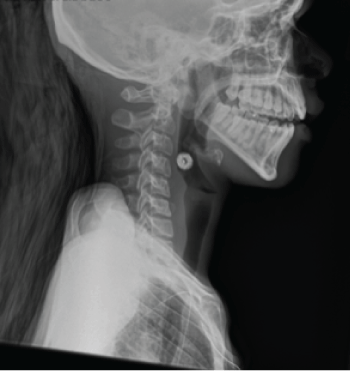
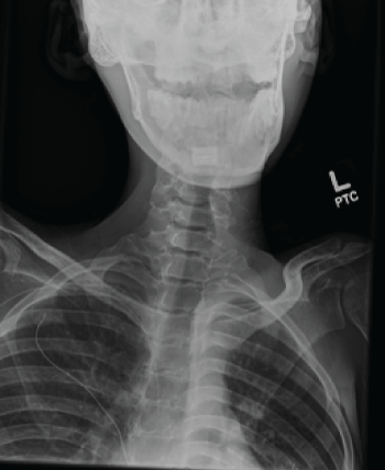
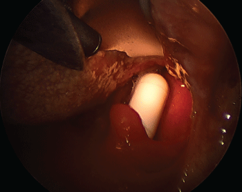
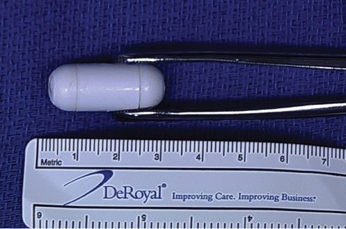
There was also a concern for an upper gastrointestinal bleed because of hematemesis and anemia on admission, and gastrointestinal medicine was consulted for evaluation. After discussing the risks of anesthesia entailed in a sedated esophagogastroduodenoscopy, the patient elected to undergo VCE, which has been reported to have some utility in acute gastrointestingal bleeding [1]. A decision was made to perform VCE, but to precede this with a patency capsule. After oral administration of the patency capsule, the patient had clear difficulty swallowing the capsule. He began choking and stated that he felt it was lodged in his throat. Shortly thereafter he became apneic and indicated that he could not breathe. An emergency was declared by the present staff, and the Heimlich maneuver was performed. Although this did not produce expectoration of the patency capsule, he was then able to breathe and phonate, and said that felt he the capsule was still lodged in his throat. Both lateral and AP and neck films were urgently performed, as seen in figures 1 and 2. He was then taken emergently to the operating room, where direct laryngoscopy revealed the foreign body to be lodged in the post-cricoid space and resting within the interarytenoid notch. Removal of the foreign body was performed with a Magill forceps, as seen in figures 3 and 4. He tolerated this well and recovered uneventfully.
.
Figure 3: Foreign body immediately viewed upon direct laryngoscopy, seen to be lying on posterior pharyngeal wall above laryngeal inlet
View Figure 3
Discussion
Since its introduction into clinical practice in 2001, VCE has become a common method of evaluation of disease of both the small and large intestine. Complications are rare, and most commonly relate to capsule retention and resultant bowel obstruction. Accordingly, VCE is contraindicated in patients with known intestinal strictures or fissures, in cases of suspected gastrointestinal obstruction, or in pregnancy. Also, individuals who have undergone multiple abdominal surgeries, or who are otherwise not surgical candidates, are also not generally candidates for VCE. Patients with risk factors for aspiration, such as swallowing disorders, neurologic disorders, a weak or absent cough, or Zenker's diverticula, are otherwise not usually candidates for VCE [2-4]. VCE is generally discouraged in children under the age of 10 years.
A more remote risk of VCE is aspiration, with an estimated prevalence of 1 out of every 800 procedures. Several reports have documented aspiration of the VCE capsule [5-9], with the potential for an aspiration to even be asymptomatic. Aspiration usually requires urgent imaging to localize the capsule, followed by bronchoscopy to retrieve it [10,11]. It has been suggested that practitioners specifically question the patient about difficulties swallowing solid foods and pills before proceeding with VCE [9]. Others have recommended a preliminary evaluation with a video esophagram in patients with a history or risk of aspiration [7].
In patients with risk factors for aspiration, directed placement of the capsule into the duodenum should be considered. This would have been the preferred method of placement for our patient, who had a known esophageal dysphagia and a diagnosis of muscular dystrophy. A real-time viewer can also be used during the ingestion of the capsule to ensure that the capsule reaches the gastrointestinal tract [3,4,12]. Only one death has been reported from aspiration during VCE, with a report of intracerebral hemorrhage resulting from capsule aspiration [4]. Lucendo et al. surmised that the capsule diameter is smaller than the tracheal luminal diameter, thus allowing adequate oxygenation after capsule aspiration in most instances [10]. Koulaouzidis et al. reported that the capsule size might be correlated with the chance of aspiration [13]. In retrospect, in our patient was probably a poor candidate for capsule endoscopy, due to his history of muscular dystrophy, esophageal dysphagia, and the fact that the origin of his bleed was likely the upper GI tract, with VCE being better indicated in cases of lower GI bleeding. We suspect that the apneic event experienced by our patient was due to temporary laryngeal obstruction, which was immediately relieved with the Heimlich maneuver. While not available at our institution, transnasal esophagoscopy under topical anesthesia would be an excellent alternative to localize the bleeding source in such a patient. As with most case reports cited previously, our patient did not have an adverse outcome, as the capsule was removed without complication.
We believe this case highlights the importance of following proper indications for VCE, assessing the risk of aspiration in patients with possible swallowing disorders prior to embarking on VCE, and the need for endoscopic placement of patency capsules in patients with swallowing disorders.
References
-
Nadler M, Eliakim R (2014) The role of capsule endoscopy in acute gastrointestinal bleeding. Therap Adv Gastroenterol 2014 7: 87-92.
-
Ali A, Santisi JM, Vargo J (2004) Video capsule endoscopy: A voyage beyond the end of the scope. Cleve Clin J Med 71: 415-425.
-
Ding NS, Hair C, De Cruz P, Watson J (2013) Education and Imaging. Gastrointestinal: symptomatic bronchial aspiration of capsule endoscope - a significant complication. J Gastroenterol Hepatol 28:761
-
Parker C, Davison C, Panter S (2012) Tracheal aspiration of a capsule endoscope: not always a benign event. Dig Dis Sci 57: 1727-1728.
-
Borman JJ, Hilbelink RT (2011) Video endoscope aspiration. Appl Radiol 40: 37-38.
-
Schneider AR, Hoepffner N, Rosch W, Caspary WF (2003) Aspiration of an M2A capsule. Endoscopy 35: 713.
-
Sinn I, Neef B, Andus T (2004) Aspiration of a capsule endoscope. Gastrointest Endosc 59: 926-927.
-
Tabib S, Fuller C, Daniels J, Lo S (2004) Asymptomatic aspiration of a capsule endoscope. Gastrointest Endosc 60: 845-848.
-
Fleischer DE, Heigh RI, Nguyen CC, Leighton JA, Sharma VK, et al. (2003) Videocapsule impaction at the cricopharyngeus: A first report of this complication and its successful resolution. Gastrointest Endosc 57: 427-428.
-
Lucendo AJ, Gonzalez-Castillo S, Fernandez-Fuente M, De Rezende LC (2011) Tracheal aspiration of a capsule endoscope: a new case report and literature compilation of an increasingly reported complication. Dig Dis Sci 56: 2758-2762.
-
Pezzoli A, Fusetti N, Carella A, Gullini S (2011) Asymptomatic bronchial aspiration and prolonged retention of a capsule endoscope: a case report. J Med Case Rep 5: 341.
-
Despott EJ, O'Rourke A, Anikin V, Davison C, Panter S, et al. (2012) Tracheal aspiration of capsule endoscopes: detection, management, and susceptibility. Dig Dis Sci 57:1973-1974.
-
Koulaouzidis A, Douglas S, Plevris JN (2011) Tracheal aspiration of capsule endoscopes: completing a cases compilation. Dig Dis Sci 56: 3101-3102.
