

Obstetrics and Gynaecology Cases - Reviews
An Incidental Diagnosis of the Cystadenofibroma of the Ovary in the Cesarean Section: A Case Report
Hasan Onur Topçu*, Zeynep Asli Oskovi, Ali Irfan Güzel, Irfan Özer, Mahmut Kuntay Kokanali and Sabri Cavkaytar
Zekai Tahir Burak Women's Health Education and Research Hospital, Ankara, Turkey
*Corresponding author: Hasan Onur Topçu, Zekai Tahir Burak Women Health Education and Research Hospital, Ankara, Turkey, Tel: +90 532 635 95 38; Fax: +90 312 306 59 17; E-mail: dronurtopcu@gmail.com
Obstet Gynecol Cases Rev, OGCR-1-002, (Volume 1, Issue 1), Case Report; ISSN: 2377-9004
Received: September 02, 2014 | Accepted: October 24, 2014 | Published: October 27, 2014
Citation: Topcu HO, Oskovi ZA, Güzel AI, Özer I, Kokanali MK, et al. (2014) An Incidental Diagnosis of the Cystadenofibroma of the Ovary in the Cesarean Section: A Case Report. Obstet Gynecol Cases Rev 1:002. 10.23937/2377-9004/1410002
Copyright: © 2014 Topcu HO, et al. This is an open-access article distributed under the terms of the Creative Commons Attribution License, which permits unrestricted use, distribution, and reproduction in any medium, provided the original author and source are credited.
Abstract
Ovarian cystadenofibromas are relatively rare epithelial ovarian tumors which contain fibrous stroma besides the epithelial proliferation. They may mimic malignant neoplasms with their gross appearance or by imaging modalities. In our case, an incidental ovarian mass was detected in a 41 year-old patient during cesarean section with an appearance like a malignant tumor. We did not have the chance for frozen-section diagnosis because the emergency cesarean section was performed in night shift conditions. Right salpingo-oophorectomy was performed and final pathology was reported as an ovarian serous cystadenofibroma. The awareness of the surgeons of this entity is particularly important to prevent the patient from unnecessary management in the operation in emergency conditions when frozen diagnosis is not possible.
Introduction
Ovarian cystadenofibromas are relatively rare epithelial ovarian tumors which contain fibrous stroma besides the epithelial proliferation. In a study, the incidence of ovarian cystadenofibroma is reported as 1.7% of all ovarian tumors while the age of the patients ranged between 12-85 years, with a median age of 52 years [1]. The classification of cystadenomas is based on the epithelial types of the tumor as serous, endometrioid, mucinous, clear cell and mixed category [2]. The behavior of the tumor is described as benign, borderline and malignant depending on the degree of epithelial proliferation [2]. Ovarian cystadenofibromas have a gross appearance of multicystic solid mass which may mimic ovarian malignancy. In this report, we present a case of serous cystadenofibroma of the ovary which was incidentally found in cesarean section.
Case
A 41 year-old female presented to our hospital with complaints of labor contractions. She had 39 weeks of gestation and her previous delivery was performed with cesarean section. The patient did not have any complaint of pelvic pain before or during pregnancy. She had admitted to other hospitals during the pregnancy follow-up, and any suspicious adnexal findings were detected at the ultrasonographic examination before the delivery at our clinic due to the difficulty of the imaging adnexa in women with term pregnancy. A cesarean section was performed because of the continuous regular contractions and a healthy 3230 gr baby was delivered. Exploration in the surgery revealed a 70x60x80mm mass with cystic and solid components on the right ovary (Figure 1). The appearance of the mass gave an impression of a malignant neoplasm; therefore, right salpingo-oophorectomy was performed (Figure 2). Frozen section diagnosis was not possible at the time of the operation due to the night shift conditions. The final result of pathology was reported as a serous cystadenofibroma of the ovary.
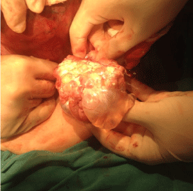 Figure 1: Exploration in the surgery revealed a 70x60x80mm mass with cystic
and solid components on the right ovary.
View Figure1
Figure 1: Exploration in the surgery revealed a 70x60x80mm mass with cystic
and solid components on the right ovary.
View Figure1
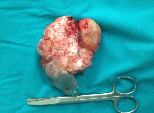 Figure 2: The appearance of the mass as an impression of a malignant
neoplasm.
View Figure2
Figure 2: The appearance of the mass as an impression of a malignant
neoplasm.
View Figure2
Discussion
Ovarian cystadenofibromas are rare and mostly asymptomatic epithelial tumors which can be seen both in premenopausal and postmenopausal period that may give a malignant tumor impression by a macroscopic analysis or with radiological imaging modalities [3]. Tumor sizes range between 2.5 and 20 cm, with a mean 8.7cm [1].They can be purely cystic or complex cystic with solid components [1].
Ovarian masses can be evaluated by using simple ultrasound rules for presumption of malignity. An irregular solid tumor, presence of ascites, more than three papillary structures, irregular multilocular solid tumor with largest diameter >100 mm, increased blood flow are ultrasound criteriadescribed for increased risk of malignity while unilocular ovarian cysts, presence of solid components with a largest diameter of less than 7 mm, presence of acoustic shadows and smooth multilocular tumor with a largest diameter smaller than 100 mm are criteria that are useful for prediction of benign lesions [4]. CT and MR are useful modalities for characterizing complex adnexal masses.
Sonographic features of ovarian serous cystadenofibromas were described by Alc�zar et al. [3] as uniocular or multilocular cystic structures which may be purely anechoic or contain septations, papillary projections or solid nodules.Blood flow can be detected by color Doppler in half of the cases usually on peripheral cyst wall and in solid areas and sometimes the blood flow impedance can be low and give false-positive results [3].
Computed Tomography (CT) has a limited value in evaluation of cystadenofibromas as it can cause preoperative misdiagnosis of malignancy [1]. MRI has been described as a reliable modality for evaluation of complex adnexal masses; the presence of very low signal intensity on T2 weighted images of fibrous tissue of the solid componentsis described as MRI characteristics of ovarian cystadenofibromas [5- 7]. Differential diagnosis of ovarian cystadenofibroma includes some other benign tumors with fibrous components like fibroma, fibrotecoma and Brenner tumor which have similar MRI findings, and malignant tumors like clear cell carcinoma and granulosa cell tumor because of their cystic and solid appearance [1, 5, 6].
Ovarian cystadenofibromas are usually benign tumors since the borderline variants are very rare [1]. However due to the malignant potential, these tumors tend to be removed surgically. MRI features may be beneficial to distinguish ovarian cystadenofibromas from malignant tumors while planning appropriate management for surgery [8]. Although the gross appearance of ovarian cystadenofibromas may resemble a malignant tumor, a frozen-section diagnosis in elective cases may save the patient from unnecessary extensive surgery. In our case we did not have the chance for frozen-section diagnosisbecause the emergency cesarean section was performed in night shift conditions. The awareness of the surgeons of this entity is particularly important in emergency conditions when frozen diagnosis is not possible, to prevent the patient from unnecessary management in the operation.
References
-
Cho SM, Byun JY, Rha SE, Jung SE, Park GS, et al. (2004) CT and MRI findings of cystadenofibromas of the ovary. Eur Radiol 14: 798-804.
-
Serov SF, Scully RE, Sobin LH (1973) Histological Typing of Ovarian Tumours: By S. F. Serov and R. E. Scully in Collaboration with L. H. Sobin and Pathologists in Ten Countries. Geneva: World Health Organization.
-
Alc�zar JL, Errasti T, M�nguez JA, Gal�n MJ, Garc�a-Manero M, et al. (2001) Sonographic features of ovarian cystadenofibromas: spectrum of findings. J Ultrasound Med 20: 915-919.
-
Joshi AA, Bradoo RA (2003) A 4. Timmerman D, Ameye L, Fischerova D, Epstein E, Melis GB, et al. (2010) Simple ultrasound rules to distinguish between benign and malignant adnexal masses before surgery: prospective validation by IOTA group. BMJ 341: c6839.
-
Wasnik A, Elsayes K (2010) Ovarian cystadenofibroma: A masquerader of malignancy. Indian J Radiol Imaging 20: 297-299.
-
Byun JY (2006) MR imaging findings of ovarian cystadenofibroma: clues for making the differential diagnosis from ovarian malignancy. Korean J Radiol 7: 153-155.
-
Outwater EK, Siegelman ES, Talerman A, Dunton C (1997) Ovarian fibromas and cystadenofibromas: MRI features of the fibrous component. J Magn Reson Imaging 7: 465-471.
-
Shimizu S, Okano H, Ishitani K, Nomura H, Nishikawa T, et al. (2009) Ovarian cystadenofibroma with solid nodular components masqueraded as ovarian cancer. Arch Gynecol Obstet 279: 709-711.
