

Clinical Medical
Reviews and Case Reports
Right Sided Approach for a Pacemaker Insertion in the presence of Persistent Left Sided Superior Vena Cava: A Pacing Challenge
Edupuganti Mohan Mallikarjuna Rao*, Hakan Paydak and Jawahar Mehta
University of Arkansas for Medical Sciences, USA
*Corresponding author:
Edupuganti Mohan Mallikarjuna Rao, University of Arkansas for Medical Sciences, Little Rock, Arkansas, USA, Tel: 2053812026; E-mail: mmedupuganti@gmail.com
Clin Med Rev Case Rep, CMRCR-2-031, (Volume 2, Issue 5), Case report; ISSN: 2378-3656
Received: April 24, 2015 | Accepted: May 23, 2015 | Published: May 25, 2015
Citation: Rao EMM, Paydak H, Mehta J (2015) Right Sided Approach for a Pacemaker Insertion in the presence of Persistent Left Sided Superior Vena Cava: A Pacing Challenge. Clin Med Rev Case Rep 2:031. 10.23937/2378-3656/1410031
Copyright: © 2015 Rao EMM, et al. This is an open-access article distributed under the terms of the Creative Commons Attribution License, which permits unrestricted use, distribution, and reproduction in any medium, provided the original author and source are credited.
Abstract
Persistent Left sided superior vena cava is a rare congenital anomaly that can complicate a variety of cardiac procedures. We present a patient where a persistent superior vena cava was encountered unexpectedly during routine pacemaker insertion. The condition was diagnosed on the table and greatly lengthened the operating time. Given the situation and an absence of a pre procedure diagnosis the surgeon had to be innovative and invent approaches to navigate the complex anatomy on the spot. Fortunately the procedure was able to be completed without complications. We illustrate how this anomaly was diagnosed on the operating table, describe the approach that were employed and review the literature on the topic and summarize the steps that can be employed in this situation.
Keywords
Persistent left sided superior vena cava, Pacemaker insertion, Venogram
Introduction
A persistent left sided superior vena cava is an uncommon congenital anomaly that can occur either alone or in association with other congenital anomalies. The prognosis dependson the associated congenital anomalies. Normally the right subclavian vein and the rightinternal jugular vein form the right innominate or brachiocephalic vein. The left subclavianvein and the left internal jugular vein drain into the left brachiocephalic vein or leftinnominate vein, both the innominate veins join together to form the right superior venacava. Typically a left sided SVC is present alongside a small right sided SVC and there is no left sided brachiocephalic vein, the left sided SVC drains directly into the coronary sinus. It is very rare to encounter a persistent left sided superior vena cava with an absent right sided vena cava.
Case Presentation
A 71 y/o Caucasian male was referred to the University of Arkansas for Medical Sciences (UAMS) for consideration for a permanent pacemaker dual chamber pacemaker insertion inview of a symptomatic high grade AV block. The past medical history was significant for hypertension, diabetes, dyslipidemia and kidney disease. He was taking a combination of aspirin, simvastatin, hydrochlorothiazide and glimepiride. Examination revealed a well built and nourished elderly male with normal arterial and venous pulses andheart sounds. Laboratory examination was unremarkable. An EKG showed a normal sinusrhythm with complete heart block and a ventricular escape rhythm. Echocardiogram revealed a normal LV function without regional wall motion abnormalities.
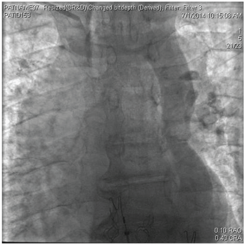
A persistent left sided vena cava was discovered intraoperatively during an unsuccessful attempt at a pacemaker implantation at an outside facility and he was referred to University of Arkansas for Medical Sciences UAMS. Aright sided approach was undertaken. During the procedure there were difficulty advancingthe leads which took an abnormal course to the left heart border. An intraoperative venogram showed an absent right sided superior vena cava and the right brachiocephalicvein draining into the persistent left superior vena cava (Figure 1). A left sided approach was considered but was not tried to minimize the risk for infection due to the recent procedureon the left side. An extended hook coronary sinus guiding catheter was used and with the help of a glide wire, entrance was made into the right ventricular apex. A Medtronic pace sense bipolar lead was then inserted through the guide catheter into the right ventricularapex under fluoroscopy. The following number were obtained RV sensing - 7mV, impedance- 675ohms and pacing threshold -1.0 volt at 0.5msec. A long Medtronic right atrial lead was inserted into the right atrial appendage under fluoroscopy without difficulty. P wave sensing was 3mV, impedance was 565 ohms and patient threshold was 1.0 volt at 0.5msec.
.
Figure 1: Venogram showing a catheter in the right subclavian vein and a persistent left sided superior vena cava
View Figure 1
.
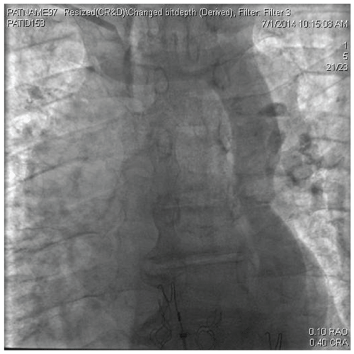
Figure 2: Posteroanterior chest radiograph demonstrating the final lead position, note the abnormal course taken by the leads to reach the right side of the heart
View Figure 2
Discussion
Persistent left sided superior vena cava is a rare congenital anomaly, and is a part of the spectrum of anomalies of the development of the great veins [1-3]. Its prevalence is estimated to be 0.3% in the general population and 4.4% in patients with congenital heart disease [4]. Persistent left SVC with an absent right SVC is even more uncommon and occurs in 0.07-0.13% of patients with congenital heart disease and viscera atrial situs solitus [5]. The condition is usually benign unless it is accompanied by other congenital developmental abnormalities. The situation usually poses a problem during procedures suchas pacemaker insertion [6-8], insertion of jugular venous lines or cardiothoracic surgery such as heart transplant. Other anomalies [9,10] and associated congenital heart defects [11] are mentioned in Tables 1 and 2 respectively.
![]()
Table 1: Anomalies of the vena cava
View Table 1
![]()
Table 2: Other congenital heart defects
View Table 2
Clues to the possibility of an abnormal venous drainage include a posteroanterior chest X-ray showing a widening of the aortic shadow, a paramediastinal bulging, para mediastinal stripe or a low density line along the upper left margin of the heart [11]. The presence of a large coronary sinus on echo, relative ease with which a coronary sinus catheter can be passed into the OS of the coronary sinus during an EP study and an abnormal course of the catheter are some other clues. When a left subclavian approach is used the catheter can be seen coursing parallel to the left side of the spine before it enters the coronary sinus [12]. A diagnosis can usually be established with imaging techniques such as a contrast echocardiogram, CT venogram and MRI. The diagnosis can be confirmed with a bubble study, when agitated saline is injected into a left arm vein the bubbles can be seen to enter the coronary sinus before opacifying the right atrium [11].
A persistent left sided superior vena cava offers unique challenges to the implantation of a permanent pacemaker from a left sided approach; it becomes even more difficult when the procedure is approached from the right side in view of the many acute angle bends encountered on the way [9]. Inserting the atrial lead into the right atrium is usually not difficult. Once in the right atrium the straight stylet can be exchanged for a curved J tip stylet and a clockwise torque can be applied to deliver the lead into the anteriorly positioned right atrialappendage. The OS of the coronary sinus is directed in such a manner that the leadentering the right atrium is directed at the lateral free wall rather than the inlet of the rightventricle making the insertion of the lead into the right ventricle challenging. Theventricular lead can be advanced to the free wall of the right atrium and reflected off of itand looped in the right atrium to enter the inlet of the right ventricle. Other methodsinclude adding a curve to the RV stylet and using a long curved venous access sheath suchas the one used for cannulating the OS of the coronary sinus. We used an extended hook coronary sinus guiding catheter andentered the right ventricular apex with ease with a glide wire.
Conclusion
Persistent Left sided superior vena cava is a rare congenital anomaly and can be encountered unexpectedly during minimally invasive procedures. When the leads take an unexpected course during a procedure the operator is often concerned about a procedural complication such as perforation. However knowledge of this condition can minimize anxiety and enable the surgeon to use novel approaches to successfully perform the procedure and avoid surgery.
References
-
Tak T, Crouch E, Drake GB (2002) Persistent left superior vena cava: incidence, significance and clinical correlates. Int J Cardiol 82: 91-93.
-
Schreve-Steensma AM, van der Valk PH, Ten Kate JB, Kofflard MJ (2008) Discovery of a persistent left superior vena cava during pacemaker implantation. Neth Heart J 16: 272-274.
-
Kilickap M, Altin T, Akyurek O, Karaoguz R, Akgun G, et al. (2005) DDD pacemaker implantation in a patient with persistent left superior vena cava and absent right superior vena cava: a four-year follow-up report. Can J Cardiol 21: 1221-1223.
-
Biffi M, Boriani G, Frabetti L, Bronzetti G, Branzi A (2001) Left superior vena cava persistence in patients undergoing pacemaker or cardioverter-defibrillator implantation: a 10-year experience. Chest 120: 139-144.
-
Uaar O, Paayaoaylu L, Ciaekaioaylu H, Vural M, Kocaoaylu I, et al. (2010) Persistent left superior vena cava with absent right superior vena cava: a case report and review of the literature. Cardiovasc J Afr 21: 164-166.
-
Lappegard KT, Prytz JF, Haug B (2004) Pacemaker implantation in patients with persistent left superior vena cava. Heart Vessels 19: 153-154.
-
Cardozo Zepeda CM, Guyomar Y, Heuls S, Graux P (2005) Implantation of a pacemaker through persistent left superior vena cava. Arch Cardiol Mex 75: 316-319.
-
Corbisiero R, DeVita M, Dennis C (2003) Pacemaker implantation in a patient with persistent left superior vena cava and absent right superior vena cava. J Interv Card Electrophysiol 9: 35-37.
-
Rizkallah J, Burgess J, Kuriachan V (2014) Absent right and persistent left superior vena cava: troubleshooting during a challenging pacemaker implant: a case report. BMC Res Notes 7: 462.
-
Bhatti S, Hakeem A, Ahmad U, Malik M, Kosolcharoen P, et al. (2007) Persistent left superior vena cava (PLSVC) with anomalous left hepatic vein drainage into the right atrium: role of imaging and clinical relevance. Vasc Med 12: 319-324.
-
Innasimuthu AL, Rao GK, Wsong P (2007) Persistent left-sided superior vena cava--a pacing challenge. Acute Card Care 9: 252.
