

International Journal of Pathology and Clinical Research
Hypercholesterolemic Diet: Its Effect on Colorectal Precancerous Lesions Induced by Dimethylhydrazine
Franciane da Silva França, Rodrigo Zeni Corá, Helena Cristina Ferreira Franz and Flávia Martinello*
Department of Clinical Analysis, Health Sciences Center, Federal University of Santa Catarina, Brazil
*Corresponding author:
Flávia Martinello, Departamento de Análises Clinicas, Centro de Ciências da Saúde, Campus Reitor João David Ferreira Lima, Universidade Federal de Santa Catarina, sala 109, Trindade, Florianópolis, Santa Catarina, 88040-970, Brazil, Tel: +55 4837213477, E-mail: flavia.martinello@ufsc.br
Int J Pathol Clin Res, IJPCR-2-040, (Volume 2, Issue 2), Short Communication; ISSN: 2469-5807
Received: May 18, 2016 | Accepted: June 25, 2016 | Published: June 30, 2016
Citation: França FDS, Corá RZ, Franz HCF, Martinello F (2016) Hypercholesterolemic Diet: Its Effect on Colorectal Precancerous Lesions Induced by Dimethylhydrazine. Int J Pathol Clin Res 2:040. 10.23937/2469-5807/1510040
Copyright: © 2016 França FDS, et al. This is an open-access article distributed under the terms of the Creative Commons Attribution License, which permits unrestricted use, distribution, and reproduction in any medium, provided the original author and source are credited.
Abstract
Objective: Evaluate the effects of dietary cholesterol, without the addition of cholic acid in the initial stage of carcinogenesis and on the bifidobacteria into the colon.
Methods: Rats were grouped according to their diets and dimethylhydrazine treatment (DMH): standard diet (CN); CN + cholesterol 1% (CHOL); CN + DMH (CNDMH) and CN + cholesterol 1% + DMH (CHOLDMH). After eight weeks of treatment were analyzed weight gain, faecal pH, lipid profile, hepatic and faecal cholesterol, faecal bifidobacteria, cell proliferation and the pre-neoplastic colorectal lesions (aberrant crypt foci (ACF)).
Results: Groups not treated with DMH didn't develop aberrant crypt. There weren't significant differences between DMH-groups in the number of ACF and in the multiplicity of ACF/foci. However, cell proliferation evaluated by PCNA-labeling index was significantly higher in the CHOLDMH group when compared to the CNDMH. The highest amount of cholesterol present in the feces of the animals treated with supplemented diet didn't influence the ACF and number of crypts per focus, but increased cell proliferation.
Conclusion: Diet supplemented with cholesterol or DMH did not influence the amount of bifidobacteria found, suggesting that the mechanism of action of these factors in the initiation of carcinogenesis does not involve change in intestinal microbiota. An increase in the fecal pH was observed in CHOLDMH group, and this association may be a factor that can influence the development of the colorectal precancerous lesions.
Keywords
Cholesterol, Aberrant crypt foci, PCNA, Bifidobacteria
Introduction
Colorectal cancer (CRC) is one of the most common cancers worldwide. Although its cause is not fully known, genetic susceptibility and diet are major risk factors [1], and fat intake has been the main nutritional influence on its development [2]. However, the involvement of dietary cholesterol in the etiology of the CRC is controversial [3]. Whilst some studies have reported increased risk with high cholesterol intake [4-9], others studies indicate that it may even diminish the development of induced tumors, when administered in the initiation phase of carcinogenesis [10,11]. Cholesterol may influence colorectal carcinogenesis through several mechanisms such as oxidative stress, lipid, glucose and bile acids metabolism or alteration on membrane properties and cell signaling [12-16]. The intestinal microbiota is also recognized as an important player in illness such as CRC. Some studies have suggested that diet can modulate the formation of preneoplastic lesions in the colon, possibly by altering the micro ecology and microbial activities on carcinogenesis [17]. A number of investigations, mainly using in vitro and animal models, have demonstrated a wide range of possible anti-CRC mechanisms by which certain strains from the genera of lactobacilli and bifidobacteria can act. Effects including competitive exclusion of pathogenic intestinal flora, alteration of intestinal microflora enzyme activity, reduction of carcinogenic secondary bile acids, binding of carcinogens and mutagens, increasing short chain fatty acids production, reduction of DNA damage and suppression of aberrant crypt foci formation and modulation of intestinal inflammation are some anti-CRC effects of probiotics on intestinal mucosa [18]. In this context, considering the influence of diet on colon health and the relation between fat intake and the risk of CRC development, this study aims to evaluate the effect of dietary cholesterol (without addition of cholic acid) on the initial phase carcinogenesis and on the amount of colonic bifidobacteria.
Materials and Methods
Rats, diet and dimethylhydrazine (DMH)
This study was approved by animal ethics committee of the Federal University of Santa Catarina (UFSC). Thirty-two lean adults (30 days old)male Wistar rats (Rattus norvergicus variety Albicanus, Rodentia) were randomly divided into four groups in eight colony cages (four rats per cage): standard diet (CN); standard diet + cholesterol 1% (CHOL); standard diet + DMH (CNDMH) and standard diet + cholesterol 1% + DMH (CHOLDMH). After one-week acclimatization period, DMH was administered intraperitoneally (150 mg/kg weigh body), and treatment was initiated with different diets ad libitum. To prepare the cholesterol diet, the regular chow (Nutrilabor, Brazil) was ground, cholesterol (Sigma-Aldrich, USA) in concentration of 1% (w/w) was added and the mixture was pelletized again. Weight gain analysis was performed by weighing the animals in the first and last day of the treatment. After eight weeks of treatment, stool samples were collected to determine the fecal pH, bifidobacteria, and cholesterol. Blood samples were collected to analyze the lipid profile. The animals were euthanized by exsanguination. Colons were removed for histological analysis. Livers were removed, weighed and the hepatic cholesterol was determined.
Amount of bifidobacteria and fecal pH
Fresh feces were collected from each animal separately. Each animal was placed in an empty cage and was awaited until it evacuate. Feces were weighed and diluted in sterile deionized water for pH determination. Each sample was also weighed and diluted in 9 ml of 0.31 mM phosphate buffer. From this dilution (10-1), serial dilutions were performed up to 10-7 and 100 μl of each dilution was spread plated on selective medium for bifidobacteria BIM-25 [19]. The plates were incubated at 37°C for 72 hours under anaerobic conditions (Anaerobac system, Probac, São Paulo, São Paulo, Brazil). At a later stage, the bacterial population (bifidobacteria) count was calculated as colony-forming units (CFU) per gram of fecal sample. Gram stain, catalase test and fructose-6-phosphate phosphoketolase (F6PPK) test according to Orban and Patterson [20] were performed to confirm Bifidobacterium spp. genus.
Biochemical parameters
Hepatic and fecal cholesterol measurements were performed according to Melo et al. [21]. Serum triglycerides, total cholesterol and (HDL)-cholesterol levels from each animal were measured using commercial enzymatic kits (Labtest, Montes Claros, Minas Gerais, Brazil).
Histological Analysis
Distal colon was cut, washed with 0.9% saline solution, opened longitudinally and fixed in buffered formalin 10% (pH 6.9-7.1) for 24 hours. Part of the distal colon was stained with methylene blue (0.02%) for five minutes immediately prior to analysis. For each part, 50 sequential fields were analyzed in microscope (100x magnifications) and the number of aberrant crypt foci (ACF) was counted and multiplied by foci (expressed as aberrant crypts per focus (AC/ACF). ACF were characterized by thick epithelial layer which stains more intense with methylene blue, luminal opening elongated, tortuous and increased in relation to the surrounding normal crypts [22,23].
Another part of the distal colon was dehydrated, diaphanized, embedded in paraffin to obtain 3 μm longitudinal histological sections. To estimate colonic cell proliferation in all animals, the longitudinal sections were immunostained for proliferative cellular nuclear antigen (PCNA) using an antibody against PCNA (Thermo Scientific, Pittsburgh, Pennsylvania, USA) [24-26]. The labeling index (LI) was determined as the ratio of the number of stained nuclei and total number of nuclei along each crypt. Ten crypts were counted per section/animal (x400). Rat colon (Wistar, male) specimens were used as a positive control and as negative control primary antibody suppression.
Statistical Analysis
The data was analyzed using GraphPad Prism V.4 (GraphPad Software, Inc., San Diego, CA). Verification of symmetric distribution of data was performed using Levene's normality (Shapiro-Wilk test). The statistical significance of differences between data means was determined by using analysis of variance one way (ANOVA), followed by post hoc testing by Tukey’s test. A value of P < 0.05 was considered as statistically significant. Results are given as means ± standard error, n = 8.
Results
Body weight gain
Initial and final weight, weight gain, serum triglycerides, total cholesterol, HDL-cholesterol, hepatic cholesterol, fecal cholesterol, bifidobacteria and pH, number of ACF/field, AC/ACF and PCNA labeling index are shown in table 1.
![]()
Table 1: Results of serum, fecal, liver and colonic samples analysis from different experimental groups.
View Table 1
There were no significant differences between groups in initial weight, gain weight or final weight. Likewise, cholesterol diet and DMH alone did not influence the amount of bifidobacteria and fecal pH.
The serum levels of triglycerides in the CNDMH and CHOLDMH groups were significantly lower than the respective groups treated with the same diet without DMH. However, the addition of 1% cholesterol in the diet was not sufficient to increase total cholesterol levels. Moreover, fecal cholesterol level in CHOL group was significantly higher than the CN group. The same finding was observed when compared CHOLDMH and CNDMH groups.
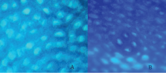
Among the precancerous lesions markers, no significant differences in ACF/field and AC/ACF were observed between groups (Table 1 and Figure 1). Otherwise, PCNA labeling index was significantly higher in CHOLDMH than the CNDMH group (Table 1).
.
Figure 1: Photomicrograph of colon stained with methylene blue.
A) Microscopic field without ACF; B) Microscopic field with ACF (aberrant crypt foci) with 4 AC (aberrant crypt). 100 × magnification.
View Figure 1
Discussion
Interactions between fatty acids and intestinal microbiota have shown to be related to colon cancer. Some fatty acids, but not all, are potent antimicrobial agents and some bacteria exhibit different sensitivities to distinct fatty acids [27]. These data suggest that fatty acids may influence the biodiversity of the intestinal microbiota and the CRC. In this regard, it is important to evaluate the effect of cholesterol, which is also a common dietary lipid, on colorectal carcinogenesis and bifidobacteria.
After eight weeks of treatment with different diets, triglycerides level was similar between groups not treated with DMH, but was lower in DMH treated groups. Although the mechanism by which DMH reduce triglycerides level is not known, this fact was reported by Nauss et al. [28] to evaluate the effect of the amount of dietary lipids on DMH-induced colonic tumorigenesis in rats for four weeks. On the other hand, Barton et al. [29] observed increased triglycerides levels in late stages of colon carcinogenesis induced by DMH.
Experimentally, diet plus 1% cholesterol was not enough to significantly increase the serum cholesterol and liver cholesterol levels. This can be explained by the higher conversion to bile acids in the liver and higher fecal cholesterol excretion [21,30]. Endogenous cholesterol is excreted into bile as free cholesterol or converted by the liver into bile salts [31]. The highest fecal cholesterol level in groups treated with cholesterol diet may have been due to increased excretion or probably due to poor absorption of dietary cholesterol, not causing, thereby, hypercholesterolemia.
Furthermore, in vivo studies using rodents, hypercholesterolemia is usually achieved with addition of cholic acid in the diet enriched with cholesterol [32,33].
Evidences have shown that the greater contact of harmful agents such as bile salts, with the colonic mucosa may determine important epithelial alterations, and may lead to the development of colorectal cancer [34,35]. Contrary to other studies, our study was conducted without addition of cholic acid in the diet to assess the effect of cholesterol alone on the development of ACF and bifidobacteria content.
The experimental model of quantifying ACF is considered an excellent parameter to evaluate the protective or risk factor for colorectal cancer [36]. In this context, it was observed that the group not treated with DMH did not develop aberrant crypt. This indicates that the cholesterol alone, in this concentration on diet, did not exert initiator action of colon carcinogenesis. The presence of higher content of cholesterol in the feces of the animals treated with cholesterol did not influence the amount of ACF and the number of crypts per focus, perhaps due to the short treatment period (60 days). However, false-positive or false-negative results can occur due to various factors of the natural process of colonic carcinogenesis [37]. On the other hand, the highest amount of fecal cholesterol increased the cell proliferation, which is an important prerequisite for colorectal carcinogenesis [37,38]. This result corroborates with studies that reported increased risk of CRC in diets with high cholesterol content [4-7] and it might be due to the high production of bile acids in the intestine.
Therefore, although no significant difference in the parameters ACF/field and AC/ACF, the largest cell proliferation (PCNA) observed in groups treated with cholesterol, and possibly caused by increased of fecal cholesterol, will not allow to discard the hypothesis that a high-cholesterol diet can influence the colorectal cancer etiology if analyzed in a later stage of this cancer. This hypothesis corroborates with Tseng et al. [32] that suggested that cholesterol is a non-carcinogenic agent that can potentiate the carcinogenicity of DMH through increased lipid peroxidation and decreased peroxidase activity of the colon. These researchers when examine the influence of hypercholesterolemic diet (0 to 2%) and cholic acid (0.25%) for 18 weeks observed that rats treated with DMH, cholic acid and higher cholesterol concentration (2%) in the diet, develop 50% more colon adenomas than rats just treated with DMH.
The results showed that dietary cholesterol or DMH does not influence the amount of bifidobacteria. It can suggest that the mechanism of action of these factors in the initiation of carcinogenesis does not involve change in intestinal microbiota as proposed by others researchers who observed a reduction on bifidobacteria in experimental colorectal carcinogenesis [39,40]. Although no effect on the amount of bifidobacteria, DMH showed a tendency to cause an increase in fecal pH, which was significant in CHOLDMH and this association may be a factor which can influence the CRC development, in agreement with other studies [41].
References
-
Tanaka T (2009) Colorectal carcinogenesis: Review of human and experimental animal studies. J Carcinog 8: 5.
-
Rao CV, Hirose Y, Indranie C, Reddy BS (2001) Modulation of experimental colon tumorigenesis by types and amounts of dietary fatty acids. Cancer Res 61: 1927-1933.
-
Ross JA, Kasum CM (2002) Dietary flavonoids: bioavailability, metabolic effects, and safety. Annu Rev Nutr 22: 19-34.
-
Kendall CW, Koo M, Sokoloff E, Rao AV (1992) Effect of dietary oxidized cholesterol on azoxymethane-induced colonic preneoplasia in mice. Cancer Lett 66: 241-248.
-
Kendall CW, Janezic SA, Friday D, Rao AV (1992) Dietary cholesterol enhances preneoplastic aberrant crypt formation and alters cell proliferation in the murine colon treated with azoxymethane. Nutr Cancer 17: 107-114.
-
De Stefani E, Mendilaharsu M, Deneo-Pellegrini H, Ronco A (1997) Influence of dietary levels of fat, cholesterol, and calcium on colorectal cancer. Nutr Cancer 29: 83-89.
-
Järvinen R, Knekt P, Hakulinen T, Rissanen H, Heliövaara M (2001) Dietary fat, cholesterol and colorectal cancer in a prospective study. Br J Cancer 85: 357-361.
-
Llaverias G, Danilo C, Mercier I, Daumer K, Capozza F, et al. (2011) Role of cholesterol in the development and progression of breast cancer. Am J Pathol 178: 402-412.
-
Hu J, La Vecchia C, de Groh M, Negri E, Morrison H, et al. (2012) Dietary cholesterol intake and cancer. Ann Oncol 23: 491-500.
-
Cohen BI, Raicht RF, Fazzini E (1982) Reduction of N-methyl-N-nitrosourea-induced colon tumors in the rat by cholesterol. Cancer Res 42: 5050-5052.
-
el-Sohemy A, Kendall CW, Rao AV, Archer MC, Bruce WR (1996) Dietary cholesterol inhibits the development of aberrant crypt foci in the colon. Nutr Cancer 25: 111-117.
-
Du Q, Wang Q, Fan H, Wang J, Liu X, et al. (2016) Dietary cholesterol promotes AOM-induced colorectal cancer through activating the NLRP3 inflammasome. Biochem Pharmacol 105: 42-54.
-
Nkondjock A, Shatenstein B, Maisonneuve P, Ghadirian P (2003) Specific fatty acids and human colorectal cancer: an overview. Cancer Detect Prev 27: 55-66.
-
Roynette CE, Calder PC, Dupertuis YM, Pichard C (2004) n-3 polyunsaturated fatty acids and colon cancer prevention. Clin Nutr 23: 139-151.
-
Kimura Y (2006) Fish, n-3 polyunsaturated fatty acid and colorectal cancer prevention: a review of experimental and epidemiological studies. Nihon Koshu Eisei Zasshi 53: 735-748.
-
Chapkin RS, Davidson LA, Ly L, Weeks BR, Lupton JR, et al. (2007) Immunomodulatory effects of (n-3) fatty acids: putative link to inflammation and colon cancer. J Nutr 137: 200S-204S.
-
Pattananandecha T, Sirilun S, Duangjitcharoen Y, Sivamaruthi BS, Suwannalert P, et al. (2016) Hydrolysed inulin alleviates the azoxymethane-induced preneoplastic aberrant crypt foci by altering selected intestinal microbiota in Sprague-Dawley rats. Pharm Biol 21: 1-10.
-
Chong ES (2014) A potential role of probiotics in colorectal cancer prevention: review of possible mechanisms of action. World J Microbiol Biotechnol 30: 351-374.
-
Muñoa FJ, Pares R (1988) Selective medium for isolation and enumeration of Bifidobacterium spp. Appl Environ Microbiol 54: 1715-1718.
-
Orban JI, Patterson JA (2000) Modification of the phosphoketolase assay for rapid identification of bifidobacterias. J Microbiol Methods 40: 221-224.
-
Melo SS, Silveira BM, Stefanes FB, Tomio TA, Tischer CA (2008) Efeito da goma arabica nas concentraçoes de colesterol hepatico, gastrico e fecal de ratos alimentados com semente de linhaça, oleo de linhaça e colesterol sintetico. Alim Nutr 19: 133-144.
-
Bird RP (1987) Observation and quantification of aberrant crypts in the murine colon treated with a colon carcinogen: preliminary findings. Cancer Lett 37: 147-151.
-
Gupta AK, Pretlow TP, Schoen RE (2007) Aberrant crypt foci: what we know and what we need to know. Clin Gastroenterol Hepatol 5: 526-533.
-
Yamada Y, Yoshimi N, Hirose Y, Kawabata K, Matsunaga K, et al. (2000) Frequent B-catenin gene mutations and accumulations of the protein in the putative preneoplastic lesions lacking macroscopic aberrant crypt foci appearance, in rat colon carcinogenesis. Cancer Res 60: 3323-3327.
-
Hsu SM, Raine L, Fanger H (1981) Use of avidin-biotin-peroxidase complex (ABC) in imunoperoxidase techniques: comparison between ABC and unlabeled antibody (PAP) procedures. J Histochem Cytochem 29: 577-589.
-
Garcia SB, Barros LTC, Turatti A, Martinello F, Modiano P, et al. (2005) The anti-obesity agent Orlistat is associated to increase in colonic preneoplastic markers in rats treated with a chemical carcinogen. Cancer Lett 240: 221-224.
-
Juste C (2005) Dietary fatty acids, intestinal microbiota and cancer. Bull Cancer 92: 708-721.
-
Nauss KM, Locniskar M, Newberne PM (1983) Effect of alterations in the quality and quantity of dietary fat on, 2-dimethylhydrazine-induced colon tumorigenesis in rats. Cancer Res 43: 4083-4090.
-
Barton TP, Cruse JP, Lewin MR (1987) Changes in serum lipids related to the presence of experimental colon cancer. Br J Cancer 56: 451-454.
-
Machado DF, Ferreira CLLF, Costa NMB (2003) Evaluation of the probiotic effect in the modulation of the levels of seric cholesterol and in the weight of the liver of mices fed with rich diet in cholesterol and colic acid. Cienc Tecnol Aliment 23: 270-275.
-
Choi MS, Do KM, Park YS, Jeon SM, Jeong TS, et al. (2001) Effect of naringin supplementation on cholesterol metabolism and antioxidant status in rats fed high cholesterol with different levels of vitamin E. Ann Nutr Metab 45: 193-201.
-
Tseng TH, Hsu JD, Chu CY, Wang CJ (1996) Promotion of colon carcinogenesis through increasing lipid peroxidation induced in rats by a high cholesterol diet. Cancer Lett 100: 81-87.
-
Ramadan MF, Hassan NA, Elsanhoty RM, Sitohy MZ (2013) Goldenberry (Physalisperuviana) Juice Rich in Health-Beneficial Compounds Suppresses High-Cholesterol Diet-Induced Hypercholesterolemia in Rats. J Food Biochem 37: 708-722.
-
Glade MJ (1999) Food, nutrition, and the prevention of cancer: a global perspective. American Institute for Cancer Research/World Cancer Research Fund, American Institute for Cancer Research, 1997. Nutrition 15: 523-526.
-
Powolny A, Xu J, Loo G (2001) Deoxycholate induces DNA damage and apoptosis in human colon epithelial cells expressing either mutant or wild-type p53. Int J Biochem Cell Biol 33: 193-203.
-
Deeptha K, Kamaleeswati M, Senfottuvelan M, Nalini N (2006) Dose dependent inhibitory effect of dietary caraway on 1,2-dimethylhydrazine induced colonic aberrant crypt foci and bacterial enzyme activity in rats. Invest New Drugs 24: 479-488.
-
Dias MC, Spinardi-Barbisan AL, Rodrigues MA, de Camargo JL, Terán E, et al. (2006) Lack of chemopreventive effects of ginger on colon carcinogenesis induced by, 2-dimethylhydrazine in rats. Food Chem Toxicol 44: 877-884.
-
Kulmar VK, Vennila S, Nalini N (2010) Inhibitory effect of morin on DMH-induced biochemical changes and aberrant crypt foci formation in experimental colon carcinogenesis. Environ toxicol pharmacol 29: 50-57.
-
Cani PD, Neyrinck AM, Fava F, Knauf C, Burcelin RG, et al. (2007) Selective increases of bifidobacteria in gut microflora improve high-fat-diet-induced diabetes in mice through a mechanism associated with endotoxaemia. Diabetologia 50: 2374-2383.
-
Hekmatdoost A, Feizabadi MM, Djazayery A, Mirshafiey A, Eshraghian MR, et al. (2008) The effect of dietary oils on cecal microflora in experimental colitis in mice. Indian J Gastroenterol 27: 186-189.
-
Samelson SL, Nelson RL, Nyhus LM (1985) Protective role of faecal pH in experimental colon carcinogenesis. J R Soc Med 78: 230-233.
